






Plant Extracellular Vesicle Proteomics: From Vesicle Isolation to LC-MS/MS Analysis
Plant Extracellular Vesicle Proteomics: From Vesicle Isolation to LC-MS/MS Analysis
Direct Answer
Plant extracellular vesicle proteomics follows a two-stage path: first isolate and characterize the vesicle preparation, then analyze its proteins by LC-MS/MS. The proteomics step identifies proteins present in the vesicle fraction and, when groups are compared, determines which proteins differ in abundance.
Isolation quality strongly affects the final protein profile because cell debris, soluble proteins, and co-isolating particles can enter the same preparation. Before LC-MS/MS, define the plant source and matrix, isolation route, comparison groups, and available vesicle characterization information.
For comparative studies, all experimental groups should follow the same isolation and sample-processing workflow so that technical variation does not obscure biological differences.
What Plant Extracellular Vesicle Proteomics Can Show
Plant cells release nanoscale extracellular vesicles that can contain proteins, lipids, and nucleic acids. Terms such as extracellular vesicles, nanovesicles, and exosome-like vesicles are sometimes used in the literature, but they do not automatically indicate the same biogenesis pathway.
For proteomics, the practical question is which proteins are identified in the vesicle preparation that was isolated.
Plant extracellular vesicle proteomics can be used to:
-
Identify proteins recovered from a plant vesicle preparation
-
Compare vesicle-associated protein abundance across genotypes, tissues, treatments, or growth conditions
-
Prioritize candidate vesicle-associated proteins for further validation
Proteomics alone does not prove that a detected protein is a definitive EV marker, that the preparation is completely free of contaminants, or that a vesicle-associated protein is functionally delivered to another cell. These conclusions require additional evidence.
Figure 1. Plant vesicle isolation and characterization precede protein preparation and LC-MS/MS analysis.
Vesicle Isolation Directly Affects the Proteomics Result
Plant vesicle preparations can originate from extracellular sources such as apoplastic fluid, root exudates, or culture medium. Preparations obtained after tissue disruption or juicing require more cautious interpretation because intracellular membrane-derived particles may also be released during tissue processing.
Matrix properties such as viscosity, debris content, and soluble protein background can influence vesicle recovery and downstream protein identification.
Common approaches for plant-derived vesicle enrichment include differential centrifugation, density gradient centrifugation, ultrafiltration, polymer precipitation, immunoaffinity capture, and size-exclusion chromatography. The appropriate approach depends on the sample matrix and study objective.
For comparative proteomics, the same isolation method, buffers, handling conditions, and processing sequence should be maintained across groups. Changing the isolation procedure between groups can introduce technical differences that may be mistaken for biological changes.
Particle size measurements and morphology imaging can help describe a vesicle preparation before proteomics. However, these measurements alone do not establish vesicle purity, biological origin, or a specific EV subtype.
Storage conditions and freeze-thaw history should also be recorded because sample degradation or repeated freeze-thaw cycles can affect downstream protein analysis.
Sample Planning Before LC-MS/MS
Plant EV proteomics should be planned at two levels: the plant material used to obtain the vesicle preparation and the vesicle-associated protein available for LC-MS/MS.
The amount of starting material depends on the plant source, sample matrix, isolation strategy, and expected vesicle recovery. It should therefore be evaluated for the specific project rather than estimated directly from general plant proteomics sample requirements.
| Planning Item | What to Define Before LC-MS/MS |
|---|---|
| Plant source and matrix | Apoplastic fluid, root exudate, culture medium, or another clearly defined plant-derived vesicle preparation |
| Isolation route | The same method and buffer system across comparison groups |
| Preparation characterization | Available particle size, morphology, and other relevant characterization information |
| Comparison groups | Treatment, genotype, tissue, condition, or time-point contrasts defined before isolation |
| Biological unit | Independent plants, cultures, fruits, or experimental batches rather than repeated aliquots from one isolate |
The amount of vesicle-associated protein recovered for LC-MS/MS varies substantially between matrices and isolation workflows. Starting tissue weight alone should not be used to predict a universal protein input.
For low-yield or unfamiliar plant matrices, expected input requirements should be evaluated before large-scale sample collection.
Figure 2. Define the source matrix, isolation route, preparation characterization, and biological comparison before LC-MS/MS.
From Vesicle Protein Preparation to LC-MS/MS
Once comparable vesicle preparations are available, the proteomics workflow generally includes protein recovery or cleanup, enzymatic digestion, peptide separation, LC-MS/MS analysis, and database-based protein identification.
Identification-focused analysis is useful when the objective is to characterize the proteins present in a vesicle preparation. Quantitative proteomics is more appropriate when the study aims to compare vesicle-associated protein abundance between predefined groups.
Both DDA and DIA can support plant EV protein identification and quantitative analysis. The acquisition strategy and mass spectrometry platform should be selected according to the study objective, sample number, quantitative requirements, sample characteristics, and overall project design.
The experimental question should determine the analysis strategy rather than selecting a method solely because it is commonly used in proteomics.
How to Interpret Plant EV Proteomics Results
A typical plant EV proteomics dataset may contain identified proteins, quantitative comparisons, and downstream functional annotations.
Depending on the study design and available species annotation, downstream analysis may include functional classification, pathway enrichment, and protein interaction context. These analyses help organize the protein list and prioritize candidates for follow-up studies.
Interpretation should remain consistent with the evidence level of the experiment.
A protein identified in a vesicle preparation can be described as present or vesicle-associated in that preparation. Detection alone does not prove that the protein:
-
Is exclusively localized to extracellular vesicles
-
Represents a universal plant EV marker
-
Originates from a specific EV biogenesis pathway
-
Is actively delivered into recipient cells
-
Directly causes a biological phenotype
Similarly, a differential protein indicates an abundance difference between the compared vesicle preparations, not automatically a regulatory mechanism.
Related Services
Supports LC-MS/MS-based protein identification and quantitative comparison for plant-derived samples and vesicle preparations.
Plant-derived Exosome Isolation and Development Service
Applicable when plant-derived vesicles need to be isolated and characterized before downstream proteomics.
Frequently Asked Questions
Are plant exosomes and plant extracellular vesicles the same?
Not exactly. Extracellular vesicle is the broader term. Exosome-like vesicle may be used when vesicles show characteristics associated with exosomes, but morphology and size alone do not prove a specific biogenesis pathway.
Can plant tissue be analyzed directly without vesicle isolation?
Not for an EV-specific proteomics study. Proteomics of crude plant tissue or tissue homogenate measures a much broader protein population. If the objective is to analyze vesicle-associated proteins, a vesicle isolation or enrichment step is required first.
Does detecting a protein prove that it is an EV marker?
No. Detection shows that the protein is present in the analyzed vesicle preparation. Marker status requires additional evidence demonstrating consistent association or enrichment and usually orthogonal validation.
How much starting plant material is required?
The required amount depends on the plant source, matrix, isolation method, and expected vesicle recovery. Starting-material requirements should therefore be evaluated according to the specific project, especially for low-yield matrices.
What information should be defined before starting plant EV proteomics?
Important information includes the plant source and matrix, vesicle isolation status or planned isolation route, comparison groups, sample storage history, and the intended protein readout.
Conclusion
Plant extracellular vesicle proteomics is most informative when vesicle isolation and LC-MS/MS are planned as a single experimental path. A matrix-appropriate isolation strategy, consistent processing across groups, suitable preparation characterization, and a clearly defined comparison are essential for generating interpretable protein data.
Proteins identified or quantified in a vesicle preparation should initially be treated as vesicle-associated candidates. Claims involving EV markers, exclusive localization, biological delivery, or functional mechanisms require additional validation.
Researchers planning plant EV proteomics can review the plant source, vesicle isolation status, experimental groups, and expected protein readout with MtoZ Biolabs before sample collection or submission.
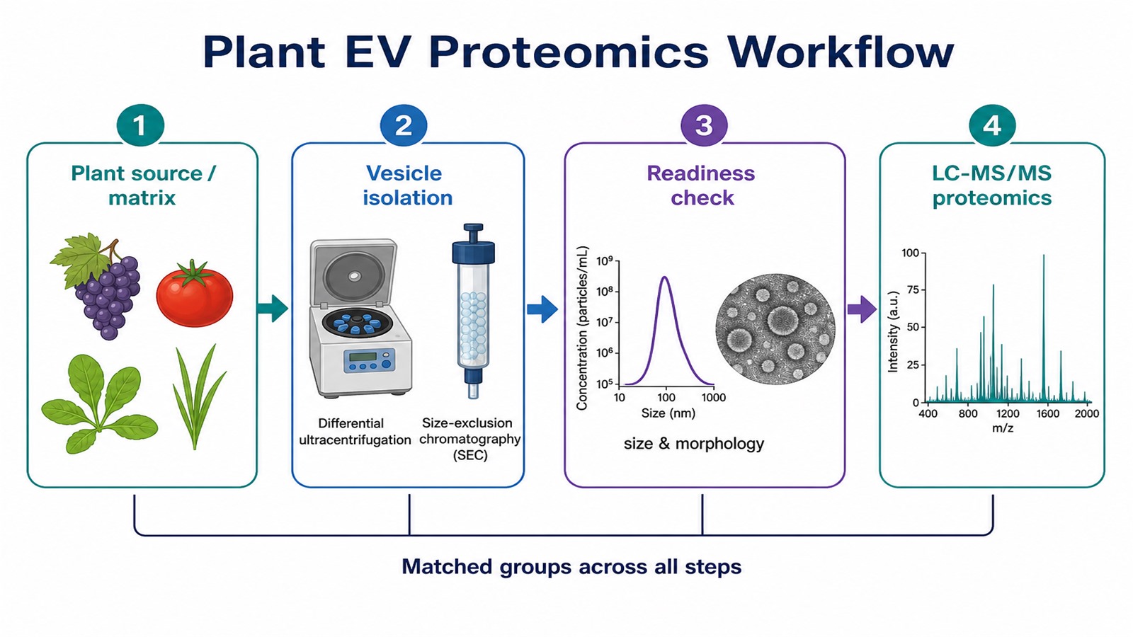
Figure 1. Isolate and check the vesicle preparation first, then move to digestion and LC-MS/MS for protein analysis.
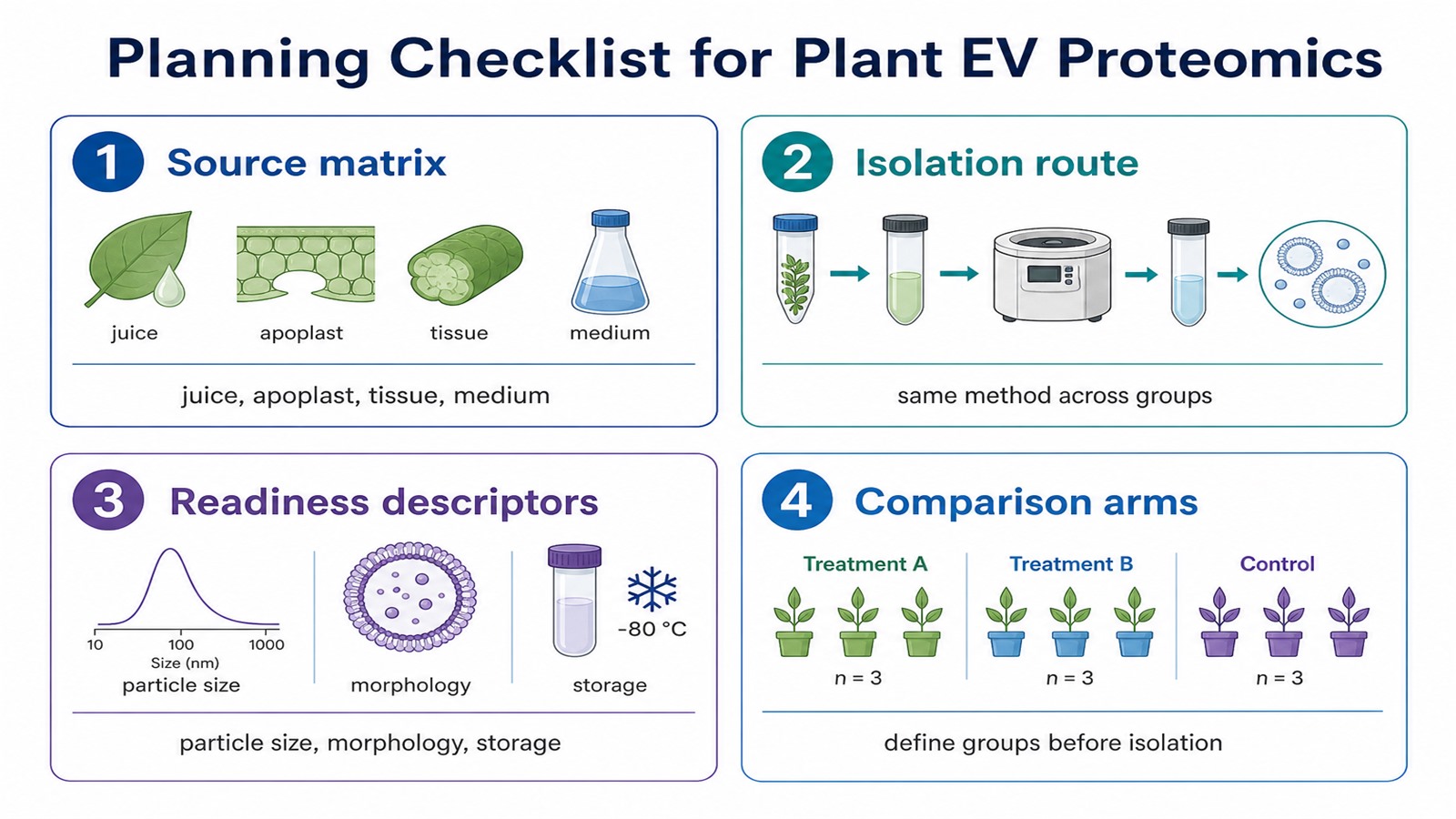
Figure 2. Lock source matrix, isolation route, readiness descriptors, and matched groups before LC-MS/MS.
How to order?




