


Phosphorylated Protein-Based Biomarker Discovery: From Identification to Validation
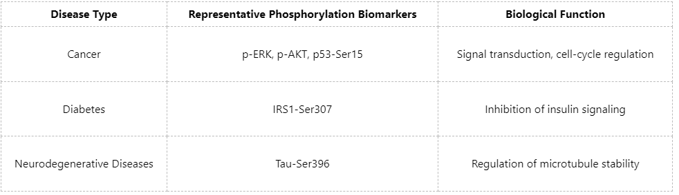
Within the highly orchestrated regulation of biological systems, protein phosphorylation is among the most prevalent post-translational modifications (PTMs) and serves a dual function as a regulatory switch and a signaling relay node. A growing body of evidence indicates that aberrant phosphorylation patterns are tightly associated with diverse diseases, including cancer, metabolic disorders, and neurodegenerative diseases, positioning phosphorylation as a high-value focal point for biomarker discovery. In particular, in the era of precision medicine, phosphorylated protein-based biomarkers can provide earlier, more specific, and more dynamic pathological information, and thus hold substantial promise for disease prediction, molecular stratification, treatment-response assessment, and individualized therapy.
Why Choose Phosphorylated Proteins as Biomarkers?
1. The Biological Distinctiveness of Phosphorylation
(1) Rapid responsiveness: as an immediate-response mechanism, phosphorylation can capture early pathological events more sensitively than changes in expression abundance.
(2) Strong signaling relevance: extensive studies support that dysregulated kinase activity in disease states drives characteristic alterations in specific phosphorylation sites.
(3) Site specificity: phosphorylation at distinct sites on the same protein can be linked to different functional outcomes, enabling more precise, mechanism-informed biomarker interpretation.
2. Practical Applications in Clinical Research
Phosphorylated protein-based biomarkers not only facilitate discovery of disease-associated signaling changes but may also serve as drug targets to support efficacy prediction and stratified therapeutic strategies.
Biomarker Discovery Workflow: From Proteomics Data to Candidate Molecules
1. Sample Collection and Study Design
Discovery efforts typically involve comparisons between disease and control groups. Fresh tissues, frozen cells, or biofluids (e.g., serum and cerebrospinal fluid) are recommended, together with appropriate group design (e.g., time-course sampling and drug-treatment arms).
2. Enrichment and Mass Spectrometry Analysis
(1) Enrichment: IMAC or TiO₂ is commonly used to selectively enrich low-abundance phosphopeptides.
(2) Mass spectrometry acquisition: DDA is applied for comprehensive spectral library construction, whereas DIA is well suited for high-throughput quantitative analysis.
3. Criteria for Candidate Biomarker Prioritization
Typical prioritization criteria include:
(1) Differentially phosphorylated sites (fold change & p-value)
(2) Significance of pathway enrichment (GO/KEGG)
(3) Kinase prediction (kinase-substrate analysis)
(4) Literature support or existing database records (e.g., PhosphoSitePlus)
A multidimensional screening framework enables refinement toward high-confidence biomarker candidates.
In-Depth Bioinformatic Analysis: Enabling Data-Driven Inference
1. Construction of Kinase-Substrate Networks
Tools such as Motif-X and NetPhorest can be used to infer upstream kinases regulating specific phosphorylation events and to identify potential signaling nodes.
For example, if p-AKT is elevated in a tumor sample, network-based analysis may reveal significant activation of its upstream kinase Akt1, suggesting potential involvement of the PI3K/AKT pathway in pathogenesis.
2. Machine Learning-Assisted Biomarker Panels
By integrating algorithms such as LASSO and random forests, multi-marker panel models can be constructed to:
(1) distinguish disease subtypes
(2) predict therapeutic response
(3) estimate risk of disease progression
Validation: From Computational Inference to Experimental Support
1. Technical Validation
Following candidate prioritization, validation should be performed in larger cohorts:
(1) Western blot: validate site-specific phosphorylation levels (requiring phosphorylation-specific antibodies).
(2) ELISA: enable high-throughput validation in biofluid samples.
(3) Targeted MS (PRM/SRM): directly quantify target phosphopeptides with high sensitivity and specificity.
2. Biological Validation
(1) Perturb kinase activity or mutate phosphorylation sites in cell lines or animal models;
(2) Assess signaling and/or phenotypic changes to strengthen mechanistic interpretability.
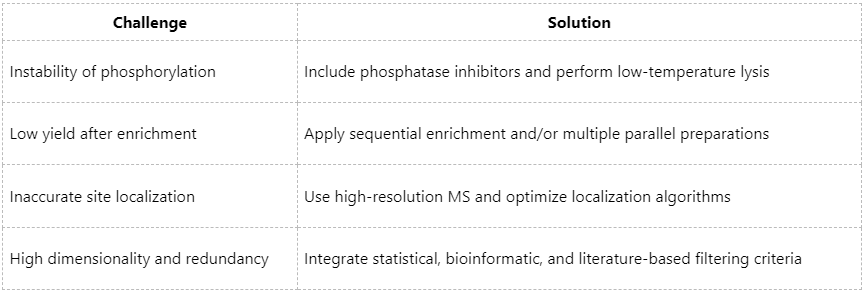
Common Challenges and Practical Solutions
Phosphorylated protein-based biomarker discovery is not only a scientific challenge but also a powerful approach to addressing clinically relevant questions. From mechanistic exploration to precise target localization and functional validation, each step demands rigorous methodology and deep biological insight. If you are seeking high-quality and clinically translatable phosphorylation biomarkers, please contact MtoZ Biolabs. With a research-driven perspective and clinically informed thinking, we are committed to helping you uncover the biological significance embedded within signaling networks.
MtoZ Biolabs, an integrated chromatography and mass spectrometry (MS) services provider.
Related Services
How to order?

