


PhIP-Seq vs Traditional Serology and Protein Arrays: Choosing the Right Antibody Profiling Method
Use PhIP-Seq when the study calls for broad peptide-level discovery across a large candidate space, especially for linear epitope mapping, cohort-scale comparative screening, or early hypothesis generation. Use traditional serology such as ELISA when the antigen list is already set and the project needs focused, easy-to-interpret readouts for a small number of targets. Use a protein array when the team wants broader multiplex antibody profiling than ELISA while still testing larger protein antigens rather than short peptides alone. Choose a staged workflow when broad discovery and downstream antigen-level confirmation are both part of the plan.
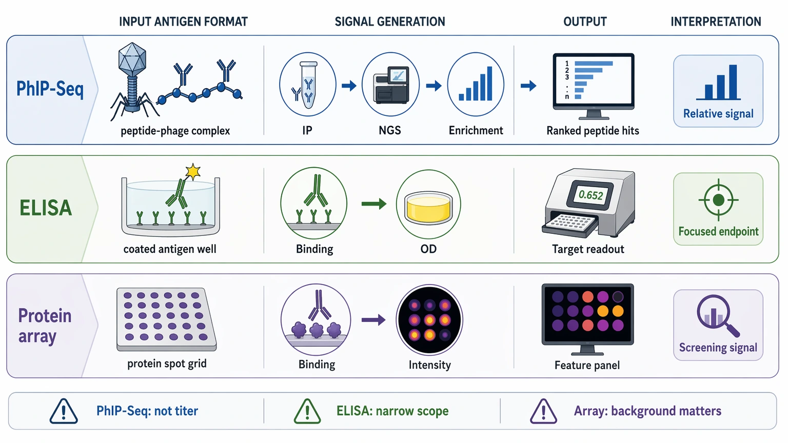
These platforms are not interchangeable. They differ in antigen representation, epitope bias, readout logic, and the amount of follow-up work they create. Phage immunoprecipitation sequencing measures sequencing enrichment from a phage-displayed peptide library after immunoprecipitation. ELISA measures binding to predefined coated antigens. A protein array screens many immobilized proteins or fragments in parallel. The right choice starts with the biological question, then narrows through sample volume, cohort size, reporting needs, and the level of orthogonal validation the team can realistically support.
Where this platform choice matters most
This decision usually comes up before a translational team commits limited serum or plasma from a cohort. At that stage, the real question may still be open: are you looking for unknown exposure signatures, autoantibody candidates, vaccine-response epitopes, or a ranked panel of known antigens? Choose the wrong platform and you may either narrow the search too soon or end up with a long candidate list that is hard to validate.
Budget and throughput matter, but they are not the whole story. The bigger issue is what form of antibody signal the study needs to capture. Some programs need peptide-level resolution across a wide discovery space. Others need cleaner measurements against a short list of known targets. The assay format shapes what can be seen and what kind of claim the study can support later.
Four comparison dimensions that drive fit-for-purpose assay selection
1. Antigen representation determines what biology is visible
PhIP-Seq relies on overlapping peptides and peptide tiling, so it is most informative when the relevant antibody signal can be represented as a linear epitope. That makes it a strong fit for discovery questions centered on peptide segments, motif sharing, or broad repertoire screening across pathogens, autoantigens, or exposure-related libraries.
ELISA and protein arrays can present larger antigen constructs, which may better preserve features needed for some conformational epitopes. But that advantage is conditional. Coating, immobilization, folding state, fragment design, and protein quality all affect what the antibody actually sees.
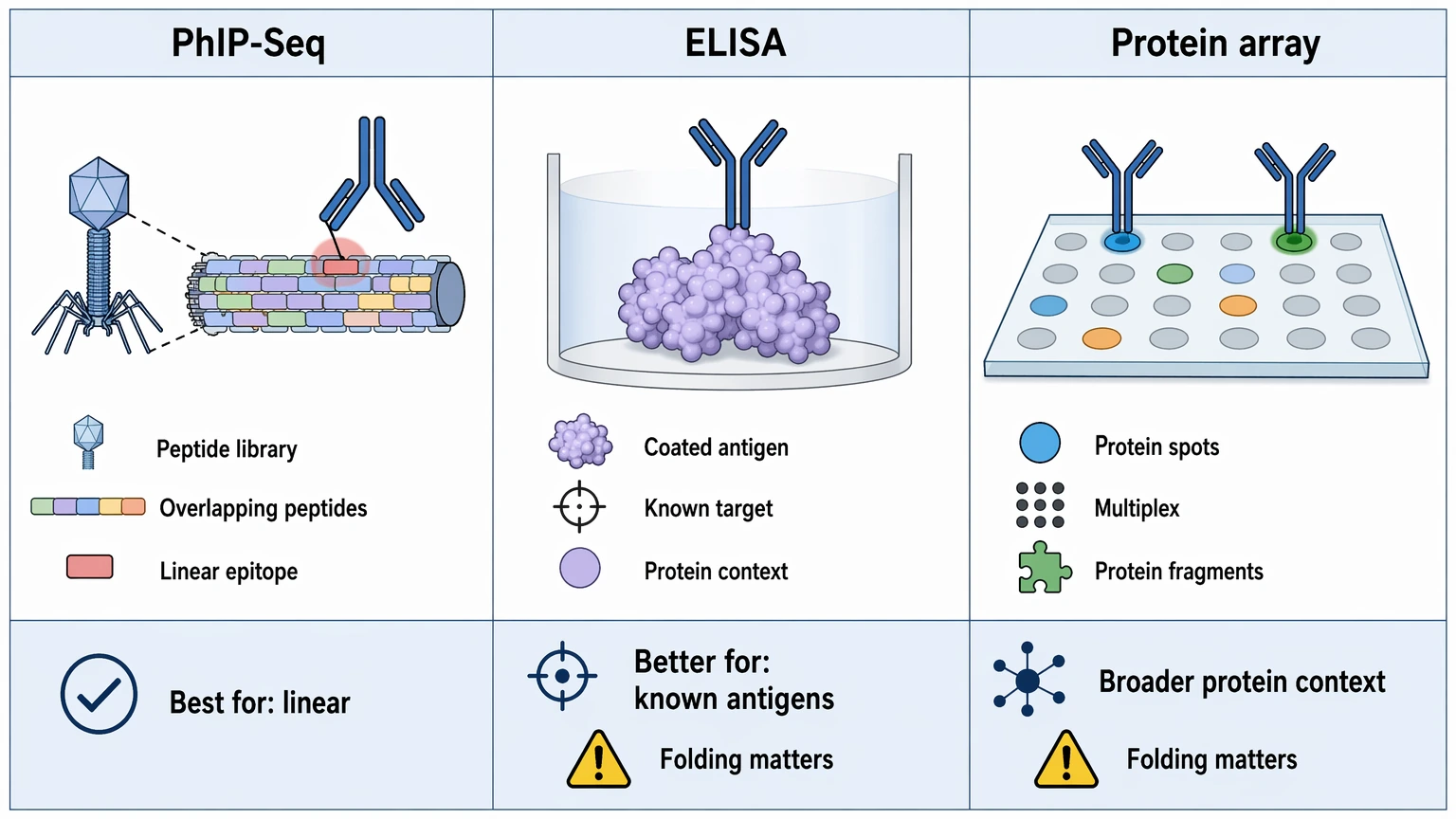
2. Readout logic changes how results should be interpreted
PhIP-Seq uses next-generation sequencing counts followed by enrichment analysis. The output is useful for relative ranking of a peptide-level hit across samples, groups, or time points. It is not a direct stand-in for antibody concentration or titer.
ELISA is usually easier to interpret when the target antigen is already known. Its outputs often map more cleanly to predefined study endpoints, especially when a project needs a focused targeted assay instead of open-ended discovery. Protein arrays sit between those two modes. They support broad screening against many proteins, but array intensity still depends on feature quality, background binding, and antigen presentation.
3. Throughput helps, but follow-up burden often decides the workflow
If a study needs to interrogate thousands of peptides across many samples, PhIP-Seq may be more defensible than running large numbers of separate serology assays. That breadth usually creates a second job, though: triaging enriched candidates for antigen-level confirmation. Breadth only pays off if the team already has a workable plan for shrinking the candidate list.
ELISA has much less discovery breadth, but it fits more naturally into a validation path. Protein arrays can screen many proteins in parallel and may narrow the gap between discovery and confirmation, although positive features still need follow-up in a secondary format when the result will drive downstream biomarker or mechanistic work.
4. Validation planning should be built into the initial platform decision
A project should not choose a platform in isolation from its endpoint. If the real goal is a shortlist for a future panel, discovery is only the first layer of evidence. If the goal is exploratory mapping of antibody reactivity across a broad peptide space, PhIP-Seq may be the most efficient first step. When the endpoint is a confirmed panel or a stronger antigen-level claim, a staged design is often the more defensible way to structure the study.
Side-by-side comparison of the main options
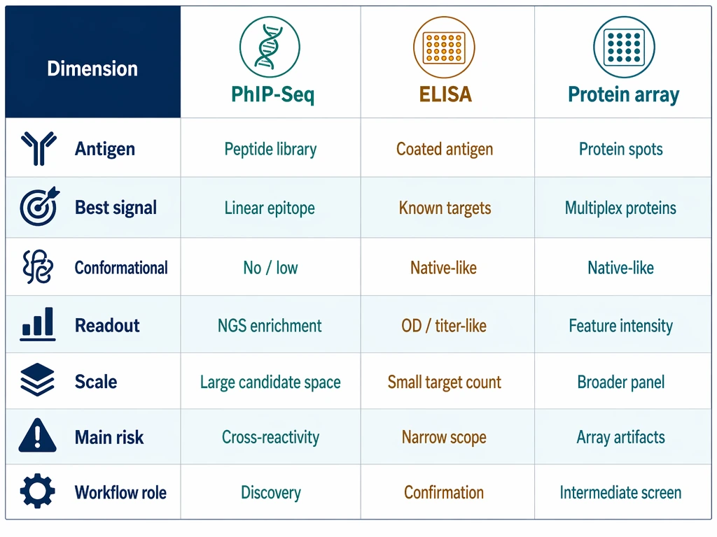
| Dimension | PhIP-Seq | Traditional serology / ELISA | Protein array |
|---|---|---|---|
| Antigen representation | Phage-displayed peptide library with overlapping peptides | Predefined coated antigen, usually one or a few targets per assay | Many immobilized proteins or fragments in parallel |
| Best signal type | Broad peptide-space discovery and linear epitope mapping | Focused testing of known antigens | Multiplex protein-level screening |
| Conformational epitope coverage | Limited when native folding is required | Can be better when antigen format is suitable | May capture more protein-context features than short peptides, but depends on array design |
| Readout logic | Sequencing enrichment after immunoprecipitation | OD, titer-like, or calibrated/semi-quantitative output | Signal intensity across many protein features |
| Cohort-scale screening | Strong when candidate space is very large | Best when target count is small | Useful for broader antigen panels |
| Main interpretation risk | Cross-reactivity, phage amplification bias, and peptide-level overinterpretation | Narrow target scope | Variable protein quality and array-specific artifacts |
| Best role in workflow | Discovery workflow | Focused confirmation or hypothesis testing | Intermediate screening or panel refinement |
A useful checkpoint is this: if your question starts with “which peptides or motifs are enriched across the cohort,” PhIP-Seq is usually the better match. If it starts with “are antibodies present against these known antigens,” serology is often the better fit. If it starts with “which protein antigens should move forward from a broader shortlist,” a protein array may be the stronger middle option.
If your team is deciding between peptide-discovery-driven screening and antigen-confirmation-driven testing, MtoZ Biolabs can evaluate your project around library scope, antigen representation, enrichment analysis criteria, and confirmatory assay planning before limited cohort samples are committed.
When each platform is the most defensible choice
Choose PhIP-Seq for broad peptide-space discovery
PhIP-Seq is a strong option when the project needs large-scale screening of peptide repertoires, such as infectious exposure profiling, vaccine epitope mapping, comparative cohort screening, or exploratory autoantibody work. It is especially useful when peptide tiling is part of the study logic itself. The tradeoff is interpretive: a strong enrichment signal supports prioritization, not automatic biological confirmation.
This platform is less suitable when the key biology depends on intact protein folding, multimeric structure, or a clearly predefined antigen list. In those cases, peptide-level breadth can turn into noise instead of an advantage.
Choose ELISA-style serology for predefined antigen questions
ELISA is usually the better option when the study already has a short list of targets and needs a focused serology readout. It fits well for validation panels, cohort subgroup comparisons against known analytes, and studies where the reporting objective is narrow and prespecified.
Its main limitation is straightforward: it cannot discover what it was not designed to test. If the underlying uncertainty is still high, targeted serology may return a clean answer to the wrong question.
Choose a protein array for broader antigen panels with protein-level context
A protein array becomes attractive when the study needs more breadth than ELISA but does not want to reduce the biology to short peptides alone. This can be useful in exploratory screening where larger antigen constructs matter, especially when a team wants to rank a moderate-to-large panel of proteins before designing narrower validation assays.
The caution is familiar. Screening output should not be overread. Signal on an array feature still needs context, replication, and sometimes orthogonal follow-up before the team treats it as a strong antigen-level finding.
Choose a staged workflow when discovery and confirmation are both required
For many translational programs, the most defensible design is sequential rather than single-platform. Start with PhIP-Seq to identify enriched peptide candidates, then move selected findings into ELISA, protein-level assays, bead-based immunoassays, or other orthogonal validation formats. That staged logic often fits biomarker programs better because it separates candidate generation from confirmation instead of asking one assay to do both jobs.

For teams planning that sequence, MtoZ Biolabs can discuss the study and help you submit your requirements for cohort size, sample volume, peptide library scope, hit-prioritization rules, and downstream assay format so the workflow matches the reporting goal.
What a sequencing enrichment signal means and what it does not mean
One of the most common mistakes in phip seq interpretation is treating enrichment like direct analyte quantitation or full antigen confirmation. A sequencing-based signal shows that peptides in the library were preferentially recovered after antibody binding and immunoprecipitation relative to background or controls. That is useful evidence, but it remains evidence in the molecular context of the displayed peptide library.
It does not automatically establish binding to a full-length antigen, functional antibody activity, disease association, or suitability for clinical interpretation. Careful analysis should account for background binding, nonspecific pull-down, shared motifs, and cross-reactivity across related peptide sequences. That boundary is not a flaw in the platform. It is simply part of using the method correctly.
Comparison summary and consultation guidance
PhIP-Seq is strongest when the study needs peptide-space breadth, linear epitope discovery, and relative enrichment patterns across a cohort. Traditional serology is stronger when the targets are already known and the project needs a focused, interpretable readout. A protein array fills the middle ground by expanding protein-level screening while retaining its own antigen-format limits. When a study needs both candidate generation and defensible follow-up, a staged workflow is often the clearest choice. For limited serum or plasma, cohort-scale screening, or a planned validation panel, prepare the sample type, antigen hypothesis, intended output, existing data, and follow-up strategy before you contact us for a project discussion.
FAQ
What project details should be prepared before selecting a platform?
Bring five items to the decision: sample type, expected cohort size, whether the question is peptide-centric or antigen-centric, the desired reporting format, and the planned confirmation path. Those details usually narrow the platform choice faster than a general discussion of throughput.
Can PhIP-Seq results be aggregated from peptide-level hits to antigen-level patterns?
Yes, but the aggregation rules should be defined carefully. Teams often group peptides by source antigen, motif family, or tiled region, then compare consistency across replicates and cohorts. Even so, aggregation is still an analytical step, not the same as direct antigen-level measurement.
When does a hybrid workflow save samples rather than consume more of them?
A staged workflow can be sample-efficient when the first pass eliminates most noninformative targets and reserves follow-up testing for a short candidate list. It is less efficient when the first-stage discovery library is poorly matched to the study question and produces many low-priority hits.
How should controls be planned for PhIP-Seq studies?
Controls should help distinguish true enrichment from assay background. Common examples include negative controls, technical replicates, reference samples, and strategies for modeling nonspecific binders. Control design affects hit calling as much as library design.
Is a protein array always better than ELISA when more targets are needed?
Not always. A protein array is useful when broader antigen coverage matters, but ELISA can still be the better choice if the target list is short and the project needs cleaner assay-by-assay interpretation. More multiplexing is not automatically better if it adds ambiguity.
What usually makes a project poorly suited to PhIP-Seq?
Projects centered on intact conformational epitopes, very small predefined antigen lists, or endpoints that require immediate antigen-level confirmation are often better served by protein-based or targeted serology formats.
How to order?

