






PhIP-Seq Characterization of Serum Antibodies Using Oligonucleotide-Encoded Peptidomes
Introduction
Serum antibody analysis is often limited by one practical question: researchers may know that an immune response exists, but not which peptide regions are being recognized. This gap is common in autoimmune disease research, infectious disease serology, vaccine response studies, and antibody biomarker discovery. Conventional assays such as ELISA, Western blotting, and immunofluorescence are useful when the antigen is already known. They are less efficient when the target space is broad, the relevant epitope is unclear, or the project requires discovery across thousands to millions of peptide candidates.
Phage immunoprecipitation sequencing, widely known as PhIP-Seq, was developed for this type of discovery challenge. By combining phage-displayed peptide libraries with DNA sequencing, PhIP-Seq can characterize antibody binding patterns in serum or plasma at peptide-level resolution. When the peptidome is oligonucleotide-encoded, each peptide is linked to a readable DNA barcode, allowing enriched antibody-bound peptides to be identified through sequencing rather than one-by-one antigen testing.
If your team has serum samples but lacks a clear antigen or epitope hypothesis, an early technical consultation can help determine whether PhIP-Seq is the right discovery approach before sample volume, cohort design, or validation budget is committed. MtoZ Biolabs can support project design for antibody profiling workflows that require both discovery depth and downstream interpretability.
Related Services
|
Research Need |
Recommended Service |
|
Need broad serum antibody profiling |
|
|
Confirm antibody sequence information |
|
|
Identify peptide candidates |
|
|
Validate protein-level evidence |
|
|
Map peptide regions in biologics or proteins |
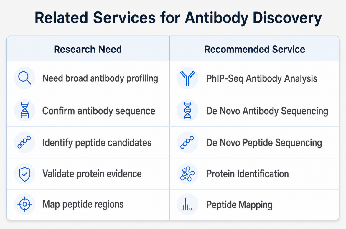
Figure 1. Related services for antibody discovery
Alt text: Related services for PhIP-Seq antibody discovery and peptide validation
What Does PhIP-Seq Characterize in Serum Antibodies?
A practical way to define PhIP-Seq is to start with the question it answers: which displayed peptides are selectively recognized by antibodies in this serum sample? This makes the method valuable when antibody reactivity is biologically meaningful, but the exact antigen, protein region, or linear epitope is not fully defined.
In a typical serum antibody profiling project, antibody-containing samples are incubated with a peptide display library. Antibodies bind to specific peptides, the antibody-bound phage particles are enriched by immunoprecipitation, and the DNA sequences associated with those phage clones are read by next-generation sequencing. Bioinformatics analysis then maps sequencing reads back to peptide identities and estimates which peptides are enriched in each sample or group.
The output is not a final diagnostic conclusion by itself. Instead, PhIP-Seq generates candidate antibody-reactive peptides, shared recognition patterns, and group-specific signals that can guide validation using targeted assays.
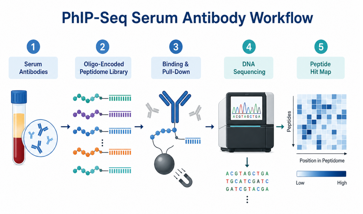
Figure 2. PhIP-Seq serum antibody workflow
Alt text: PhIP-Seq workflow for serum antibody profiling using oligonucleotide-encoded peptidomes
How Oligonucleotide-Encoded Peptidomes Work
The strength of oligonucleotide-encoded peptidomes lies in the link between phenotype and genotype. Each displayed peptide is associated with an encoding DNA sequence. After antibody binding and enrichment, researchers do not need to directly identify every peptide by protein chemistry. Instead, sequencing reads reveal which encoded peptide clones were enriched.
This design allows large peptide collections to be screened in parallel. A peptidome may represent human proteins, viral proteins, pathogen-derived sequences, custom antigen regions, or focused peptide panels. The library can be broad for discovery-oriented studies or targeted for projects with a defined biological hypothesis.
Library design is one of the most important determinants of PhIP-Seq performance. The assay can only detect antibody binding to peptides represented in the library. It is well suited for linear peptide recognition, but it may miss conformational epitopes that require folded protein structure. For this reason, researchers should match the peptidome design to the biological question, sample type, and validation strategy.
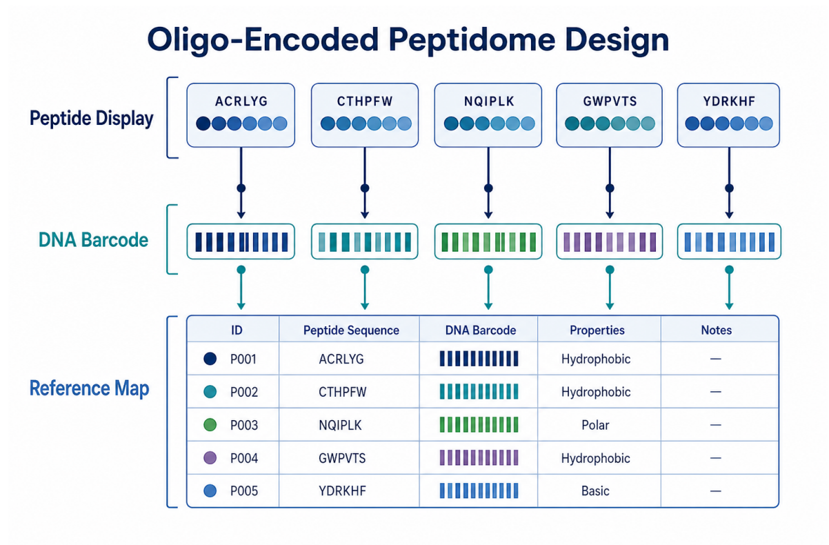
Figure 3. Oligonucleotide-encoded peptidome design
Alt text: Oligonucleotide-encoded peptidome library linking peptide display to DNA barcodes
When Researchers Should Use PhIP-Seq
PhIP-Seq is most appropriate when researchers need broad antibody profiling rather than single- antigen confirmation. It is especially useful in discovery-oriented projects where the antigen space is large or uncertain.
In autoimmune disease research, serum antibodies may recognize heterogeneous protein regions across patients. PhIP-Seq can help identify candidate autoantigens or shared antibody- reactive peptides that would be difficult to capture with a narrow predefined panel.
In infectious disease and vaccine studies, researchers may need to compare antibody recognition across many pathogen-derived peptides. PhIP-Seq can reveal peptide-level patterns that support immune history analysis, vaccine response profiling, or candidate antigen prioritization.
In biomarker discovery, PhIP-Seq can compare antibody enrichment patterns between disease and control groups, treatment groups, or longitudinal time points. These results should be treated as candidates until validated in independent cohorts.
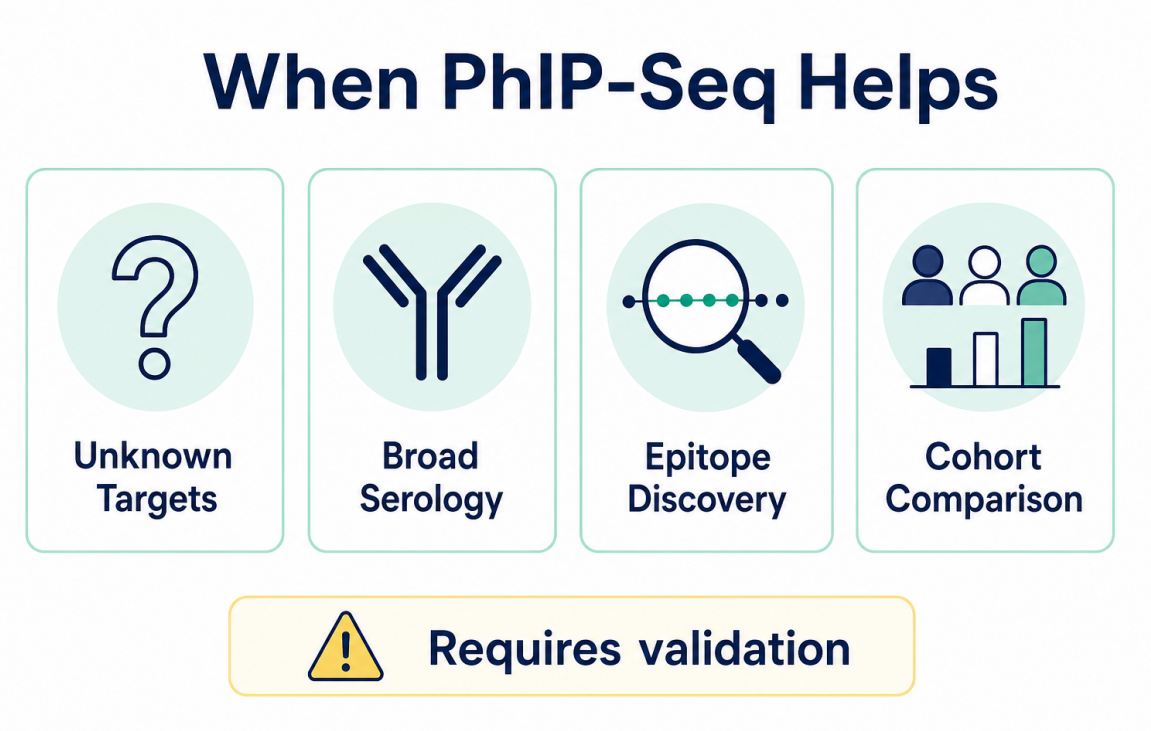
Figure 4. When PhIP-Seq helps
Alt text: PhIP-Seq use cases for unknown targets, broad serology, epitope discovery, and cohort comparison
When PhIP-Seq May Not Be the Best Choice
PhIP-Seq is not necessary for every antibody project. If the research question involves one known antigen and the goal is simple confirmation, ELISA or Western blotting may be more direct and cost-effective. If the key antibody response depends on conformational epitopes, folded protein assays or structural methods may be required.
Researchers should also be cautious when sample size is too small, cohort grouping is weak, metadata are incomplete, or downstream validation is not planned. PhIP-Seq can produce rich discovery data, but interpretation depends on experimental design. Without appropriate controls and validation, enriched peptides should not be overstated as disease markers or definitive epitopes.
Key Factors Before Starting a PhIP-Seq Project
Before beginning a PhIP-Seq serum antibody study, researchers should define the biological question clearly. Is the goal to discover unknown antibody targets, compare immune recognition across cohorts, evaluate vaccine response, or prioritize peptide candidates for validation? Each goal affects library selection, sample grouping, and analysis strategy.
Sample quality is also important. Serum or plasma samples should be collected, stored, and shipped under consistent conditions. Freeze-thaw cycles, hemolysis, inconsistent storage time, and incomplete metadata can introduce noise that complicates group comparison.
Bioinformatics planning should not be postponed until sequencing is complete. Researchers should define comparison groups, enrichment thresholds, statistical methods, and candidate ranking logic before analysis begins. A well-planned workflow makes the final peptide hit list easier to interpret and validate.
PhIP-Seq vs Traditional Antibody Detection Methods
Traditional antibody assays and PhIP-Seq answer different questions. ELISA is useful when a specific antigen is known. Western blotting supports protein-level confirmation. Immunofluorescence provides cellular localization context. Peptide arrays can test defined peptide panels.
PhIP-Seq is different because it is designed for broad discovery. It can screen large peptide libraries in parallel and identify candidate antibody-reactive sequences from serum samples. In many studies, these methods are complementary. PhIP-Seq identifies candidates, while targeted assays confirm selected findings.
FAQ
1. Is PhIP-Seq suitable for serum antibody profiling?
Yes. PhIP-Seq is commonly used with antibody-containing samples such as serum or plasma. It is most useful when researchers want to discover antibody-reactive peptides rather than test a single known antigen.
2. Can PhIP-Seq identify conformational epitopes?
Usually not directly. PhIP-Seq is strongest for linear peptide recognition. Conformational epitopes may require folded protein assays, structural biology methods, or complementary validation.
3. How important is the peptidome library design?
Library design is critical. PhIP-Seq can only detect peptides represented in the library. A human proteome library, viral peptidome, pathogen library, or custom antigen-focused library will answer different questions.
4. Do PhIP-Seq results require validation?
Yes. Enriched peptides should be treated as candidates. Follow-up validation using ELISA, peptide arrays, Western blotting, or other targeted methods is recommended.
Conclusion
PhIP-Seq characterization of serum antibodies using oligonucleotide-encoded peptidomes provides a scalable way to connect antibody binding with peptide identity. It is especially valuable when the antigen space is broad, the target is unknown, or researchers need cohort-level antibody profiling. Its value depends on thoughtful library design, consistent sample handling, appropriate controls, and a clear validation plan.
For teams planning antibody discovery, epitope mapping, or serum antibody biomarker projects, MtoZ Biolabs can help evaluate whether PhIP-Seq, peptide sequencing, protein identification, or peptide mapping is the most suitable next step. Contact our technical team to discuss sample type, research objective, library strategy, and validation workflow before starting your project.
How to order?




