


Particle Size Distribution Analysis Service | Cryo-EM
Particle size distribution analysis is a high-resolution imaging and quantitative analysis service based on Cryogenic Electron Microscopy (Cryo-EM), designed to accurately measure the particle size distribution of nanoscale samples. This technique rapidly freezes samples under liquid nitrogen conditions, preventing aggregation and deformation caused by drying or staining, and captures high-contrast two-dimensional images under low-dose electron beam conditions. Image processing algorithms are then applied to precisely measure and statistically analyze particle sizes.
Particle size distribution analysis service is suitable for various types of samples, including liposomes, extracellular vesicles (EVs), virus-like particles (VLPs), polymer nanoparticles, and protein complexes. It is widely used in fields such as nanomedicine development, biopharmaceutical quality control, vaccine structure evaluation, and new material characterization. Compared to traditional methods such as Dynamic Light Scattering (DLS), Cryo-EM particle size analysis offers higher spatial resolution and structural fidelity, providing a more accurate reflection of the sample’s true morphology and heterogeneity.
Services at MtoZ Biolabs
Based on an advanced cryogenic electron microscopy platform, MtoZ Biolabs offers a particle size distribution analysis service based on Cryo-EM, enabling rapid freezing and label-free imaging of samples under ultra-low temperature conditions. Utilizing high-resolution, low-dose imaging technology combined with professional image processing and statistical analysis, this service accurately measures particle size distribution, size uniformity, and potential heterogeneity features. Standardized outputs such as particle size distribution curves, average particle size, and polydispersity index (PDI) are provided, ensuring results with nanometer-level resolution and high structural fidelity, suitable for particle size evaluation of various types of samples.
Analysis Workflow
1. Sample Vitrification
Rapidly cool the sample to liquid nitrogen temperature, forming amorphous ice to maximally preserve the native morphology and size features of the particles.
2. Low-Dose Imaging Acquisition
Capture high-resolution images of a large number of particles under low-dose conditions using cryogenic electron microscopy, minimizing radiation damage.
3. Image Preprocessing and Particle Identification
Denoise, align, and automatically identify particles from raw images, extracting target particle regions to ensure the accuracy of subsequent size measurements.
4. Particle Size Measurement and Distribution Statistics
Measure the size of extracted particles, plot particle size distribution histograms, and calculate key parameters such as average diameter, median diameter, standard deviation, and polydispersity index (PDI).
5. Data Output and Report Generation
Provide raw images, particle size statistical data, particle size distribution curves, and a professional analysis report to support formulation development, quality control, and stability assessment needs.
Service Advantages
1. Accurate in Situ Particle Size Measurement
By rapid vitrification and low-dose imaging, avoid size deviations caused by drying, staining, or other treatments, truly reflecting the native dimensions of the particles.
2. High-Resolution Data Support
Relying on an advanced Cryo-EM platform, capture clear images at nanometer resolution to ensure accuracy and completeness of particle size measurements.
3. Standardized Statistical Analysis
Combine automated image processing with professional particle size extraction algorithms to provide standardized statistical data, including size distribution, average diameter, and median diameter.
4. Fine-Grained Distribution Analysis
Capable of precisely identifying multimodal size distributions within samples, supporting subpopulation analysis and feature classification of heterogeneous samples.
Applications
1. Particle Size Evaluation for Vaccines and Viral Vectors
Particle size distribution analysis service can be used to determine the size distribution of virus-like particles (VLPs) and recombinant viral vectors, supporting the assessment of assembly uniformity and formulation quality.
2. Nanomedicine Carrier Development
This service supports the analysis of particle size characteristics for lipid nanoparticles, polymeric nanoparticles, and other delivery systems, helping optimize drug loading capacity, delivery efficiency, and stability.
3. Extracellular Vesicle and Exosome Research
Through particle size distribution analysis, reveal the size heterogeneity of extracellular vesicles (EVs) and exosome samples, providing data support for biomarker development and mechanistic studies.
4. Characterization of Novel Nanomaterials
Particle size distribution analysis service can be applied to analyze the size and distribution characteristics of novel nanoparticles or self-assembled systems, aiding in material performance optimization and functional development.
Case Study
1. Biophysical Characterization of Polydisperse Liposomal Adjuvant Formulations
This study employed cryogenic electron microscopy (Cryo-EM) to analyze the particle size distribution characteristics of polydisperse liposomal adjuvant formulations, focusing on two liposome formulations containing monophosphoryl lipid A (MPLA) and saponin QS-21. Through low-temperature Cryo-EM imaging, the particle size, morphology, and distribution features of the liposomes were directly observed. The results showed that both formulations exhibited broad particle size distributions, and the QS-21-containing samples had a higher proportion of small-sized and irregular vesicles, suggesting that formulation components significantly influence particle size and structural stability. Cryo-EM revealed size heterogeneity that could not be distinguished by conventional methods such as DLS. The study demonstrated that Cryo-EM is a critical technique for evaluating the microstructure of polydisperse liposomal formulations and optimizing adjuvant performance.
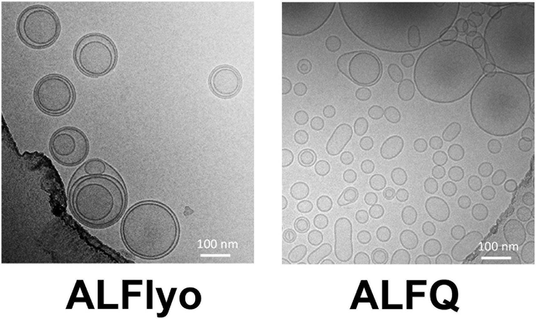
Singh, P. et al. Biochemical and Biophysical Research Communications, 2020.
Figure 1. Cryo-Electron Micrographs for (A) ALFlyo and (B) ALFQ.
2. Particle Size Distributions for Cellulose Nanocrystals Measured by Transmission Electron Microscopy: An Interlaboratory Comparison
This study aimed to evaluate the consistency and reliability of measuring cellulose nanocrystals (CNCs) particle size distributions using cryogenic transmission electron microscopy (Cryo-TEM). The study focused on uniformly prepared CNCs samples and involved multiple laboratories collaborating to perform Cryo-TEM imaging and particle size analysis. Variations were observed among laboratories in sample vitrification, imaging parameter settings, and particle size measurement methods. The results showed that, although there were some fluctuations in particle size measurements across different laboratories, the overall distribution trends remained consistent. Cryo-TEM was able to accurately capture the in situ size information of CNCs with higher resolution compared to conventional TEM. The study concluded that Cryo-TEM, combined with standardized operating procedures, improves the accuracy and comparability of CNCs particle size distribution data, making it a reliable method for nanoparticle characterization.
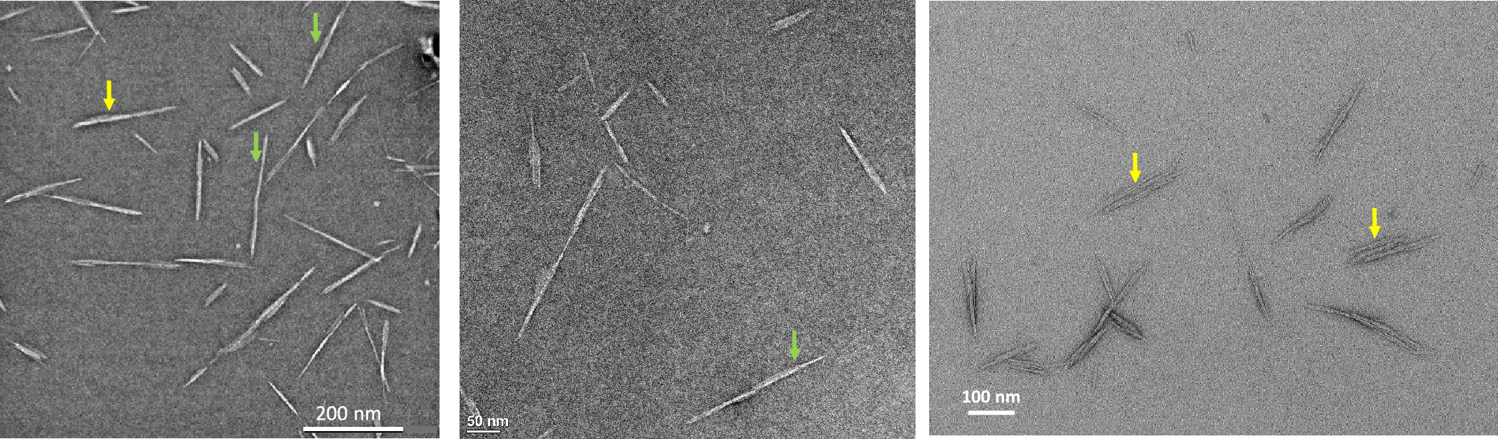
Meija, J. et al. Analytical Chemistry, 2020.
Figure 2. Representative TEM Images from Labs T3 (Left), T6 (Center), and T9 (Right).
FAQ
Q1: Can Both Unilamellar and Multilamellar Particles Be Analyzed Simultaneously?
A1: Yes. Cryo-EM can not only measure particle size but also visualize membrane layers and structural features, enabling the distinction between unilamellar, multilamellar, or more complex particles.
Q2: Can Cryo-EM Analyze Highly Heterogeneous Samples?
A2: Yes. Cryo-EM can clearly resolve and classify particles with different sizes and shapes, making it particularly suitable for samples with broad size distributions and high heterogeneity.
Q3: What Are the Advantages of Cryo-EM Particle Size Analysis Compared to Dynamic Light Scattering (DLS)?
A3: Cryo-EM provides direct imaging of individual particles, accurately reflecting particle size and heterogeneity, whereas DLS offers an overall intensity-weighted average and cannot distinguish between different size subpopulations.
How to order?

