


Nano-Flow Cytometry (nFCM) Service
Nano-Flow Cytometry (nFCM) Service enables multidimensional, real-time, high-throughput analysis of nanoparticles ranging from 30 to 1000 nm, providing more precise particle characterization and biological function analysis. Traditional flow cytometry (FCM) is limited by its resolution, making it difficult to accurately detect particles smaller than 200 nm. With the expanding applications of subcellular nanoparticles—such as exosomes, microvesicles, and viral particles—in disease diagnosis, drug delivery, and biomarker discovery, there is an increasing demand for high-sensitivity, high-resolution characterization techniques for nanoparticles. nFCM has been developed in this context as a flow cytometry platform specifically for nanoparticles. It utilizes enhanced optical systems and sensitive fluorescence detectors to simultaneously analyze multiple parameters at the single-particle level, including particle size, concentration, surface marker expression, and content. By combining the high-throughput advantage of FCM with the capability to detect nanoparticles, nFCM is an important tool for analyzing the heterogeneity and exploring the functions of nanoparticles such as exosomes. MtoZ Biolabs offers the Nano-Flow Cytometry (nFCM) Service for high-precision single-particle analysis of nanoparticles (such as exosomes and viral particles), helping to deeply elucidate nanoparticle heterogeneity, functionality, and application potential.
Mao C. et al. Talanta. 2020.
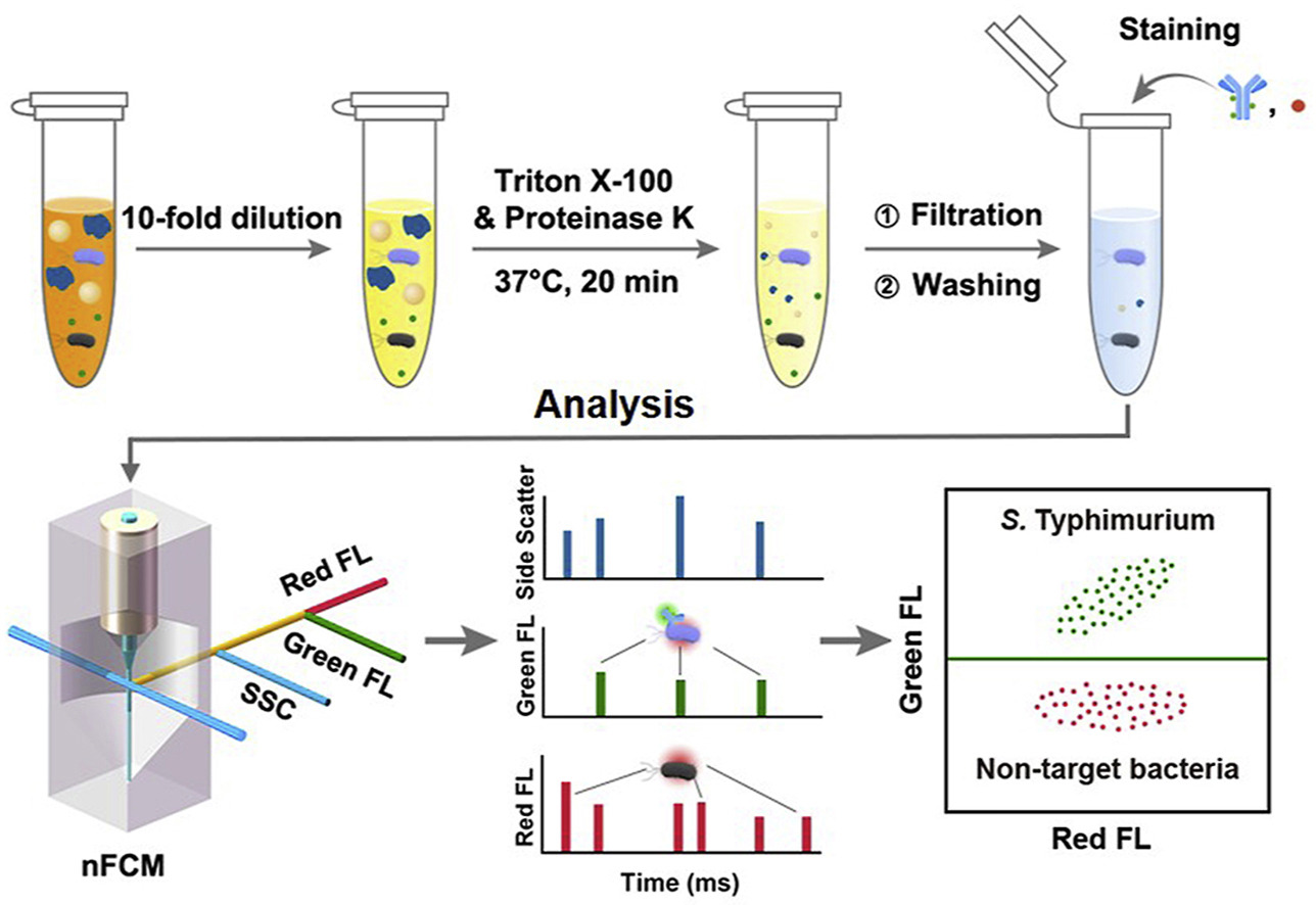
Figure 1. nFCM Quantification of Bacteria.
Technical Principles
Nano-Flow Cytometry (nFCM) is a high-sensitivity flow cytometry technique that enables precise single-particle analysis based on enhanced laser scattering detection and multi-channel fluorescence signal acquisition. Utilizing a high-power laser and an optimized optical system, nFCM records scattered light information to measure particle size and concentration while employing fluorescent labeling combined with high-sensitivity detectors to analyze surface markers and intracellular contents. Additionally, nFCM uses standardized nanoparticles for calibration, ensuring accurate quantification of nanoparticle size, concentration, and fluorescence signals. This capability enables a deeper understanding of nanoparticle heterogeneity and biological functions.
Service Advantages
1. High Sensitivity: Capable of detecting low refractive index nanoparticles and the fluorescence of single phycoerythrin molecules.
2. Multiparametric Analysis: Enables simultaneous detection of multiple parameters of individual nanoparticles, revealing diverse biochemical characteristics through multicolor fluorescence.
3. Low Sample Requirement: Suitable for analyzing precious or limited samples.
4. High Throughput: Offers rapid analysis comparable to techniques like TEM.
5. One-Time-Charge: MtoZ Biolabs' pricing is transparent, no hidden fees or additional costs.
Applications
Microbiological Research
Nano-Flow Cytometry (nFCM) is widely applied in microbiology for high-throughput, multiparameter quantitative analysis at the single-particle level. It is used for detecting pathogenic bacteria in food, environmental water samples, and clinical specimens, as well as for characterizing bacterial stress responses to antibiotics.
Multi-parameter Detection of Mitochondria
Compared to whole-cell analysis or Western blot, nFCM not only reveals individual particle variations but also simplifies complex samples, making experimental data analysis clearer and more straightforward.
Virology Research
By enhancing laser power density and reducing the detection volume, nFCM enables single-particle scattering detection of particles as small as 27 nm, including small viruses such as MS2. In addition, single-particle analysis eliminates interference from contaminant particles, making nFCM particularly suitable for analyzing polydisperse or mixed samples. It has been successfully applied in virus purity assessment and dynamic monitoring of viral genome release.
Detection of Extracellular Vesicles (EVs)
Most extracellular vesicles (EVs) are smaller than 100 nm, with extremely low and heterogeneous expression of specific proteins. nFCM shows significant advantages in characterizing EV size distribution, concentration, and biochemical properties such as nucleic acids. Its high side-scatter sensitivity allows for single-particle, multiparameter quantitative analysis of EVs with diameters as small as 40 nm.
FAQ
Q. How to Ensure Comparability of Particle Size and Concentration Measurements in Nano-Flow Cytometry (nFCM) Analysis?
To ensure the comparability of particle size and concentration data in nFCM analysis, standardization and instrument calibration are critical steps. First, the instrument must be calibrated using standard nanoparticles with known sizes and fluorescence intensities to establish a calibration curve correlating particle size and scattering signals, ensuring accurate size measurement. Additionally, concentration calibration should be performed using particles of known concentrations to correct for variations in flow rate and detection sensitivity.
Furthermore, all sample batches should be diluted to a consistent and appropriate concentration range to avoid signal deviations caused by overly concentrated or overly dilute samples. To minimize inter-experimental variability, it is recommended to run standard particles before and after each experiment and perform background signal subtraction and normalization during data analysis. These measures help guarantee cross-batch and cross-study comparability of nFCM results.
Q. How to Address Spectral Overlap between Fluorescence Channels When Analyzing Multiple Surface Markers?
When performing multiplex labeling analysis, spectral overlap (spillover) is a major factor affecting data accuracy, so strict fluorescence compensation is essential. First, when selecting fluorophores, it is important to avoid combinations with overlapping emission spectra. Priority should be given to dyes with distinct excitation and emission peaks (e.g., choosing FITC and APC rather than FITC and PE). Second, single-stain controls must be included in the experimental design—these are samples labeled with only one fluorophore—to calculate the fluorescence spillover matrix between channels. Finally, using compensation matrices, appropriate compensation algorithms should be applied in flow cytometry analysis software to correct spectral spillover in real-time, ensuring independent and accurate quantification of each channel’s signal. For very low-abundance markers, it is also recommended to optimize antibody concentration and incubation conditions and apply background subtraction to avoid false positives or negatives caused by compensation errors.
Case Study
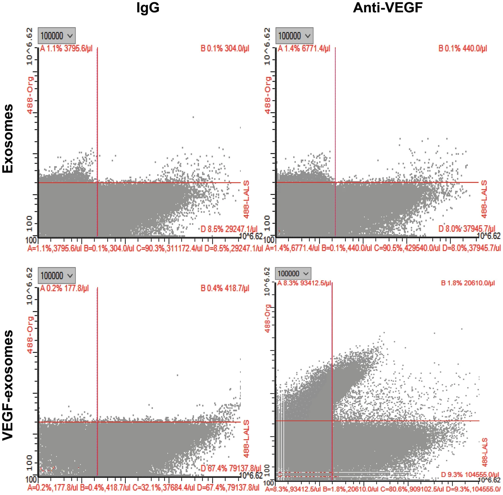
This study utilized nFCM to perform single-particle fluorescence labeling and quantitative analysis of VEGF molecules within exosomes, aiming to confirm the true presence and expression levels of VEGF in exosomes. The results demonstrated that exosomes released by tumor cells exhibited significantly high levels of VEGF. Moreover, these VEGF-enriched exosomes showed enhanced pro-angiogenic activity in subsequent endothelial cell tube formation assays and in vivo angiogenesis experiments.
Zeng Y. et al. Methods in molecular biology. 2022.
Figure 2. Nano-flow Cytometry Analysis of Exosomes and VEGF-positive Exosomes Labeled by Anti-VEGF Antibody.
How to order?

