


MRI-based Exosome Quantification Service
MRI-based exosome quantification is an advanced method for non-invasive and real-time quantification of exosomes using magnetic resonance imaging (MRI). This technique typically involves labeling exosomes with magnetic nanoparticles or other MRI contrast agents, enabling them to generate specific signals in MRI imaging, thereby allowing precise visualization and quantification of exosomes. Compared to traditional exosome detection methods, MRI offers high sensitivity and spatial resolution while also providing real-time tracking of exosome distribution and dynamic changes in vivo.
MRI-based exosome quantification service is widely applied in the fields of oncology, neurodegenerative diseases, cardiovascular diseases, and inflammation, particularly in studying exosomes as drug delivery vehicles, biomarkers, or therapeutic agents. Through MRI imaging, researchers can dynamically observe the distribution of exosomes in different organs and tissues, assess their targeting capabilities and delivery efficiency, and obtain intuitive and reliable data to support the application of exosomes in disease diagnosis, therapy, and precision medicine.
Services at MtoZ Biolabs
Based on high-resolution magnetic resonance imaging (MRI) systems and combined with magnetic nanoparticles and other magnetic probes, MtoZ Biolabs offers the MRI-based exosome quantification service that enables precise quantification and real-time tracking of labeled exosomes. Utilizing advanced MRI instruments, this service provides high-resolution imaging data on the distribution, localization, and dynamic migration of exosomes both in vivo and in vitro, offering a clear visualization of exosome spatial distribution and accumulation. A comprehensive quantitative analysis and imaging report is provided, supporting applications in drug delivery, disease mechanism studies, and biomarker development.
Service Advantages
1. High Sensitivity and Accurate Quantification
By combining MRI with magnetic nanoparticle labeling technology, high-sensitivity detection and precise quantification of exosomes are achieved, capturing the distribution dynamics of trace amounts of exosomes.
2. One-Time-Charge
Our pricing is transparent, no hidden fees or additional costs.
3. Non-invasive Visualization
MRI imaging offers the advantage of being non-invasive and non-damaging, allowing repeated monitoring of exosome distribution in vivo, ensuring experimental continuity and animal welfare.
4. Professional Customized Solutions
We provide a one-stop service for exosome labeling, imaging parameter optimization, and data analysis based on different research needs, ensuring efficient and reliable results to meet various applications in basic research and translational medicine.
Applications
1. Exosome in Vivo Distribution and Targeting Evaluation
The MRI-based exosome quantification service can trace the distribution path of exosomes in vivo, assess their targeting effect in specific tissues or organs, and support the development of drug delivery systems and gene therapies.
2. Drug Delivery Effect Monitoring
By observing the in vivo transport and release process of exosomes loaded with drugs, the service helps evaluate the drug delivery efficiency and therapeutic effects, aiding in the design of new nano drug carriers.
3. Disease Model Mechanism Research
In disease models such as cancer, neurodegenerative diseases, and inflammation, the MRI-based exosome quantification service can dynamically monitor the behavior and mechanisms of exosomes, exploring their roles in disease initiation and progression.
4. Therapeutic Exosome Verification
The MRI-based exosome quantification service can be used for in vivo delivery, distribution, and efficacy monitoring of therapeutic engineered exosomes, ensuring their safety and effectiveness, and supporting clinical translation research.
Case Study
1. Magnetic Resonance Imaging of Melanoma Exosomes in Lymph Nodesc
This study aims to use magnetic resonance imaging (MRI) to visualize and track the distribution of melanoma cell-derived exosomes in lymph nodes, in order to explore the role of exosomes in tumor metastasis. The study focused on exosomes secreted by melanoma cells, which were labeled with superparamagnetic iron oxide nanoparticles (SPIONs) to enable MRI imaging. In terms of methodology, after labeling the exosomes in vitro, they were injected into an animal model, and high-resolution MRI was used to dynamically image and quantify the distribution of exosomes in the lymph nodes. The results showed that SPION-labeled exosomes could be clearly detected by MRI after injection, and a significant signal change was observed in the lymph node area, indicating that exosomes can accurately accumulate in the lymph nodes. The study concluded that MRI imaging technology effectively allows non-invasive in vivo tracking of exosomes, revealing their potential role in melanoma lymph node metastasis and providing a new tool for research on exosome-related disease diagnostics and drug delivery systems.
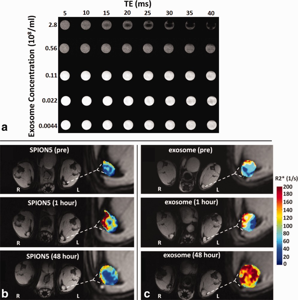
Hu, L Z. et al. Magnetic Resonance in Medicine, 2014.
Figure 1. MRI of SPION5 Loaded Melanoma Exosomes in Vitro and in Vivo.
FAQ
Q1: How Are Exosomes Labeled for MRI Imaging?
A1: Exosomes are typically labeled with magnetic nanoparticles, such as superparamagnetic iron oxide nanoparticles (SPIONs), to enable MRI visualization. Optimized labeling methods ensure that the exosome structure and function are not affected while maintaining high-quality imaging results.
Q2: What Are the Advantages of MRI-based Exosome Quantification Compared to Traditional Exosome Quantification Methods?
A2: MRI imaging allows for non-invasive, dynamic, and real-time monitoring in vivo, providing a clear visualization of exosome distribution and migration. This overcomes the limitations of traditional methods that cannot track live animals, making it particularly suitable for drug delivery and targeted therapy scenarios.
How to order?

