


Morphology & Shape Analysis Service | Cryo-EM
Morphology & shape analysis is a high-resolution imaging and structural analysis technique based on cryogenic electron microscopy (Cryo-EM), specifically designed to observe and characterize the morphological features and shape variations of various samples such as nanoparticles, liposomes, extracellular vesicles (EVs), exosomes, and viral particles. This service captures the three-dimensional morphological characteristics of samples directly, providing detailed visualization of particle contours, boundary clarity, and surface microstructures, making it particularly suitable for analyzing subtle differences and morphological heterogeneity.
Morphology & shape analysis service is widely applied in areas such as the design and optimization of nano drug carriers, vesicle structure evaluation, viral particle morphology research, novel material development, and monitoring of sample uniformity and stability. Through high-resolution imaging and quantitative analysis, this service can deliver critical data including particle size distribution, shape classification (such as spherical, ellipsoidal, or flattened forms), surface feature descriptions, and deformation rate assessments, providing essential structural support for scientific research, process improvement, and quality control.
Services at MtoZ Biolabs
Based on an advanced cryogenic electron microscopy platform, the morphology & shape analysis service based on Cryo-EM provided by MtoZ Biolabs enables high-precision characterization of sample morphology and structural features. Utilizing low-dose imaging techniques, the service offers direct visualization of sample boundary contours, surface structures, and overall morphological characteristics without the need for staining or drying treatment. It is suitable for structural evaluation of various membrane vesicles, polymer nanoparticles, and viral vectors. This service delivers high-resolution, low-interference data to support morphology consistency validation and structural optimization studies.
Analysis Workflow
1. Sample Vitrification
Rapidly cool the sample to liquid nitrogen temperature to form amorphous ice, preserving the original morphology and microstructure to the greatest extent.
2. Low-Dose Imaging Acquisition
Perform low-dose, high-contrast imaging under cryogenic electron microscopy to capture clear, artifact-free two-dimensional morphology images.
3. Image Processing and Particle Identification
Denoise, align, and select particles from the acquired raw images to extract target regions for quantitative analysis.
4. Morphological and Structural Feature Analysis
Assess particle size distribution, shape classification (such as spherical, ellipsoidal, irregular), surface contours, and degree of deformation.
5. Data Output and Report Generation
Provide raw images, quantitative analysis data, and morphology feature description reports to support research and development, quality control, and product optimization applications.
Service Advantages
1. High-Fidelity Morphology Preservation
Through rapid freezing and low-dose imaging, the native morphology of the sample is fully preserved, avoiding structural changes caused by drying or staining.
2. Nanoscale Resolution Observation
Leveraging an advanced Cryo-EM platform, clear imaging at the nanoscale is achieved, finely presenting particle shapes, boundaries, and surface features.
3. Accurate Morphology Classification and Measurement
Supports multidimensional quantitative analysis including particle size distribution, shape classification, surface smoothness, and deformation rate, delivering standardized data outputs.
4. Comprehensive Structural Feature Analysis
Simultaneously analyzes particle contours, shape heterogeneity, and localized deformation, comprehensively revealing the microstructural characteristics of the sample.
Applications
1. Nanodrug Carrier Development and Optimization
Morphology & shape analysis service can be used to evaluate particle size, morphological uniformity, and surface features of drug carriers such as liposomes and polymer nanoparticles, guiding delivery system design and process improvement.
2. Extracellular Vesicle and Exosome Research
High-resolution imaging analysis of extracellular vesicles (EVs) and exosomes supports studies on morphological heterogeneity and integrity, aiding in disease biomarker development and functional mechanism exploration.
3. Viral Particle Structural Evaluation
Morphology & shape analysis service assists in observing the morphology and analyzing the particle size distribution of virus-like particles (VLPs) or gene therapy viral vectors, ensuring quality stability for vaccines and gene medicines.
4. Characterization of Novel Nanomaterials
Used to characterize the morphological features of new nanomaterials or self-assembled nanostructures, revealing microstructural differences and supporting material performance optimization and application development.
Case Study
1. Characterization of VSV-GP Morphology by Cryo-EM Imaging and SEC-MALS
This study aimed to analyze the morphology and shape characteristics of recombinant viral vector VSV-GP through cryo-electron microscopy (Cryo-EM) imaging. The study focused on VSV-GP particles prepared from different batches, with direct imaging performed under cryogenic conditions to assess overall morphology, structural integrity, and size consistency. The results showed that VSV-GP particles maintained a typical bullet-shaped structure with smooth surfaces and clear contours. Different batches exhibited high consistency in particle size, shape, and structural stability, with no significant morphological abnormalities or fragmentation observed. The study demonstrated that Cryo-EM can accurately capture the native morphological details of VSV-GP particles, making it an important tool for evaluating morphological uniformity and manufacturing quality of viral vectors, and providing reliable structural support for the application of VSV-GP in vaccine and gene therapy development.
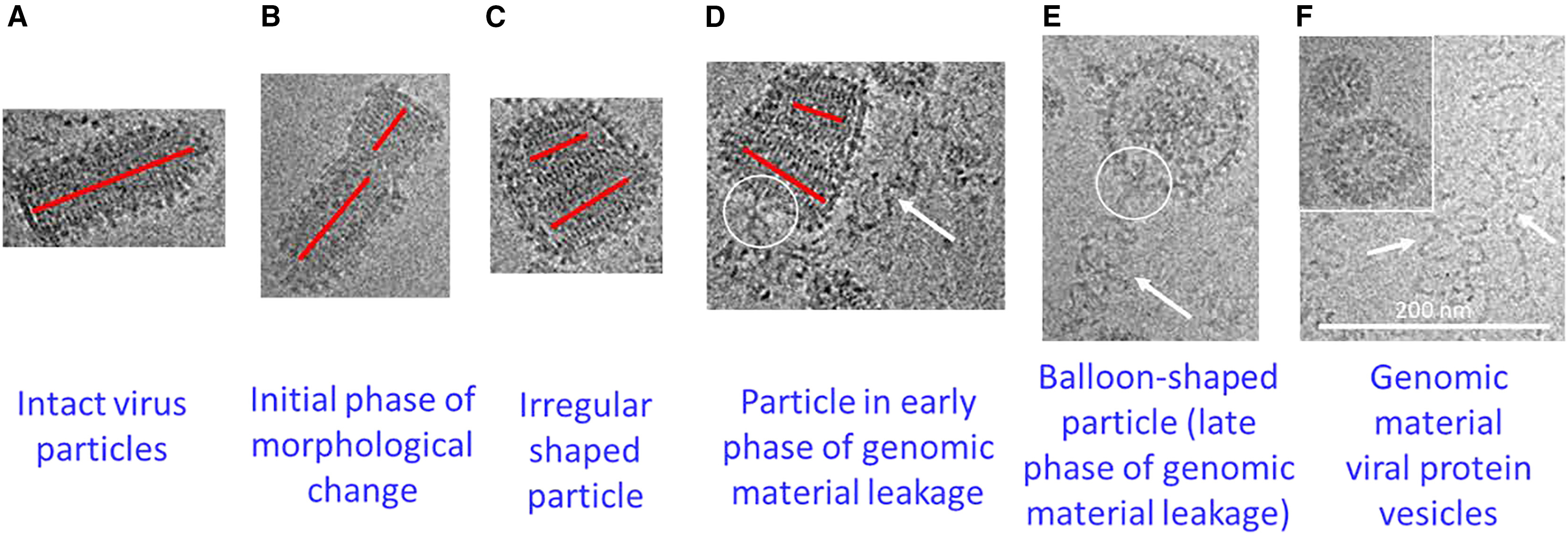
Xin, D Y. et al. Molecular Therapy - Methods & Clinical Development, 2025.
Figure 1. A Spectrum of VSV-GP Subpopulations in Sample B.
2. Analysis of Nanoparticle Scale Rules and Shape Heterogeneity
This study aimed to analyze the morphology and shape heterogeneity of nanoparticles with the assistance of cryo-electron microscopy (Cryo-EM). The study focused on nanoparticles of various sizes and materials. Initial particle size measurements were conducted using size exclusion chromatography (SEC) coupled with multi-angle light scattering (MALS), followed by direct imaging of each size fraction using Cryo-EM to systematically assess the true variations in particle shape. The results revealed significant morphological diversity among particles of similar sizes, including spherical, ellipsoidal, and irregular structures, with Cryo-EM effectively capturing these microstructural differences. The study concluded that the combination of SEC-MALS and Cryo-EM provides a comprehensive approach to revealing the scale and morphology characteristics of nanoparticles, offering important structural analysis tools for nanoparticle design, formulation development, and quality control.
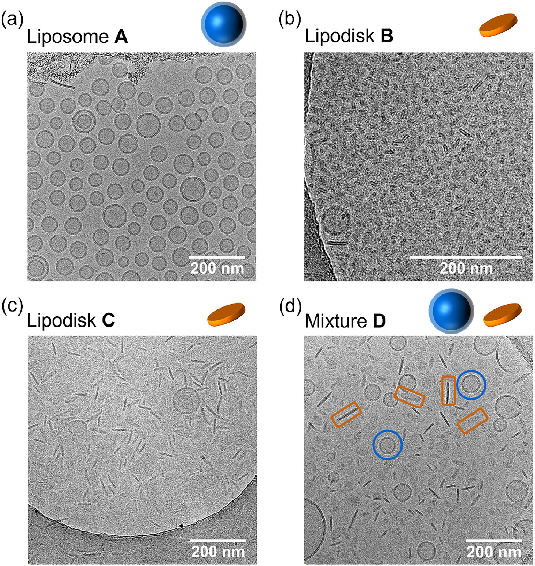
Tang, S J. et al. Journal of Chromatography A, 2024.
Figure 2. Representative cryo-EM images of liposome and lipodisk formulations A–D.
FAQ
Q1: Can Different Particle Shapes Be Distinguished?
A1: Yes. Cryo-EM imaging combined with quantitative analysis can clearly distinguish spherical, ellipsoidal, flattened, or irregularly shaped particles, and provides statistical classification.
Q2: Does the Service Include Particle Size Distribution Analysis?
A2: Yes. The service includes particle size measurement and distribution statistics, which can be used to assess sample uniformity, stability, and process consistency.
How to order?

