


Microscopy Imaging Service
Microscopy imaging is a central technique in life science research, enabling visualization of biomolecules at both cellular and subcellular levels. It provides critical insights into protein assembly, signal transduction, and molecular interactions within the natural cellular environment. These capabilities offer direct evidence to advance the understanding of cellular function and disease mechanisms.
MtoZ Biolabs offers Microscopy Imaging Service combining optical, fluorescence, confocal, electron, Raman, FLIM, and FRET platforms. Our integrated solutions cover experimental design, sample preparation, imaging acquisition, and data analysis, delivering high-quality results that support both scientific research and industrial applications.
Services at MtoZ Biolabs
MtoZ Biolabs provides a full spectrum of imaging options tailored to diverse research needs:
Optical microscopy for cellular and tissue morphology studies in routine biology.
Fluorescence microscopy for specific localization and real-time tracking of biomolecules.
Confocal microscopy for high-resolution three-dimensional imaging of cellular structures.
Electron microscopy for ultrastructural observation at the nanoscale.
Raman microscopy for label-free chemical imaging based on molecular spectra.
FLIM and FRET for probing conformational changes, molecular interactions, and signaling dynamics.
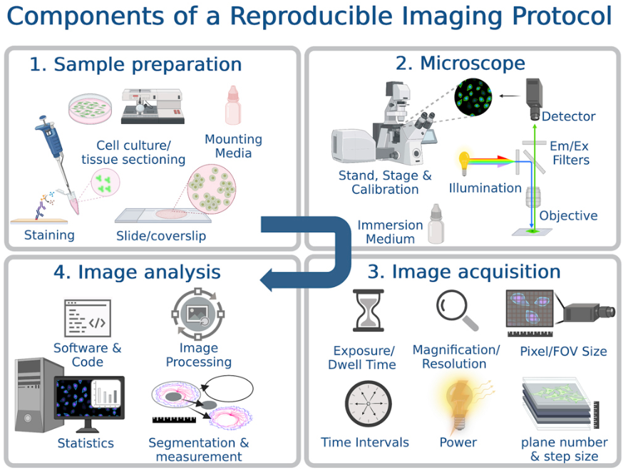
Analysis Workflow
1. Project Evaluation and Design
Define objectives and select the most suitable imaging technology.
2. Sample Preparation
Apply fixation, staining, labeling, or live-cell culture depending on project requirements.
3. Image Acquisition
Capture high-resolution two-dimensional, three-dimensional, or time-lapse datasets.
4. Data Processing
Perform image stitching, noise reduction, 3D reconstruction, trajectory tracking, and quantitative analysis.
5. Result Delivery
Provide comprehensive imaging data, visualizations, and a detailed report.
Larsen, D. D. et al. STAR Protocols. 2023.
Why Choose MtoZ Biolabs?
✅ Comprehensive Imaging Platforms: Integration of optical, fluorescence, confocal, electron, Raman, FLIM, and other advanced modalities.
✅ Live-cell and Dynamic Imaging: Real-time tracking of molecular behavior in physiologically relevant conditions.
✅ Tailored Experimental Design: Customized imaging strategies based on sample type and research objectives.
✅ Stringent Quality Control: Standardized procedures and rigorous validation ensure reproducible, high-quality data.
✅ One-Time-Charge: Our pricing is transparent, no hidden fees or additional costs.
Sample Submission Suggestions
For our Microscopy Imaging Service, we accept a wide range of samples including cells, tissue sections, organoids, viral particles, and nanomaterials. To achieve optimal imaging outcomes, samples should be appropriately processed, such as fixed or fluorescently labeled, and carefully preserved during storage and shipment. Fixed samples may be shipped at room temperature, while live or sensitive specimens should be transported at low temperature and delivered promptly. Detailed submission guidance is available from MtoZ Biolabs.
Applications
● Structural biology for molecular and organelle conformations
● Molecular interaction studies including protein–protein, protein–nucleic acid, and protein–small molecule binding
● Cell and pathogen interaction research
● Drug development and mechanism studies on candidate compounds
What Could be Included in the Report?
1. Comprehensive Experimental Details
2. Materials, Instruments, and Methods
3. High-Resolution Imaging Data
4. Processed Data and Plots
5. Statistical Summary
6. Raw Data Files
MtoZ Biolabs Microscopy Imaging Service delivers advanced imaging capabilities supported by state-of-the-art technology and expert analysis. Whether you are investigating cellular dynamics, molecular interactions, or drug effects, we provide reliable visual data and scientific interpretation to accelerate both research and application.
FAQ
Q1: How should I choose the right microscopy technique?
Optical microscopy is best for routine observations, fluorescence and confocal microscopy are used for molecular localization and 3D imaging, electron microscopy provides nanoscale detail, while Raman microscopy and FLIM reveal conformational states and interaction dynamics. Our experts will guide you in selecting the most appropriate platform for your project.
Q2: Can you handle complex or sensitive samples?
Yes. We have experience working with challenging samples such as tissue sections, viral particles, and nanomaterials. We provide optimized preparation and imaging strategies to ensure accurate and reproducible results.
Related Services
How to order?

