



Tandem mass tag (TMT) labeling is an isobaric chemical tagging method that marks peptides from different samples with channel-specific reagents that share the same total mass at the precursor level and release distinct reporter ions after fragmentation. Digested peptides are labeled mainly at N-termini and lysine amines, mixed into one closed plex, separated by liquid chromatography, and analyzed by tandem mass spectrometry. At MS1 the same peptide from every channel appears as one precursor. Upon collision-induced dissociation (CID) or higher-energy collisional dissociation (HCD), reporter ions of different masses carry the relative channel intensities (Thompson et al. 2003). Ordinary TMT ratios support relative comparison inside that plex. They do not report calibrated protein concentration.
Researchers who already have defined samples, comparison groups, or a specific project objective can also review the MtoZ Biolabs TMT Quantitative Proteomics Analysis Service for project-specific feasibility and analysis planning.
Chemical Structure Behind the Isobaric Mechanism
A TMT reagent is built from three functional parts that must be understood together.
1. Reactive group
An amine-reactive moiety, commonly an N-hydroxysuccinimide (NHS) ester chemistry class, covalently attaches the tag to peptide N-termini and lysine side chains after digestion. Amine-rich residual buffers compete for reagent and leave unlabeled peptides that distort later ratios.
2. Mass balancer
The balancer offsets isotopic differences among channels so that the complete tag mass is effectively the same across the kit. That design keeps labeled forms of the same peptide co-eluting and co-selected as one MS1 precursor.
3. Reporter group
After fragmentation, the reporter ion of each channel appears at a distinct low m/z. Reporter intensities are summarized to peptide and protein relative abundances. The balancer is not the quantitative signal; the reporter ions are.
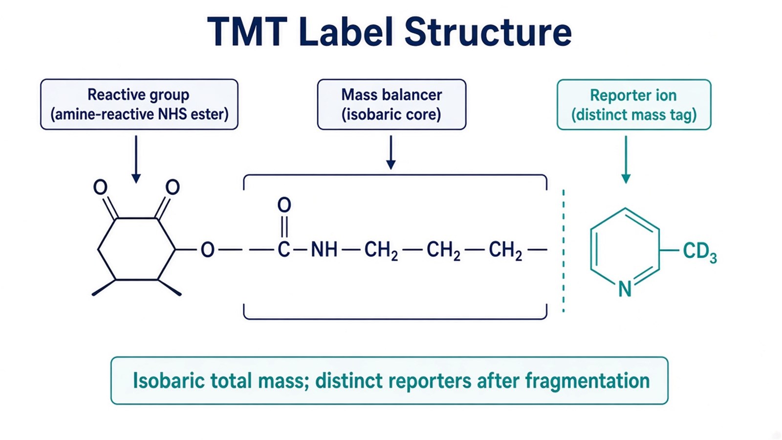
Figure 1. TMT labeling chemistry couples an amine-reactive handle, a mass balancer, and a channel-specific reporter so peptides remain isobaric until MS2 or MS3 fragmentation.
TMT labeling makes channels isobaric at MS1; quantification begins only when reporter ions are released.
From Labeling Chemistry to Reporter-Ion Readout
The mechanism proceeds in a fixed order.
Digestion creates the labeling sites
Proteins are reduced, alkylated, and digested, usually with trypsin. Incomplete digestion or unequal protein loads before labeling become artificial channel differences after mixing.
Channel-specific labeling and cleanup
Each digest receives one tag from a commercial multiplex kit. After quenching, excess reagent is removed so free tag does not dominate spectra. Incomplete labeling and residual free tag both change reporter intensities without a corresponding biological change.
Pooling makes one chromatographic path
Labeled peptides are combined. From that point, every channel shares retention time, ionization, and precursor selection. That shared path is why TMT reduces run-to-run drift for relative comparisons inside the plex, and why later enrollment requires a new kit rather than an extra injection in the same ratio matrix.
Fragmentation releases reporters
In data-dependent acquisition (DDA) TMT methods, selected precursors are fragmented and reporter ions are measured at MS2, or after a further MS3 step such as synchronous precursor selection MS3 (SPS-MS3) on compatible instruments. Co-isolated peptides can still mix reporters at MS2 and compress fold changes (Ting et al. 2011). SPS-MS3 can reduce that mixing but does not repair unmatched loads or incomplete labeling (McAlister et al. 2014).
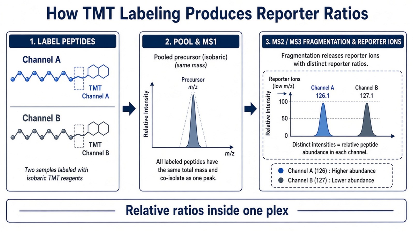
Figure 2. After TMT labeling and pooling, channels share one MS1 precursor; relative quantification comes from reporter-ion intensities released by fragmentation.
For the operational labeling-to-acquisition sequence beyond this mechanism page, researchers can refer to A Detailed Workflow of TMT-Based Quantitative Proteomics. For acquisition choices against ratio compression, see TMT MS3 and SPS-MS3: How to Mitigate Ratio Compression and Enhance Quantitative Accuracy.
Judgment Criteria: When TMT Labeling Fits
Study need |
TMT labeling often fits |
Reassess before labeling |
|---|---|---|
Comparison design |
Relative ratios across a closed multiplex set |
Open-ended enrollment after mixing |
Chemistry |
Peptides can be labeled cleanly at amines |
Amine buffers or incomplete digestion dominate |
Claim type |
Same-peptide relative change inside one plex |
Absolute concentration or calibrated amount |
Acquisition |
DDA methods that read reporters (MS2 or MS3) |
Treating DIA as a TMT labeling step |
Matrix scale |
Groups and replicates fit one commercial kit |
Sample number forces many disconnected plexes without bridging |
Prefer TMT labeling when the cohort can be frozen in one kit and the claim is relative same-run comparison. Prefer unlabeled LFQ or DIA when samples must stay as independent injections. Prefer an absolute AQUA or SIS assay when concentration units are required.
Researchers comparing design-level trade-offs can also refer to Advantages and Disadvantages of TMT-Based Quantitative Proteomics Analysis.
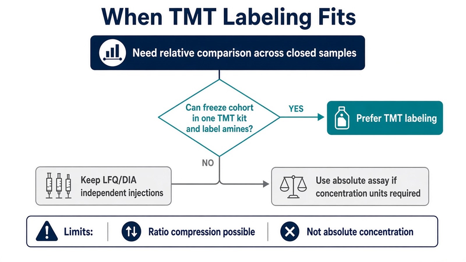
Figure 3. TMT labeling fits a closed relative multiplex design; keep independent unlabeled runs or absolute assays when the claim or enrollment rules differ.
Limits and Application Boundaries
TMT labeling does not invent absolute protein amounts. Ratio compression can underestimate true fold changes in complex digests. Channel count is finite. Cross-plex comparison needs a bridging design. Incomplete amine labeling, residual free tag, and swapped channel maps remain chemistry failures that acquisition cannot erase.
Typical applications are multi-condition drug studies, genotype or treatment panels, time-course designs that fit one kit, and biomarker discovery cohorts that need relative ranking inside a frozen plex. Diagnostic cutoffs and clinical decision rules sit outside ordinary research TMT reporting unless a separately validated assay framework is in place.
Once the channel map, labeling chemistry constraints, and relative-ratio claim are defined, the project can be evaluated as a TMT labeling study. Researchers can review the MtoZ Biolabs TMT Quantitative Proteomics Analysis Service for sample evaluation, feasibility assessment, and workflow planning.
Frequently Asked Questions
1. Why are TMT-labeled peptides isobaric at MS1?
The reporter and balancer masses are designed so that complete tags share effectively the same total mass. Channels separate only after fragmentation releases distinct reporters.
2. Which peptide sites are labeled?
Primarily peptide N-termini and lysine residues after digestion. Chemistry that leaves amines blocked or competes with residual buffer amines reduces labeling completeness.
3. Is the balancer the quantitative signal?
No. Quantitative information comes from reporter-ion intensities. The balancer keeps the precursor isobaric across channels.
4. Does TMT labeling provide absolute quantification?
No. Ordinary TMT reporters support relative ratios inside one plex. Absolute amount needs matched standards and a calibration model.
5. Does MS3 change the labeling mechanism?
No. MS3 or SPS-MS3 changes where reporters are read after labeling and pooling. It does not change the amine-tagging chemistry itself.
Reference
- A. Thompson, J. Schafer, K. Kuhn, S. Kienle, J. Schwarz, G. Schmidt, T. Neumann, C. Hamon (2003). Tandem mass tags: a novel quantification strategy for comparative analysis of complex protein mixtures by MS/MS. Anal. Chem., 75, 1895-1904. https://doi.org/10.1021/ac0262560
- L. Ting, R. Rad, S.P. Gygi, W. Haas (2011). MS3 eliminates ratio distortion in isobaric multiplexed quantitative proteomics. Nat. Methods, 8, 937-940. https://doi.org/10.1038/nmeth.1714
- G.C. McAlister, D.P. Nusinow, M.P. Jedrychowski, M. Wuhr, E.L. Huttlin, B.K. Erickson, R. Rad, W. Haas, S.P. Gygi (2014). MultiNotch MS3 enables accurate, sensitive, and multiplexed detection of differential expression across cancer cell line proteomes. Anal. Chem., 86, 7150-7158. https://doi.org/10.1021/ac502040v
