






Lysosome Isolation and Proteomics Service: Support from Cells and Tissues to LC-MS/MS Analysis
Whether cells or tissues can support lysosome-focused proteomics depends on more than the presence of lysosomes in the starting material. Organelle recovery, membrane integrity, tissue or cellular matrix, and consistency across experimental groups all influence whether the resulting fraction can support protein identification or quantitative comparison.
Lysosome isolation should therefore be designed together with downstream LC-MS/MS rather than treated as an independent preparation step. Starting-material assessment, enrichment strategy, quality evaluation, protein recovery, and analytical objectives need to be aligned from the outset.
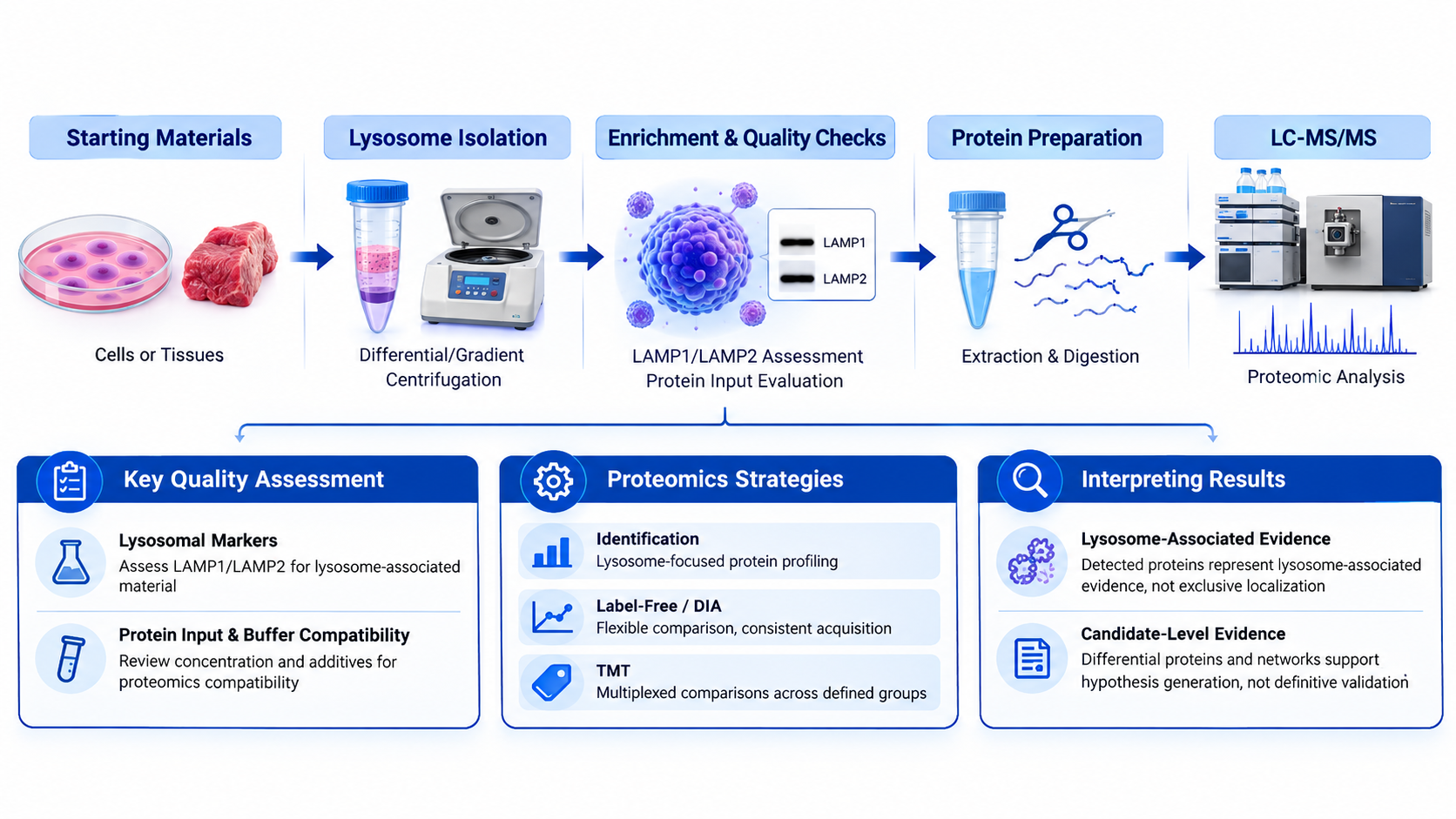
Figure 1. Integrated Workflow from Cells or Tissues to LC-MS/MS-Based Lysosomal Proteomics.
Evaluating Cells and Tissues as Starting Materials
1. Cell-Based Starting Materials
(1) Sample State and Cellular Integrity
Cell collection and storage should preserve enough cellular structure for organelle recovery. Degradation, contamination, uncontrolled lysis, or repeated freeze-thaw exposure may alter membrane integrity and protein composition before lysosome isolation begins, reducing recovery or introducing handling-related signals.
(2) Consistency Across Experimental Groups
Cell number, culture conditions, treatment duration, collection timing, washing, pelleting, and storage should be aligned across groups. Otherwise, differences in enrichment efficiency may be misinterpreted as condition-dependent changes in the lysosomal proteome.
2. Tissue-Based Starting Materials
(1) Sampling Location and Tissue Handling
Tissues introduce spatial and cellular heterogeneity. Sampling position, necrotic content, blood contamination, ischemic interval, preservation, and storage can influence the recovered profile. Comparison groups should use matched anatomical regions and consistent handling whenever the design permits.
(2) Matrix Complexity and Homogenization
Fibrous, lipid-rich, soft, and vascularized tissues respond differently to homogenization. Insufficient disruption can limit organelle release, whereas excessive force can damage lysosomal membranes and increase mixing with other cellular components. Processing conditions should reflect the tissue matrix.
3. Feasibility Before Isolation
(1) Species, Available Material, and Storage History
Feasibility assessment should establish the species, starting material, available amount, sample state, and storage history. These factors influence expected recovery, downstream protein input, and analytical planning. Uncommon species or limited material may require additional review.
(2) Matching Starting Material to the Downstream Objective
Protein identification asks which proteins and peptides are detected in an enriched fraction. Comparative quantification also requires balanced groups, biological replication, consistent handling, and adequate input across samples because uneven material can weaken abundance comparisons.
Connecting Lysosome Isolation to LC-MS/MS
1. Isolation, Enrichment, and Purification
(1) Distinguishing the Three Terms
Lysosome isolation separates a lysosome-containing fraction from the starting sample. Lysosome enrichment means lysosome-associated material has increased relative to the original mixture. Purification makes a stronger claim about removal of other components and should be used only when supported by appropriate evidence.
(2) Why Enrichment Quality Affects Proteomic Coverage
Residual proteins from other compartments can consume analytical capacity, while lysosome loss can reduce detection of lower-abundance proteins. Variation in enrichment between groups may also change measured abundance independently of the biological condition.
2. Quality Assessment Before Proteomics
(1) Lysosomal Marker Assessment
LAMP1 and LAMP2 are commonly used to assess whether a prepared fraction contains lysosome-associated material. Positive marker evidence and adequate protein input support progression to proteomics, but marker detection alone does not establish complete purity or exclude other subcellular components.
(2) Protein Input and Buffer Compatibility
Protein concentration and buffer composition should be reviewed before digestion. Detergents, reducing agents, stabilizers, preservatives, salts, and other additives may interfere with cleanup, digestion, labeling, chromatography, or ionization when present at incompatible levels.
3. Preparing the Enriched Fraction for LC-MS/MS
(1) Protein Extraction and Enzymatic Digestion
Extraction should recover proteins from the enriched fraction while remaining compatible with digestion. Membrane-associated proteins may require stronger solubilization than soluble proteins, but the workflow must still support cleanup and protease activity before peptide analysis by LC-MS/MS.
(2) Controlling Protein Loss and Analytical Interference
Low-input fractions are vulnerable to losses during transfer, precipitation, cleanup, and digestion. Surface adsorption, incomplete resolubilization, or removal of incompatible components can reduce peptide yield, so processing steps should match the available input and analytical objective.
Choosing the Proteomics Readout
1. Protein Identification or Comparative Quantification
(1) Identification for Lysosome-Focused Protein Profiling
Protein identification establishes which proteins and peptides are detected under the selected analytical conditions. It supports lysosome-focused profiling and candidate generation, but detection in an enriched fraction does not prove exclusive lysosomal localization.
(2) Quantification for Defined Group Comparisons
Quantitative proteomics compares relative protein abundance across predefined groups, genotypes, treatments, or time points. The resulting differences are comparative evidence that requires biological interpretation; they do not directly confirm a mechanism.
2. Matching the Quantitative Strategy to Study Design
(1) Label-Free and DIA Workflows
Label-free analysis supports flexible comparison without multiplex labeling, while data-independent acquisition supports consistent acquisition across multiple samples. Selection should consider sample number, input, quantitative consistency, missing-value tolerance, and downstream analysis. Neither approach corrects inconsistent isolation or group design.
(2) TMT for Multiplexed Comparisons
Tandem mass tag quantification combines labeled samples within a multiplexed design. It can support coordinated comparison across defined groups, but feasibility depends on sample number, channel arrangement, protein input, labeling compatibility, and the intended statistical contrasts.
3. Interpreting Lysosomal Proteomics Results
(1) Lysosome-Associated Evidence Versus Organelle Localization
A detected protein represents lysosome-associated evidence within the analyzed preparation. Routine proteomic data do not independently distinguish lysosomal membrane proteins from luminal proteins, define membrane topology, or demonstrate exclusive subcellular localization.
(2) Differential Proteins as Candidate-Level Evidence
Differential proteins, enrichment results, and interaction networks support candidate prioritization and hypothesis development. They do not independently validate a biomarker, drug target, or causal pathway. Follow-up experiments should match the candidate and the claim being tested.
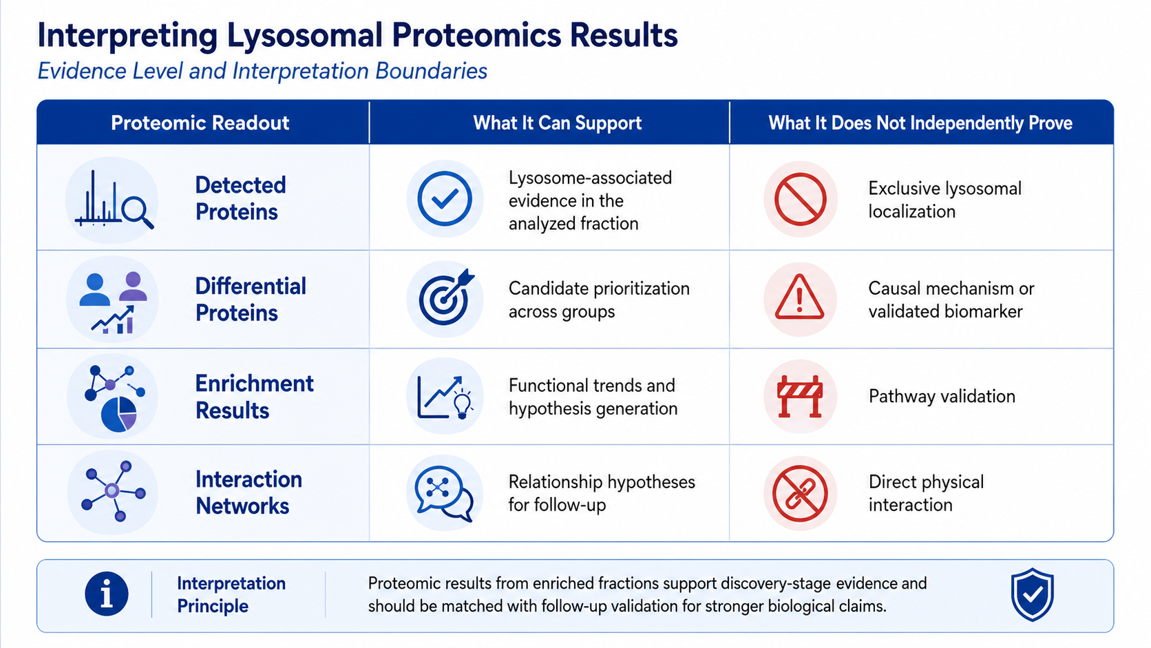
Figure 2. Evidence Levels and Interpretation Boundaries in Lysosomal Proteomics.
Preparing for Project Evaluation
1. Sample Information Needed for Route Selection
(1) Sample Type, State, and Preparation History
Project assessment should document the cell or tissue type, species, treatment, collection method, fresh or frozen status, and previous processing. For prepared fractions, the isolation procedure, buffer composition, protein concentration, and available quality records are also relevant.
(2) Available Material and Handling Records
Available material, collection consistency, storage temperature, transport, and freeze-thaw history influence route selection. These records help separate biological variation from preanalytical variation and show whether comparison groups can supply reasonably matched input.
2. Study Design Information Needed for Analysis Selection
(1) Groups, Controls, and Biological Replicates
Groups, controls, treatments, time points, and biological replicates should be defined before selecting an analytical workflow. A clear design supports balanced processing and determines which comparisons are interpretable. Proteomic analysis cannot recover a biological contrast absent from the sampling plan.
(2) Research Goal and Expected Result Type
The intended result should be defined as protein identification, comparative quantification, differential-protein screening, or candidate prioritization. These objectives require different analytical decisions and evidence thresholds, reducing the risk of treating discovery-stage findings as validated conclusions.
The route from cells or tissues to LC-MS/MS depends on sample condition, enrichment feasibility, available input, group design, and the intended evidence level. MtoZ Biolabs can evaluate eligible starting materials and connect lysosome isolation, LAMP1/LAMP2 assessment, protein preparation, and LC-MS/MS-based identification or Label-free, DIA, or TMT quantification within a project-specific plan. Submit your inquiry below for project evaluation.
MtoZ Biolabs, an integrated chromatography and mass spectrometry (MS) services provider.
Related Services
How to order?




