






Lysosome-Enriched Fraction Proteomics Service: Support for Protein Identification and Quantitative Analysis
A client-prepared lysosome fraction may appear ready for proteomic analysis once separation is complete, but successful fractionation and LC-MS/MS compatibility are different questions. The submitted material still needs to be evaluated for fraction identity, protein recovery, buffer composition, storage history, quality-control evidence, and consistency across experimental groups.
These factors determine whether the preparation is suitable for protein identification, comparative quantification, or further cleanup before analysis. They also shape how confidently detected and differential proteins can be interpreted as lysosome-associated evidence.
Determining Whether the Prepared Fraction Is Ready
1. Defining the Submitted Fraction
(1) Fraction Identity and Separation Endpoint
A lysosome-enriched fraction, broader subcellular fraction, and membrane-rich fraction may differ substantially in composition. The fraction name alone does not reveal which separation steps were completed, where the material was collected, or how selectively lysosomes were enriched. Evaluation should begin with the original sample type, disruption method, fractionation sequence, collection point, and intended enrichment target.
(2) Sample Condition and Storage History
The physical condition of the submitted fraction affects protein integrity and recoverability. Relevant details include whether the material is supplied as a pellet or solution, whether it was stored fresh or frozen, how it was transported, and whether it experienced repeated freeze-thaw cycles. Visible precipitation, degradation, contamination, or prolonged storage under unsuitable conditions may alter the detectable protein profile.
2. Confirming Usable and Comparable Material
(1) Protein Concentration and Recoverable Input
A reported protein concentration is useful, but it does not by itself establish the amount available for LC-MS/MS. Measurement accuracy may depend on the assay method and buffer composition, while cleanup, solubilization, digestion, and transfer steps can reduce recoverable material. Sample evaluation should therefore consider both the reported concentration and the preparation conditions that affect usable peptide input.
(2) Consistency Across Comparison Groups
Comparative proteomics depends on consistency before mass spectrometry begins. Fractions from different groups should be prepared using comparable starting material, disruption conditions, centrifugation or separation steps, wash procedures, storage conditions, and concentration measurements. Otherwise, variation in fraction recovery or background composition may be mistaken for treatment-related or genotype-related protein changes.
Reviewing Fractionation Records and Quality Evidence
1. Documenting the Preparation Workflow
(1) Isolation Sequence and Collection Details
A complete preparation record should describe how the starting cells or tissues were processed and how the submitted fraction was obtained. Useful information includes disruption conditions, separation steps, collected layers or pellets, wash procedures, resuspension conditions, and any concentration or precipitation steps. These records help identify possible sources of protein loss, organelle disruption, and non-target background.
(2) Buffer, Detergent, and Additive Composition
The complete composition of the final sample buffer is more informative than a general buffer name. Detergents, salts, reducing agents, stabilizers, preservatives, protease inhibitors, and other additives can affect protein assays, precipitation, enzymatic digestion, labeling reactions, chromatography, or ionization. Compatibility depends on the component, its concentration, the available input, and the cleanup strategy that can be applied.
2. Interpreting Quality-Control Evidence
(1) Lysosomal Marker Evidence
LAMP1 and LAMP2 are commonly assessed as lysosome-associated markers. Their detection supports the presence of lysosomal material in the prepared fraction, especially when interpreted relative to the starting sample or another fraction. Marker positivity, however, does not demonstrate that the preparation contains only lysosomes or exclude proteins originating from other organelles, membranes, or the cytosol.
(2) Purity Evidence and Its Limits
Existing Western blot results, electrophoretic profiles, protein concentration records, and other quality data can support sample assessment, but each addresses a different question. A marker confirms the presence of a target-associated component, while a broader contamination panel provides additional information about fraction selectivity. Without sufficient purity evidence, terms such as lysosome-enriched or lysosome-associated are more appropriate than highly purified lysosomes.
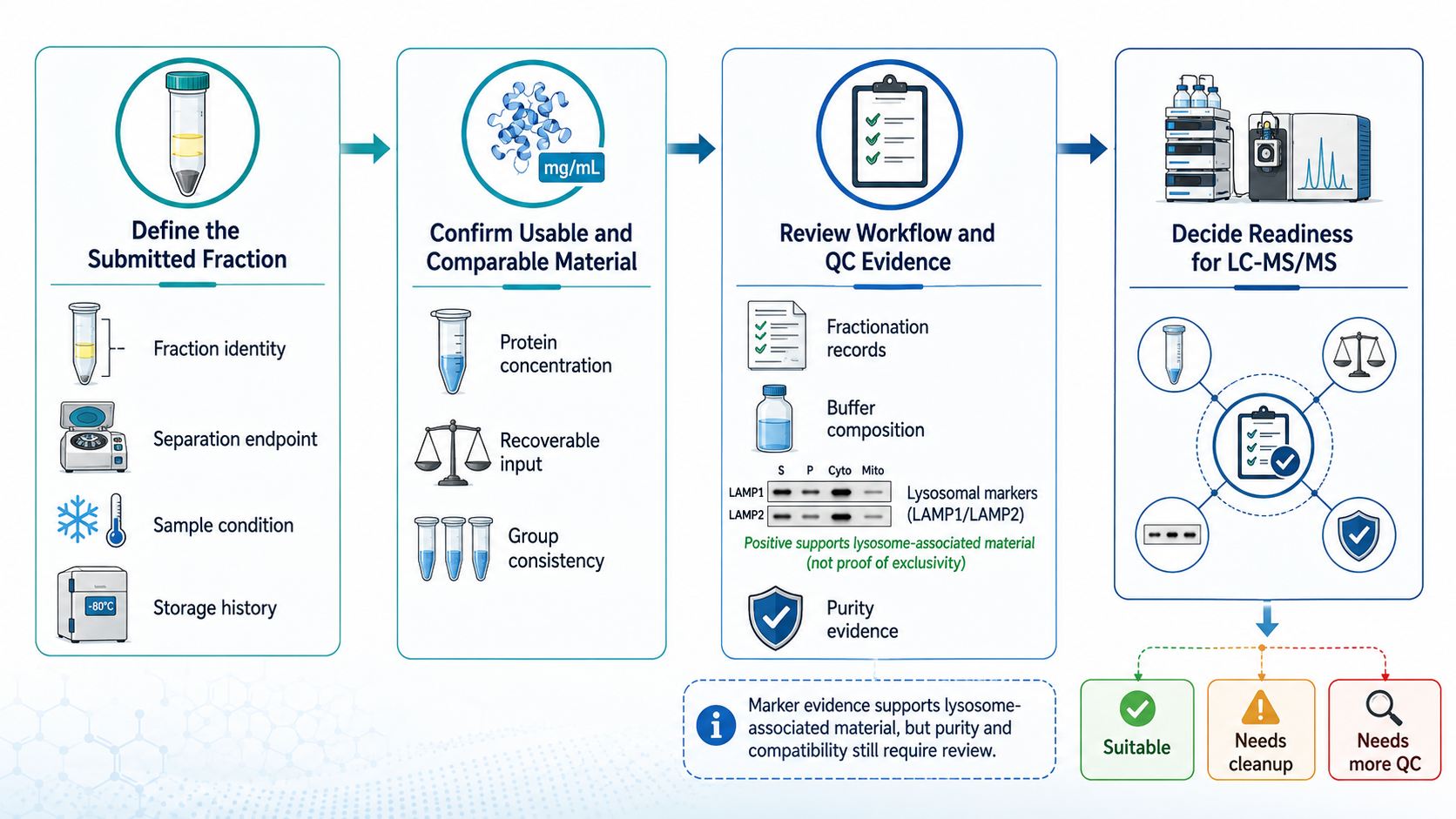
Figure 1. Assessment workflow for determining the LC-MS/MS readiness of a client-prepared lysosome fraction.
Connecting the Fraction to LC-MS/MS
1. Assessing Proteomic Compatibility
(1) Digestion, Cleanup, and Ionization Risks
A fraction may contain enough protein yet remain unsuitable for direct digestion because of its chemical environment. Incompatible detergents or additives can reduce cleanup recovery, inhibit protease activity, interfere with labeling, distort chromatographic separation, or suppress ionization. The appropriate preparation route depends on the sample composition, protein input, analytical method, and expected level of quantitative consistency.
(2) Membrane-Rich Fractions and Protein Solubilization
Membrane-rich fraction proteomics presents additional challenges because hydrophobic and membrane-associated proteins may be difficult to solubilize and digest. Stronger extraction conditions may improve recovery but can introduce components that require removal before LC-MS/MS. The workflow should balance protein extraction, peptide recovery, digestion efficiency, and downstream analytical compatibility rather than optimizing only one step.
2. Selecting the Analytical Objective
(1) Protein Identification
Protein identification determines which proteins and peptides are detected in the submitted fraction under the selected analytical conditions. This approach can establish a lysosome-focused or subcellular fraction protein profile and support candidate generation. Detection does not independently prove exclusive lysosomal localization, membrane topology, or a direct functional role in lysosomal biology.
(2) Comparative Quantification
Quantitative proteomics measures relative protein abundance across predefined groups, such as treatments, controls, genotypes, models, or time points. The resulting differences reflect both biology and upstream sample preparation. Reliable interpretation therefore requires comparable fractionation, adequate biological replication, consistent sample handling, and a statistical design aligned with the planned contrasts.
3. Matching Quantification to Study Design
(1) Label-Free and DIA Strategies
Label-free analysis supports flexible comparison across sample sets without multiplex labeling. Data-independent acquisition can improve acquisition consistency across larger comparative studies. Selection between these strategies should consider sample number, available input, expected missingness, quantitative consistency, and analysis goals. Neither strategy can remove variation introduced by inconsistent fraction preparation.
(2) TMT-Based Comparison
Tandem mass tag quantification allows multiple samples to be analyzed within a multiplexed design. It may suit studies requiring coordinated comparison across several predefined groups, but feasibility depends on sample number, channel arrangement, protein input, labeling compatibility, and balance across samples. Experimental contrasts should be planned before channel assignment rather than reconstructed after data acquisition.
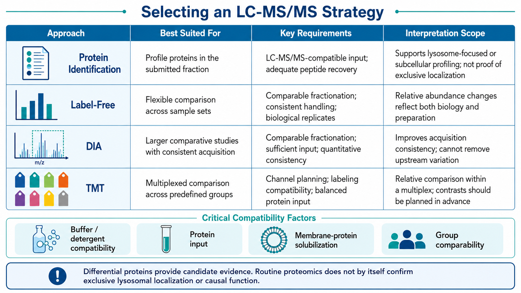
Figure 2. Comparison of LC-MS/MS strategies for client-prepared lysosome-enriched fractions.
Interpreting Results and Preparing Submission
1. Defining the Evidence Level
(1) Lysosome-Associated Detection Versus Localization
Proteins detected in a client-prepared lysosome fraction represent evidence from that specific enriched preparation. Routine proteomic results do not independently distinguish lysosomal membrane proteins from luminal proteins, confirm exclusive organelle localization, or establish that every detected protein is a resident lysosomal component. Localization claims require evidence designed specifically for that question.
(2) Differential Proteins as Candidate Evidence
Differential abundance, functional enrichment, and protein interaction analysis can support candidate prioritization and hypothesis development. These outputs remain discovery-stage or comparative evidence. They do not by themselves validate a causal mechanism, disease biomarker, therapeutic target, or functional consequence. Follow-up experiments should be selected according to the specific claim and candidate under investigation.
2. Assembling Information for Project Evaluation
(1) Fractionation, Buffer, Concentration, and QC Records
Submission assessment should include the original sample type, complete fractionation procedure, submitted fraction identity, final buffer composition, protein concentration method, storage conditions, transport history, and available quality-control records. Providing only a sample label and volume may not supply enough information to determine cleanup requirements or LC-MS/MS compatibility.
(2) Groups, Replicates, and Expected Outputs
The project description should also define experimental groups, controls, biological replicates, sample relationships, and the intended result type. Protein identification, comparative quantification, differential-protein screening, and candidate prioritization require different study designs and analytical decisions. Clarifying the expected output reduces the risk of applying a technically feasible method to a question it cannot adequately answer.
The suitability of a client-prepared fraction depends on how it was generated, what it contains, whether its buffer is compatible with downstream processing, and whether the samples are comparable across the study design. MtoZ Biolabs can evaluate client-prepared lysosome-enriched, subcellular, or membrane-rich fractions and plan LC-MS/MS protein identification or Label-free, DIA, or TMT quantification based on fractionation records, sample condition, protein input, quality evidence, experimental groups, and research objectives. Submit your inquiry below for project evaluation.
MtoZ Biolabs, an integrated chromatography and mass spectrometry (MS) services provider.
Related Services
How to order?




