






Lysosomal Proteomics Analysis Service: Support from Lysosome Isolation to LC-MS/MS and Bioinformatics
Lysosomal protein profiling measures the proteins recovered from a lysosome-enriched preparation, not the subcellular location of every detected protein. The distinction matters because enrichment efficiency, non-lysosomal background, protein recovery, and sample handling can shape the final dataset before LC-MS/MS acquisition begins.
A technically coherent lysosomal proteomics analysis therefore links the starting sample, enrichment evidence, analytical objective, mass spectrometry design, bioinformatics, and interpretation criteria. Each stage contributes a different type of evidence, and weaknesses introduced early in the workflow cannot be fully corrected during data analysis.
Establishing a Lysosome-Focused Sample Basis
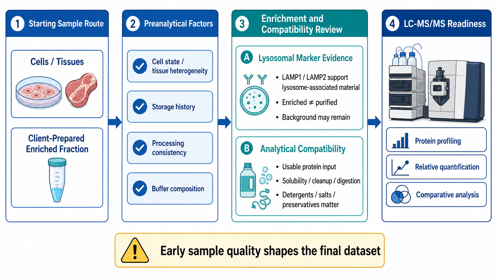
Figure 1. Sample Preparation Factors in Lysosomal Proteomics
1. Selecting the Starting Sample Route
(1) Cells and Tissues Requiring Lysosome Isolation
Cells and tissues require controlled disruption that releases intracellular organelles without causing excessive membrane damage or uncontrolled mixing of cellular contents. Cell state, tissue heterogeneity, storage history, and processing consistency influence lysosome recovery and the abundance of background proteins. Comparative studies also require matched handling across groups because differences in collection or homogenization can resemble biological remodeling in the final proteomic profile.
(2) Client-Prepared Enriched Fractions
A client-prepared lysosome-enriched, subcellular, or membrane-rich fraction represents a later starting point, but the fraction identity still requires technical review. The separation sequence, collected fraction, buffer composition, protein state, storage conditions, and available quality records determine whether the material is compatible with protein extraction, digestion, and LC-MS/MS. A fraction label alone does not establish enrichment quality or analytical readiness.
2. Evaluating Enrichment and Analytical Compatibility
(1) Lysosomal Marker Evidence and Fraction Selectivity
LAMP1 and LAMP2 measurements can support the presence of lysosome-associated material in an enriched fraction. Marker detection is a technical quality indicator rather than a complete purity assessment. Other organelles, cytosolic proteins, and membrane fragments may remain in the preparation, so the term lysosome-enriched is generally more defensible than purified lysosomes unless broader evidence supports the stronger claim.
(2) Protein Input and Chemical Compatibility
The reported protein concentration does not necessarily equal the amount that remains available after cleanup and digestion. Solubility, sample loss, detergents, salts, reducing agents, stabilizers, and preservatives can affect protein assays, protease activity, peptide recovery, chromatography, and ionization. Analytical compatibility should therefore be evaluated from both usable input and complete chemical composition rather than nominal concentration alone.
Designing the LC-MS/MS Analysis
1. Defining the Analytical Objective
(1) Protein Identification for Lysosomal Protein Profiling
Protein identification establishes which peptides and proteins are detected under defined analytical conditions. The resulting lysosomal protein profile supports composition mapping and candidate generation within the enriched preparation. It does not provide reliable relative abundance comparisons unless the experiment and data processing are designed for quantification, and it does not independently confirm exclusive lysosomal localization.
(2) Quantification for Defined Biological Comparisons
Quantitative proteomics compares relative protein abundance among predefined conditions, such as treatments, controls, genotypes, models, or time points. Biological replication, balanced groups, consistent enrichment, and a planned statistical contrast are necessary for interpretable comparisons. Observed abundance differences may reflect biological remodeling, enrichment variability, or both, so the analytical design must preserve information needed to separate these possibilities.
2. Separating Quantification and Acquisition Decisions
(1) Label-Free and TMT-Based Quantification
Label-free and tandem mass tag approaches describe different strategies for organizing quantitative comparisons. Label-free designs measure samples separately and retain flexibility when sample numbers change, whereas TMT combines labeled samples within a multiplexed experiment. Selection depends on sample number, input, batch structure, desired comparisons, and labeling compatibility. These choices concern quantification design rather than organelle specificity.
(2) DIA as an Acquisition Strategy
Data-independent acquisition describes how precursor and fragment-ion data are collected across broad mass ranges. DIA can support consistent peptide measurement across multi-sample studies, but it is not the same classification dimension as Label-free or TMT. A label-free quantitative study may use DIA acquisition, while TMT commonly relies on isobaric reporter-ion quantification. Distinguishing these dimensions prevents method names from being treated as interchangeable alternatives.
Converting Mass Spectrometry Data into Biological Evidence
1. Building Protein-Level Results
(1) Peptide and Protein Identification Evidence
Raw spectra are converted into peptide and protein evidence through database searching, identification scoring, false-discovery control, and protein inference. The resulting protein list depends on spectral quality, sequence-database coverage, peptide uniqueness, and inference rules. A reported protein should therefore be interpreted through its supporting peptide evidence rather than treated as an unqualified presence-or-absence statement.
(2) Quantitative Matrices and Differential Analysis
Quantitative matrices organize relative abundance values across samples and form the basis for statistical comparison. Data normalization, missingness, variance, replicate consistency, and the predefined comparison model influence differential results. A statistically significant difference indicates a reproducible abundance pattern under the selected model; it does not by itself establish a functional mechanism or causal relationship.
2. Adding Functional and Network Context
(1) Functional Enrichment and Pathway Analysis
Gene Ontology and pathway analysis summarize whether particular functions or biological processes occur more frequently in a selected protein set than expected from an appropriate background. Results depend on annotation coverage, background definition, candidate-selection criteria, and statistical correction. Functional enrichment supports interpretation at the group level but does not prove that every protein in the category drives the observed phenotype.
(2) Protein Interaction and Candidate Prioritization
Protein interaction networks combine known or predicted relationships with quantitative changes and functional annotations to organize candidate proteins. Network position, differential abundance, pathway membership, and consistency across replicates can support prioritization. These features remain interpretive evidence and do not substitute for direct interaction assays, localization experiments, or functional validation.
Preserving Interpretation Across the Workflow
1. Controlling Technical and Biological Variation
(1) Matched Groups and Biological Replication
Experimental groups should be matched for sample collection, storage, enrichment, protein preparation, and analytical processing. Biological replicates estimate variation among independent specimens, whereas technical controls assess specific procedural effects. Neither compensates for a confounded design in which sample condition, preparation batch, and biological group change together.
(2) Separating Enrichment Variability from Protein Remodeling
Differences between lysosome-enriched fractions may arise from altered lysosome abundance, changes in protein composition, variable recovery, membrane disruption, or residual background. Interpretation should therefore consider marker evidence, total protein recovery, fractionation consistency, and the broader biological design. Differential abundance is most informative when technical variation has been constrained and alternative explanations remain visible.
2. Defining the Final Evidence Level
(1) Lysosome-Associated Detection Versus Confirmed Localization
Detection in a lysosome-enriched preparation supports lysosome-associated evidence for the analyzed sample. Routine LC-MS/MS does not independently distinguish membrane proteins from luminal proteins, define membrane topology, or demonstrate exclusive lysosomal localization. Claims about subcellular localization require experiments specifically designed to resolve location rather than composition alone.
(2) Candidate Findings Versus Experimental Validation
Differential proteins, enriched pathways, interaction networks, and ranked candidates belong to discovery or comparative analysis. They can guide subsequent hypotheses and experimental priorities, but they do not establish a validated biomarker, confirmed mechanism, or therapeutic target. The required follow-up depends on the precise claim, the protein of interest, and the biological system.
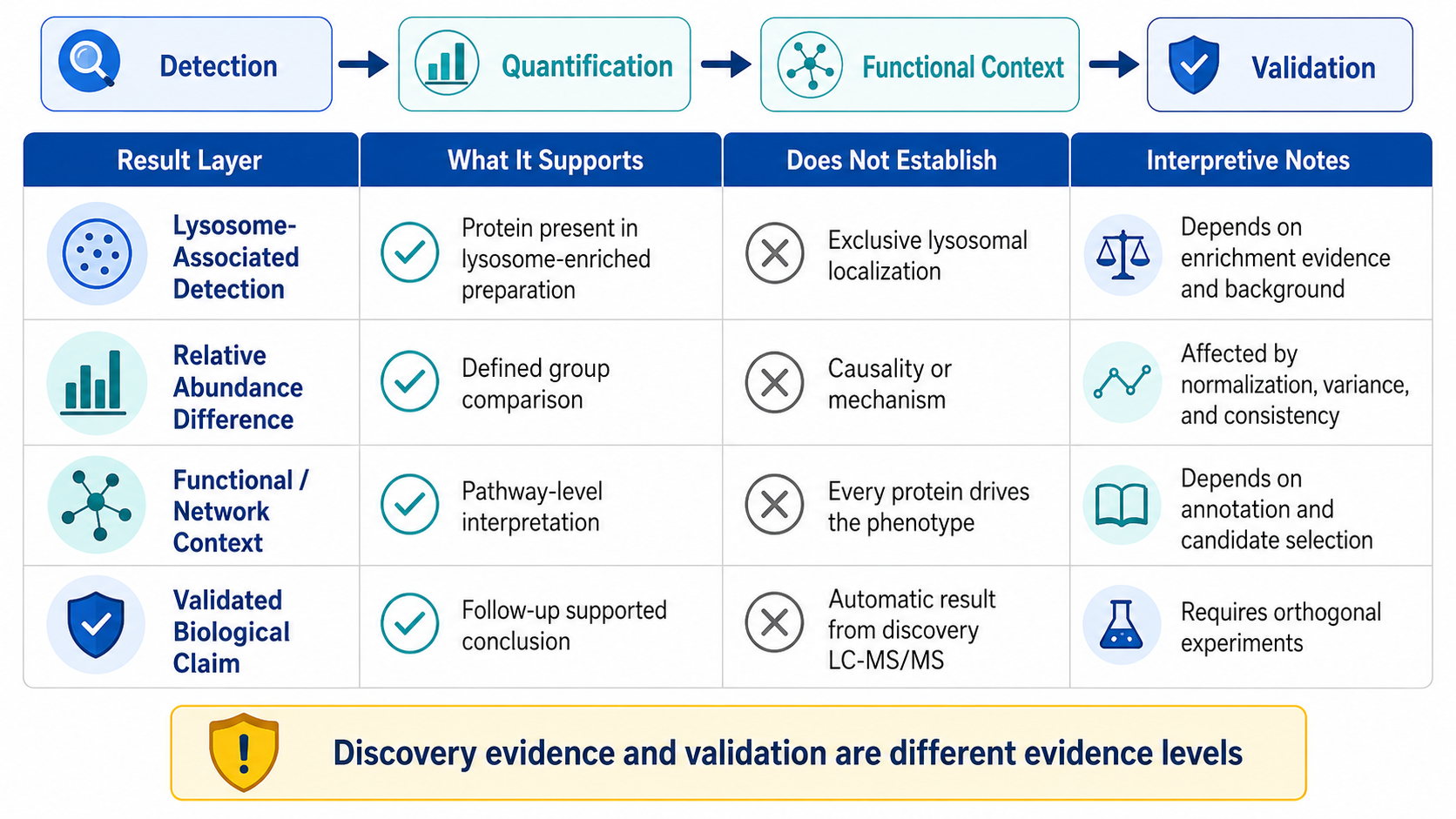
Figure 2. Evidence Boundaries in Lysosomal Protein Interpretation
A coherent lysosomal proteomics study aligns the starting material, enrichment evidence, quantitative design, LC-MS/MS acquisition, bioinformatics, and interpretation threshold. MtoZ Biolabs supports project evaluation from eligible samples or client-prepared enriched fractions through lysosomal protein identification, Label-free, DIA, or TMT analysis and bioinformatics, based on sample condition, group design, available material, and research objective. Submit your inquiry below for project evaluation.
MtoZ Biolabs, an integrated chromatography and mass spectrometry (MS) services provider.
Related Services
How to order?




