


Immuno Transmission Electron Microscopy (immuno-TEM) Analysis Service
- Submit samples as fresh as possible to minimize antigen degradation.
- Fix tissue or cell samples promptly after collection to preserve ultrastructure.
- Provide relevant background information, including sample type, source, and research objectives, which allows us to design suitable protocols.
Immuno-transmission electron microscopy (immuno-TEM) is a high-resolution imaging technique that integrates immunolabeling with transmission electron microscopy. By coupling electron-dense markers such as gold particles to specific antibodies, immuno-TEM enables precise localization of proteins or antigens at the subcellular level and reveals their spatial distribution within cells and tissues. This approach provides direct visualization of molecular targets in biological samples and is widely applied in neurobiology, nuclear protein research, immune cell typing, and studies of molecular mechanisms, offering valuable insights into cellular function and disease processes.
MtoZ Biolabs offers Immuno Transmission Electron Microscopy (immuno-TEM) Analysis Service with advanced imaging platforms and a skilled scientific team, delivering reliable datasets and expert experimental support for research at both the cellular and molecular levels.
Technical Principles
The principle of immuno-TEM is based on the specific binding of antibodies to target molecules, which are visualized using conjugated gold particles or other electron-dense probes. Under electron beam irradiation, gold particles scatter electrons strongly and appear as high-contrast signals in TEM images, thereby pinpointing molecular localization. By integrating ultrastructural morphology with molecular information, this approach provides clear and direct evidence to support the investigation of cellular functions and biological processes.
Services at MtoZ Biolabs
MtoZ Biolabs’ Immuno Transmission Electron Microscopy (immuno-TEM) Analysis Service offers a complete workflow from sample fixation and immunolabeling to TEM imaging and data interpretation. Depending on the sample type and research objectives, we apply either resin embedding or cryosectioning to preserve antigenicity and maintain ultrastructural integrity.
For labeling, we employ a diverse range of markers, including ferritin and horseradish peroxidase (HRP), tailored to different experimental requirements. Antibody concentrations, dilution conditions, and staining protocols are carefully optimized to reduce nonspecific binding and ensure high specificity and reproducibility. Whether the goal is to localize membrane proteins, cytoplasmic proteins, or antigens within subcellular structures, MtoZ Biolabs provides high-resolution images with robust molecular specificity.
Analysis Workflow
1. Sample Fixation
Cells and tissues are preserved using appropriate chemical fixatives to maintain structure.
2. Embedding and Sectioning
Samples are embedded in resin, and ultrathin sections are prepared.
3. Immunolabeling
Target molecules are labeled using antibodies conjugated with gold particles.
4. Staining and Contrast Enhancement
Heavy metal stains are applied to increase contrast.
5. TEM Imaging
High-resolution images of ultrastructure and molecular localization are acquired under transmission electron microscopy.
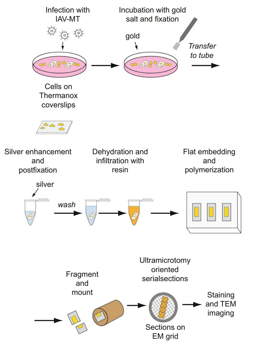
Sachse, M. et al. Methods Mol Biol. 2018.
Figure 1. Tmmuno-TEM Sample Preparation and Imaging Workflow
Why Choose MtoZ Biolabs?
✅ Flexible labeling systems with options such as ferritin and HRP, suitable for varied localization needs.
✅ High-resolution imaging that visualizes ultrastructure alongside precise antigen localization.
✅ Optimized fixation strategies that preserve both ultrastructure and antigenicity.
✅ Enhanced specificity and reliability through optimized antibody concentrations and labeling conditions.
✅ Expert technical support, offering customized experimental design and professional data interpretation.
Sample Submission Suggestions
To ensure successful analysis, clients are encouraged to follow these recommendations:
For samples with limited quantity or special requirements, please contact MtoZ Biolabs in advance for customized handling advice.
FAQs
Q1: Can Immuno-TEM Be Used for Quantitative Analysis?
A1: Immuno-TEM is primarily qualitative, providing high-resolution molecular localization. Under carefully designed experimental conditions, however, semiquantitative analysis can be achieved by evaluating the density of labeling particles. MtoZ Biolabs supports not only imaging but also experimental design optimization to improve the reliability of quantitative studies.
Q2: How Does Immuno-TEM Differ From Conventional TEM?
A2: Conventional TEM visualizes overall ultrastructure, while immuno-TEM adds molecular localization information. The two methods are complementary, allowing simultaneous structural and functional analysis. MtoZ Biolabs helps researchers integrate both datasets for comprehensive scientific insights.
Q3: What Factors Affect Immuno-TEM Performance?
A3: Key factors include the choice of labeling probes, fixative type and concentration, antigen preservation, antibody dilution, and staining procedures. MtoZ Biolabs takes all these into account during protocol design to ensure reliable and accurate results.
Choosing MtoZ Biolabs Immuno Transmission Electron Microscopy (immuno-TEM) Analysis Service ensures access to high-resolution imaging, rigorous experimental standards, and expert interpretation. Our service not only provides reliable structural insights but also supports translational applications across life sciences and biomedical research. For customized project solutions and detailed consultation, please contact MtoZ Biolabs and explore how our expertise can advance your research.
How to order?

