



Lysine lactylation (Kla) can change what a named lysine is chemically able to do at a written contact. The ε-amino group loses its positive charge when it is N-acylated. The lactyl moiety adds steric bulk and a hydroxyl that can donate or accept a hydrogen bond. Those three chemical facts are the usable answer to how lactylation can influence protein–molecule interactions. They are not proof that the protein remakes its contacts with DNA, RNA, small molecules, or other proteins, and they do not grant cell-fate, metabolic, or disease sentences.
A Kla site table is a peptide-level inventory. It is not a binding table. Antibody enrichment plus liquid chromatography–tandem mass spectrometry (LC-MS/MS) can nominate residues. It does not measure affinity, occupancy at the contact, or a new preference. Researchers who already have a lysate or a tissue pellet and need that site table can review the MtoZ Biolabs Protein Lactylation Modification Analysis Service for project-specific enrichment and site-assignment planning.
Charge, Bulk, and Hydrogen Bonds Are Chemical Possibilities
The original charge paragraph remains usable if it is read as chemistry, not as a measured electrostatic collapse. Lysine is cationic at physiological pH. Kla converts that residue to a neutral acyl-lysine. Electrostatic attraction to a phosphate backbone or to a phosphorylated partner can weaken at that residue. The word markedly is not a measurement. Disruption of an electrostatic network, loss of the original selectivity, and a new binding preference are later claims. They require a named contact and an assay that reports that contact.
The polar acyl group also occupies volume that the unmodified side chain did not occupy. If the lysine sits at a written interface or inside a packed motif, that volume can clash, tilt a loop, or leave the interface unchanged. Local conformational rearrangement is a possibility, not an automatic remodeling of the binding geometry. Exposure or burial of a second residue is likewise a possibility. Coordinated effects across multiple regulatory layers are not a granted readout.
The hydroxyl on the lactyl moiety can participate in hydrogen bonds that acetyl-lysine cannot form in the same way. A new hydrogen-bond donor or acceptor can stabilize one pose or compete with a pre-existing pair. It can also do neither. A shift in the hydrophobic–hydrophilic patch around that lysine is a surface description. It is not evidence that a substrate, a cofactor, or a protein partner now binds differently.
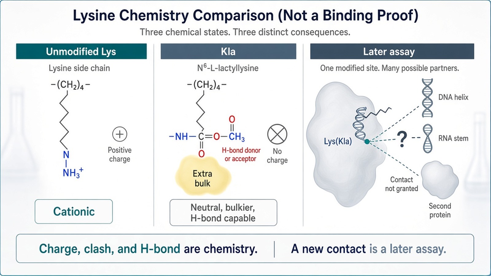
Figure 1. Unmodified lysine is cationic. Kla is a neutral acyl-lysine with extra volume and a hydroxyl. Charge, clash, and hydrogen-bond possibilities are chemistry. A new DNA, RNA, or protein contact is a later assay.
Write the lysine, the partner class, and the assay before any sentence that Kla remade the contact.
A Site Table Is Not a Binding Table
High-resolution mass spectrometry after Kla-directed enrichment can identify lactylated peptides and, when the spectrum supports it, the modified residue. Relative peak areas can rank a written contrast between two conditions. That file is the foundation for choosing a site. It is not a functional analysis of binding. Antibody specificity among acyl-lysine marks remains a design limit. A site that appears only after enrichment still has to survive an unmodified-lysine search and a localization score.
Site-directed substitutions test the lysine. They do not install Kla. K→Q removes charge and is a conventional acetyl-lysine charge mimic. It is smaller than lactyl-lysine and lacks the hydroxyl. K→E reverses charge rather than neutralizing it. K→R retains charge. Those alleles can change a pull-down, an electrophoretic mobility-shift assay (EMSA), a co-immunoprecipitation (Co-IP), surface plasmon resonance (SPR), or isothermal titration calorimetry (ITC) readout. The change reports the lysine, not the lactyl mark. For primer architecture on a complementary whole-plasmid pair, How Are Primers Designed for Site-Directed Mutagenesis in Protein Interaction Studies is the complementary oligo page. A closer chemical mimic, when the project needs the lactyl moiety itself, is a separately written incorporation method, not a default glutamine.
A reconstituted binding assay then asks whether that written site changes recovery or affinity of a named partner. Pull-down reports what a ligand retained in vitro. Co-IP reports what survived an antibody wash from a lysate. SPR and ITC report an in-vitro isotherm when both partners are soluble and the surface or cell is written. None of those formats is Kla-specific. For how a retained band is read after an immobilized ligand, How to Interpret the Results of a Pull-Down Experiment is the complementary pull-down page.
Molecular modeling and molecular-dynamics trajectories can place the lactyl group on a starting coordinate set and watch local clashes or hydrogen bonds. The trajectory is a prediction under the force field and the starting pose. It is not mechanistic insight into how Kla governs binding, and it does not replace a measured isotherm or a recovered partner.
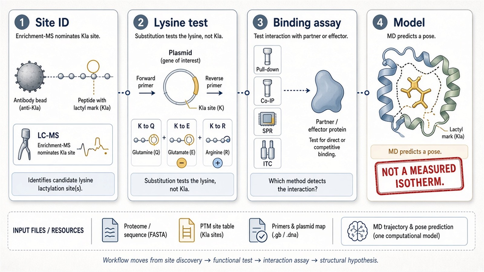
Figure 2. Enrichment-MS nominates a Kla site. A substitution tests the lysine, not the lactyl mark. Pull-down, Co-IP, SPR, or ITC then tests a named partner. A dynamics trajectory remains a model.
A Systems Sentence Cannot Borrow an Interactome
Protein–molecule contacts can be drawn as a network. Drawing them does not make Kla a cascade at multiple organizational levels. SH2 domains read phosphotyrosine. Bromodomains read acetyl-lysine. Kla may sit near those motifs on a particular protein. That proximity does not alter the recognition code of the domain as a class, and it does not grant a new phospho- or acetyl-mark reader function.
Whether a subunit remains inside a complex is a recovery or a structural question about that complex. Kla can be present on one subunit without deciding assembly. Affinity for an organelle membrane, a nucleic-acid fold, or a cytoskeletal filament is likewise a later localization or binding claim. Kla is not a finely tuned regulatory mediator that integrates structural, chemical, and spatial cues. It is a lysine mark that can be mapped and then tested at a written contact.
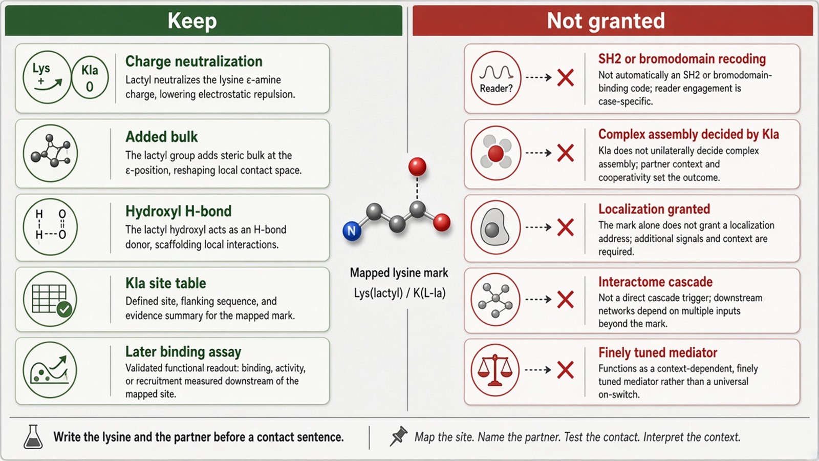
Figure 3. Keep: charge neutralization, added bulk, a hydroxyl, a site table, and a later binding assay. Not granted: SH2 or bromodomain recoding, complex assembly, localization, or a network cascade.
Experimental Limits
This page reduces the Kla-binding question to one named lysine and one named partner class. Cell-fate determination, metabolic regulation, disease progression, and a future central focus for protein functional diversity sit outside ordinary research reporting unless a separately validated phenotype is already named. Decoding complex regulatory networks is a slogan, not a step after a site list.
Project Planning and Service Scope
A usable request names the sample, the enrichment reagent, the residue that must be localized, and the orthogonal binding format that would be required before a contact sentence. MtoZ Biolabs scopes Kla enrichment and LC-MS/MS site assignment on Orbitrap-class instruments. Interpretability still follows the spectrum and the localization score, not a high-precision omics heading.
Once the next sentence is a lysate co-recovery after a named site, this Kla-chemistry page should hand off. Researchers who need that recovery table can review the MtoZ Biolabs Co-IP-MS Protein Interaction Analysis Service for project-specific sample evaluation, feasibility assessment, and workflow planning.
Frequently Asked Questions
1. Does Kla always weaken electrostatic binding to DNA or RNA?
It removes the lysine charge at that residue. Whether the contact weakens depends on whether that lysine was carrying the interaction.
2. Does a Kla site table show a new binding preference?
It shows a modified peptide. Preference is a later assay.
3. Do K→Q or K→E substitutions mimic Kla?
They test the lysine. Q is a charge-neutral substitute, not a lactyl group. E reverses charge.
4. Does molecular dynamics explain how Kla governs binding?
It predicts poses under a model. It does not measure binding.
5. Does Kla recode SH2 or bromodomain recognition?
Not as a class. A nearby Kla site still has to be tested on that protein.
Reference
- Zhang D., Tang Z., Huang H., et al. (2019). Metabolic regulation of gene expression by histone lactylation. Nature, 574, 575-580. https://doi.org/10.1038/s41586-019-1678-1
- Choudhary C., Weinert B.T., Nishida Y., Verdin E., Mann M. (2014). The growing landscape of lysine acetylation links metabolism and cell signalling. Nat. Rev. Mol. Cell Biol., 15, 536-550. https://doi.org/10.1038/nrm3841
- Huang H., Lin S., Garcia B.A., Zhao Y. (2015). Quantitative proteomic analysis of histone modifications. Chem. Rev., 115, 2376-2418. https://doi.org/10.1021/cr500491u
- Olsen J.V., Mann M. (2013). Status of large-scale analysis of post-translational modifications by mass spectrometry. Mol. Cell. Proteomics, 12, 3444-3452. https://doi.org/10.1074/mcp.O113.034181
