



Ratio compression in tandem mass tag (TMT) workflows is diagnosed when measured reporter-ion fold changes are systematically closer to one than expected biological contrasts, especially in complex digests acquired with MS2 reporters. The usual cause is co-isolation: nontarget peptides that enter the precursor window also release reporters and dilute channel differences. Synchronous precursor selection MS3 (SPS-MS3) on compatible Orbitrap Tribrid methods reduces that mixing by reading reporters after a second fragmentation of selected MS2 fragments. It does not repair unmatched channel loads, incomplete labeling, or a broken plex map. Absolute protein amount still requires matched standards outside ordinary TMT discovery tables.
Researchers who already have a closed TMT plex or a ratio-compression concern can also review the MtoZ Biolabs TMT Quantitative Proteomics Analysis Service for project-specific feasibility and acquisition planning.
Diagnose Compression Before Changing Acquisition
Treat compressed ratios as a troubleshooting tree, not as a single instrument upgrade.
Symptom or finding |
Likely cause |
First corrective check |
|---|---|---|
Fold changes smaller than orthogonal assays or known spike ratios |
MS2 co-isolation / ratio compression |
Review isolation width, fractionation depth, and MS2 vs MS3 plan |
One channel globally high or low |
Load mismatch before labeling |
Recheck protein assay and digest yields |
Many unlabeled or partially labeled peptides |
Labeling chemistry failure |
Review amine buffers, reagent excess, quench, and cleanup |
Channel map does not match sample IDs |
Mixing or annotation error |
Rebuild channel map; do not normalize away the swap |
Sparse IDs after switching to MS3 |
Cycle-time trade-off |
Expect coverage loss; adjust fractionation or gradient if needed |
SPS-MS3 is the right branch when co-isolation is the dominant problem and the instrument supports the scan type. It is the wrong first branch when upstream chemistry already confounds the plex.
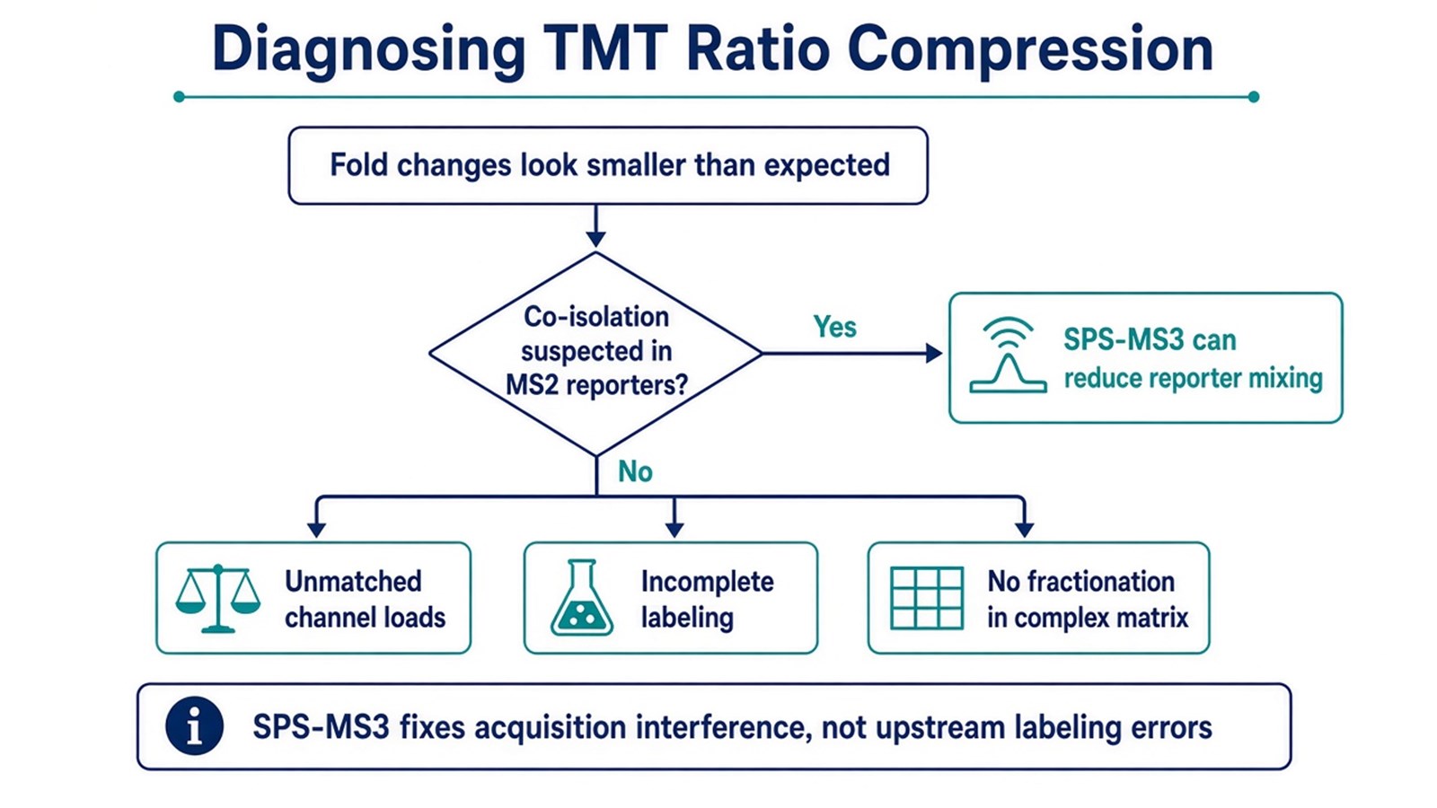
Figure 1. SPS-MS3 addresses MS2 reporter mixing; unmatched loads and incomplete labeling remain separate upstream failures.
If fold changes look compressed, confirm whether the problem is reporter isolation before retuning every search filter.
How SPS-MS3 Is Utilized Inside a TMT Workflow
SPS-MS3 changes where reporters are collected. It does not change the TMT claim: relative channel ratios inside one closed plex.
1. Keep the labeling workflow matched
Match protein loads, complete amine labeling, remove excess tag, and freeze the multiplex set before acquisition. SPS-MS3 cannot invent a biological difference that never entered the pool.
2. Reduce peptide density when the matrix is complex
High-pH reversed-phase or related fractionation lowers co-elution density and reduces how often nontarget precursors share the isolation window. Fractionation and SPS-MS3 are complementary controls, not substitutes for each other.
3. Acquire MS2 for sequence, then SPS for reporters
After precursor isolation and MS2 fragmentation, the method selects multiple high-intensity peptide fragment ions and fragments them together in one MS3 event. Reporter ions measured at MS3 come mainly from that selected fragment set rather than from every ion in the original precursor window (McAlister et al. 2014; Ting et al. 2011).
4. Review method and QC settings that decide whether the gain appears
Useful records include SPS notch count, MS3 resolution adequate for the TMT reporter set, collision energy for reporter release, isolation width, and filters such as precursor co-isolation or reporter signal-to-noise. Exact numeric setpoints depend on instrument class and plex format; copying a published NCE without local QC does not guarantee the intended accuracy gain.
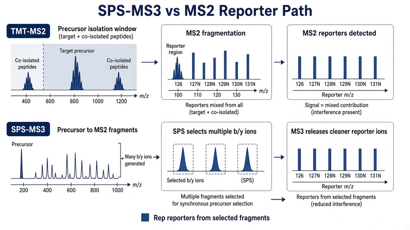
Figure 2. SPS-MS3 utilizes a second fragmentation of selected MS2 fragments so TMT reporters are less mixed by co-isolated precursors.
For a deeper comparison of MS2, MS3, and SPS-MS3 accuracy versus coverage, researchers can refer to TMT MS3 and SPS-MS3: How to Mitigate Ratio Compression and Enhance Quantitative Accuracy.
Judgment Criteria: When SPS-MS3 Fits the TMT Project
Study need |
SPS-MS3 often fits |
Reassess before switching |
|---|---|---|
Quantitative claim |
Relative fold-change magnitude matters |
Coverage or instrument hours dominate |
Matrix |
Complex tissue or biofluid digests |
Simple digest with low co-isolation |
Instrument |
Orbitrap Tribrid methods that support SPS |
Platform cannot run SPS-MS3 |
Upstream QC |
Loads matched; labeling complete; plex closed |
Chemistry or channel map already failed |
Adjacent control |
Pair with fractionation in dense samples |
Expect MS3 alone to fix unmatched digests |
Prefer SPS-MS3 when compressed MS2 ratios would hide real differentials and the cohort already fits one TMT kit. Prefer MS2 when identification depth under fixed instrument time is the priority and directional consistency across replicates is enough for triage. Prefer unlabeled independent injections when samples cannot enter a closed labeling set.
Researchers comparing labeling design limits can also refer to Advantages and Disadvantages of TMT-Based Quantitative Proteomics Analysis. For the full labeling-to-acquisition sequence, see A Detailed Workflow of TMT-Based Quantitative Proteomics.
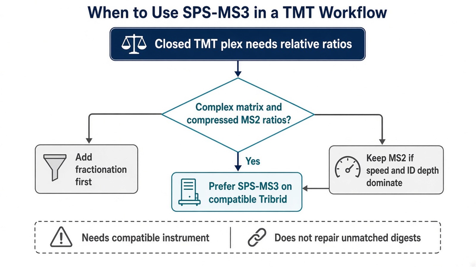
Figure 3. Use SPS-MS3 to optimize TMT reporter accuracy when co-isolation is confirmed; keep MS2 or add fractionation when coverage or platform limits dominate.
Limits and Application Boundaries
SPS-MS3 reduces, but does not erase, interference in crowded chromatographic regions. Notches can still capture nonunique fragments. Cycle-time cost usually lowers peptide and protein coverage relative to MS2 under matched run time. Cross-plex comparison still needs a bridging design; SPS-MS3 does not merge separate kits into one ratio space. Reporter ratios remain relative. Absolute concentration needs matched standards and a calibration model.
Typical applications are multi-condition drug studies, complex tissue or biofluid discovery where fold-change magnitude ranks candidates, and biomarker screens that would lose differentials under strong MS2 compression. Diagnostic cutoffs and clinical decision rules sit outside ordinary research TMT reporting unless a separately validated assay framework is in place.
Once the plex map, fractionation plan, and MS2-versus-SPS-MS3 choice are defined, the project can be evaluated as a TMT acquisition optimization study. Researchers can review the MtoZ Biolabs TMT Quantitative Proteomics Analysis Service for sample evaluation, feasibility assessment, and workflow planning.
Frequently Asked Questions
1. How do I know the problem is ratio compression rather than bad labeling?
Compression usually shrinks many true differentials toward one while identification still looks usable. Global channel skew, unlabeled peptides, or swapped sample IDs point to upstream chemistry or annotation errors first.
2. Does SPS-MS3 always restore the true fold change?
No. It reduces reporter mixing from co-isolated precursors. Residual interference, weak reporters, and upstream load mismatch can remain.
3. Can fractionation replace SPS-MS3?
Fractionation lowers peptide density and helps both MS2 and MS3 methods. It does not provide the same reporter re-isolation path as SPS-MS3. Many complex projects use both.
4. Will switching to SPS-MS3 increase protein counts?
Usually not. Extra MS3 events consume cycle time, so coverage often falls relative to MS2 under the same instrument hours. The intended gain is quantitative accuracy, not deeper identification.
5. Does SPS-MS3 provide absolute quantification?
No. TMT reporters support relative channel ratios inside the plex. Absolute amount needs matched standards and a calibration model.
Reference
- L. Ting, R. Rad, S.P. Gygi, W. Haas (2011). MS3 eliminates ratio distortion in isobaric multiplexed quantitative proteomics. Nat. Methods, 8, 937-940. https://doi.org/10.1038/nmeth.1714
- G.C. McAlister, D.P. Nusinow, M.P. Jedrychowski, M. Wuhr, E.L. Huttlin, B.K. Erickson, R. Rad, W. Haas, S.P. Gygi (2014). MultiNotch MS3 enables accurate, sensitive, and multiplexed detection of differential expression across cancer cell line proteomes. Anal. Chem., 86, 7150-7158. https://doi.org/10.1021/ac502040v
