


Extracellular Vesicles & Exosomes Structure Characterization Service | Cryo-EM
Extracellular vesicles (EVs) are membrane-bound vesicles secreted by cells and are widely present in various biological fluids. They play critical roles in intercellular communication, immune regulation, and molecular transport. Among them, exosomes are a subtype of EVs with diameters ranging from approximately 30 to 150 nm. They originate from the fusion of multivesicular bodies with the plasma membrane and are rich in proteins, lipids, and nucleic acids. Exosomes have been extensively utilized in research areas such as cancer diagnostics, immunotherapy, regenerative medicine, and targeted drug delivery.
The extracellular vesicles & exosomes structure characterization service is a high-resolution imaging service based on cryogenic electron microscopy (Cryo-EM), specifically designed to characterize the three-dimensional morphology, membrane integrity, size distribution, and internal contents of EVs and exosomes. This technique rapidly freezes samples at liquid nitrogen temperatures and performs imaging under low-dose, stain-free conditions, preserving the vesicles’ native structure to the greatest extent possible. With resolution reaching the nanometer to sub-nanometer scale, this service is widely applied in basic EV research, functional validation of delivery vehicles, quality control, and biomarker development, providing precise structural support for both mechanistic studies and product development.
Services at MtoZ Biolabs
Based on an advanced cryogenic electron microscopy (Cryo-EM) system, the extracellular vesicles & exosomes structure characterization service based on Cryo-EM provided by MtoZ Biolabs enables in situ imaging of extracellular vesicles and exosomes by rapidly freezing samples to liquid nitrogen temperatures, thereby avoiding structural alterations caused by drying or staining. The service delivers high-contrast, high-fidelity two-dimensional images that clearly reveal membrane structures, vesicle size, morphological features, and internal content distribution. With nanometer-scale resolution, this analysis is particularly well-suited for evaluating structural heterogeneity, verifying carrier stability, and assessing formulation morphology.
Analysis Workflow
1. Sample Freezing Fixation
EV or exosome samples are rapidly cooled to liquid nitrogen temperature to form vitreous ice, preserving their native state and preventing structural deformation.
2. Low-Dose Image Acquisition
High-contrast, low-dose two-dimensional images are captured using a cryogenic transmission electron microscope to ensure image quality and structural integrity.
3. Image Processing and Structural Identification
Raw images are denoised, aligned, and analyzed to identify and extract target vesicle regions for detailed structural evaluation.
4. Feature Parameter Analysis
Vesicle size distribution, membrane integrity, morphological diversity, and internal content visibility are assessed and quantified.
5. Data Output and Reporting
Original images, analytical results, and a detailed structural report are provided to support scientific publication, product development, and quality control.
Service Advantages
1. Faithful Preservation of Native Structure
Rapid freezing preserves vesicles in their aqueous environment, avoiding structural distortion caused by drying or staining.
2. High-Resolution Imaging Capability
Utilizing advanced Cryo-EM platforms, the service enables nanometer-scale imaging to clearly visualize membrane structures, internal contents, and morphological heterogeneity.
3. Stain-Free and Low-Damage Imaging
Low-dose electron beam imaging eliminates the need for staining, minimizing radiation damage and external interference while enhancing image quality.
4. Compatibility with Diverse EV Samples
Supports a wide range of sample types including plasma, urine, and cell culture supernatants, accommodating various research and clinical applications.
Applications
1. Structural and Compositional Studies
The extracellular vesicles & exosomes structure characterization service enables detailed observation of membrane structure, internal content, and morphological heterogeneity of EVs and exosomes, supporting investigations into their biological functions and mechanisms.
2. Drug Delivery Carrier Development
By evaluating the structural integrity and stability of EVs as natural nanocarriers, this service aids in the design and performance validation of delivery systems.
3. Morphological Analysis for Disease Biomarkers
The extracellular vesicles & exosomes structure characterization service supports morphological characterization of EVs derived from biological fluids such as blood and urine, contributing to early diagnostic research in cancer, neurodegenerative diseases, and more.
4. Evaluation of Novel Nanomaterials
Facilitates the development of engineered exosomes and artificial vesicles by providing visual structural support and morphological verification for innovative functional materials.
5. Formulation Quality Control and Consistency Verification
The extracellular vesicles & exosomes structure characterization service can be applied to monitor structural integrity and batch-to-batch consistency of industrial-grade EV formulations, ensuring product quality and stability.
Case Study
1. Altered Level of Plasma Exosomes in Patients with Gaucher Disease
This study utilized cryo-electron microscopy (Cryo-EM) to characterize the structural features of plasma-derived exosomes from patients with Gaucher disease (GD), aiming to evaluate their potential as biomarkers. The results revealed a significant increase in exosome size in GD patients, with Cryo-EM clearly visualizing double- or multilayered membrane structures, in contrast to the typical single-membrane vesicles observed in healthy controls. These findings were further supported by nanoparticle tracking analysis (NTA) and dynamic light scattering (DLS). Additionally, elevated levels of exosomal marker proteins such as CD9, CD63, and CD81 were detected in GD samples. The study demonstrates that Cryo-EM enables faithful visualization of exosomes in their native state and sensitively detects morphological alterations, making it a powerful tool for uncovering disease-associated structural features and supporting diagnostics and research of lysosomal storage disorders.
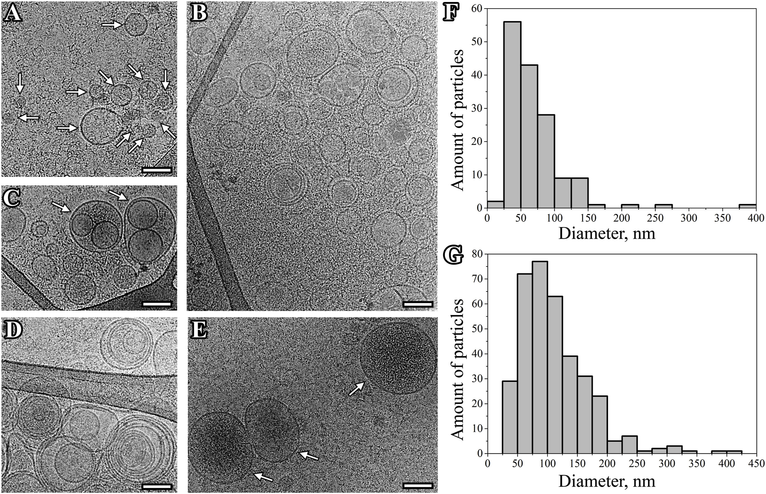
Tatiana, S. et al. European Journal of Medical Genetics, 2020.
Figure 1. Cryo-EM Images of Extracellular Vesicles (EVs) Isolated from Plasma Sample.
2. Cryo-Electron Microscopy of Extracellular Vesicles from Cerebrospinal Fluid
This study aimed to analyze the morphology of extracellular vesicles (EVs) in human cerebrospinal fluid (CSF) using cryo-electron microscopy (Cryo-EM) to explore their structural features and potential functions. EVs were isolated from CSF samples of Parkinson’s disease patients and healthy controls via ultracentrifugation and imaged at high resolution using Cryo-EM. The results revealed diverse EV structures, including single-, double-, and multilayered vesicles, with some containing electron-dense material. The vesicles ranged in size from 26 to 435 nm, with over 80% exhibiting intact lipid bilayers. Compared to conventional TEM, Cryo-EM more accurately preserved the native state of EVs, enabling clear visualization of structural heterogeneity. The study concludes that Cryo-EM is a powerful tool for investigating EV ultrastructure in CSF and holds promise for advancing the discovery of biomarkers for neurological disorders.
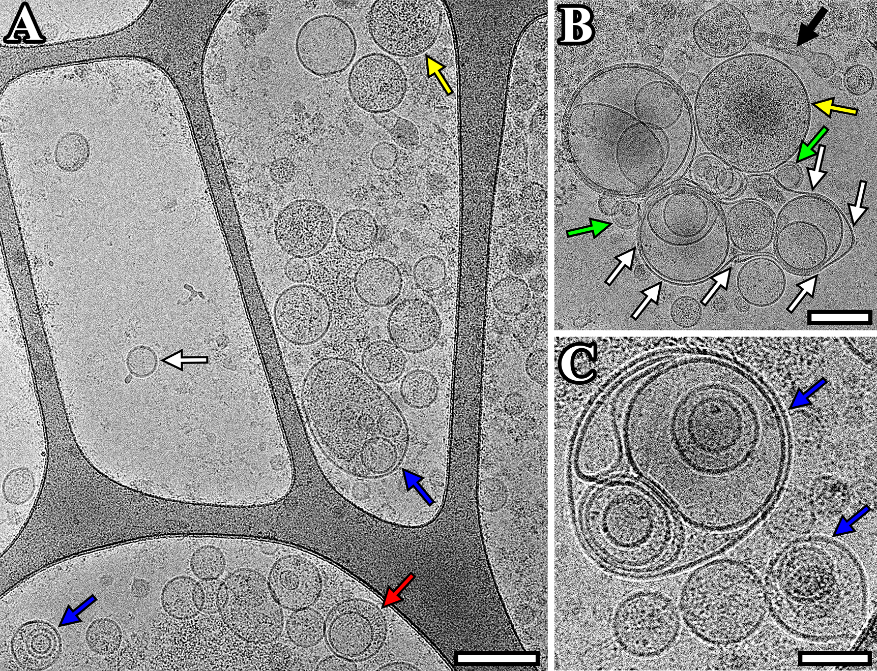
Emelyanov, A. et al. PLOS ONE, 2020.
Figure 2. Cryo-EM Images of EVs Isolated from Pooled CSF Sample from Individuals of the Comparison Group.
FAQ
Q1: Why Is Cryo-EM Suitable for Structural Analysis of EVs and Exosomes?
A1: Cryo-EM allows imaging without staining or drying, effectively preventing deformation or collapse of vesicle structures. It faithfully preserves the native morphology of EVs and exosomes, making it especially suitable for structurally delicate and environmentally sensitive samples.
Q2: Can the Internal Structures of Exosomes Be Visualized?
A2: Yes. With nanometer-scale resolution, Cryo-EM enables detailed observation of membrane bilayers, internal granules, and encapsulated contents, providing insights into the internal architecture of exosomes.
How to order?

