


Exosome Characterization Service
- Based on sample type (cell culture supernatant, serum, tissue extracts), optimized standard isolation methods (e.g., ultracentrifugation, size exclusion chromatography) are employed.
- Purity verification: Western blot analysis for exosome-specific and negative markers.
- Concentration quantification: Nanoparticle tracking analysis (NTA) or dynamic light scattering (DLS).
- Size and Distribution (NTA/DLS): Precise assessment of exosome particle size (30-150 nm) and homogeneity.
- Morphology Observation (TEM): Transmission electron microscopy to confirm membrane structure and integrity.
- Proteomics: Targeted protein validation or high-throughput mass spectrometry for differential protein identification.
- RNA Profiling: qPCR or RNA-seq to analyze exosomal miRNA, mRNA, and lncRNA.
- DNA Profiling: Cell-free DNA extraction and mutation analysis (e.g., PCR amplification).
- Structured reports: Including raw data (TEM images, particle distribution charts, molecular quantification), statistical analysis, and biological interpretation.
- Technical consultation: Recommendations for experimental design and future research directions.
- Standardization and Reproducibility: Strict adherence to ISO quality systems ensures cross-platform consistency.
- Enhanced Sensitivity: Capable of detecting trace exosome samples down to 10⁴ particles/mL for rare sample analysis.
- Flexible Compatibility: Supports diverse sample types (cells, body fluids, frozen samples) and customized marker analysis.
- Multi-omics Integration: Comprehensive analysis across proteins, nucleic acids, and lipids to elucidate molecular networks underlying exosome functions.
- Function-oriented Design: From basic characterization to mechanistic validation, supporting full-cycle research.
- Rapid Turnaround: Basic physical characterization (NTA + TEM) completed within 3–5 business days.
- Full Process Support: Expert team offering technical consultation, protocol optimization, and real-time updates on project progress.
Exosomes are nanosized extracellular vesicles with diameters ranging from 30 to 150 nm, acting as "molecular messengers" in intercellular communication. They carry essential biomolecules such as proteins, nucleic acids (e.g., mRNA, miRNA), and lipids, playing critical roles in cellular signaling, disease mechanisms, and physiological regulation. However, the inherent heterogeneity, low abundance, and complex compositions of exosomes present significant challenges for accurate characterization in research.
MtoZ Biolabs offers Exosome Characterization Service, specifically designed to meet the needs of basic research. Utilizing cutting-edge technologies and standardized workflows, we deliver comprehensive insights into the physical properties, molecular cargo, and biological functions of exosomes, helping researchers explore their roles in cellular communication and disease models.
Our Core Service Focus
1. Physical Property Analysis: Particle size distribution, concentration, and morphology characterization.
2. Component Analysis: Identification of protein markers, nucleic acids (miRNA/mRNA), and lipid species.
3. Functional Validation: Evaluation of cellular targeting, immune modulation, and drug delivery efficiency.
Dilsiz, N. Transl Oncol. 2024.
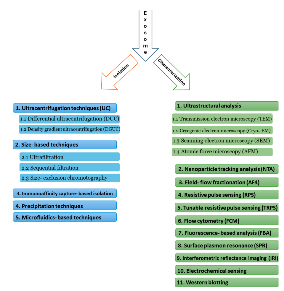
Figure 1. Isolation and Characterization Methods for Exosome
Analysis Workflow
1. Sample Pre-processing and Quality Control
2. Multidimensional Physical Characterization
3. Molecular Component Analysis
4. Data Integration and Reporting
Service Advantages
1. Addressing Research Pain Points
2. In-Depth Analysis Capability
3. Efficient and Transparent Service
Applications
1. Intercellular Communication Mechanisms: Studying exosome-mediated signaling in tumor microenvironments or stem cell paracrine regulation.
2. Disease Model Development: Identifying disease-specific exosomal biomarkers for molecular mechanism research (e.g., neurodegenerative diseases, fibrosis).
3. Engineered Exosome Development: Validating functional properties of modified exosomes (e.g., targeted peptide display).
4. Molecular Carrier Research: Evaluating exosomes as delivery vehicles for RNA interference or protein therapeutics regarding efficiency and stability.
Case Study
1. Molecular Characterization of Exosomes for Subtype-Based Diagnosis of Breast Cancer
This study developed biomimetic vesicles for subtype-specific recognition and molecular characterization of breast cancer-derived exosomes. By camouflaging catalytic DNA machinery with breast cancer cell membranes, the vesicles achieved homotypic recognition of exosomes sharing similar phenotypes and used exosomal RNA as an endogenous trigger to amplify electrochemical signals for high-sensitivity detection. Vesicles derived from MCF-7 membranes selectively recognized estrogen receptor-positive exosomes with a detection limit of 557 particles/mL using miR-375 as a marker, while vesicles from MDA-MB-231 membranes targeted triple-negative breast cancer exosomes, using PD-L1 mRNA as a biomarker. The method demonstrated high accuracy and subtype classification capability in clinical samples, showing electrochemical signals correlated with disease progression. This strategy offers a new approach for precise diagnosis and personalized treatment of breast cancer. Exosome Characterization Service provides multidimensional molecular analysis for detailed exosome subtyping, revealing their origin and functional characteristics. Applicable to various biological samples, it supports advanced research on disease classification, mechanism exploration, and biomarker discovery.
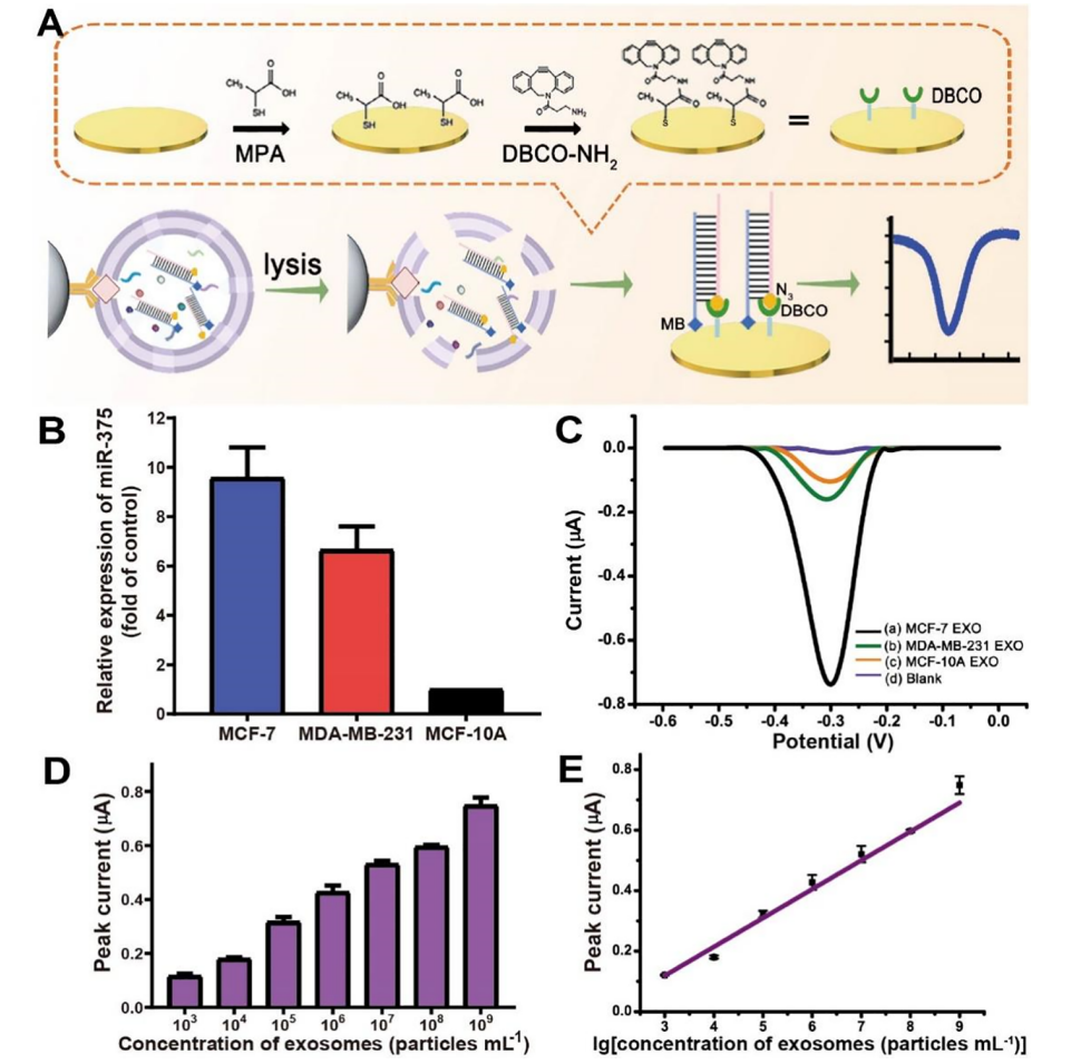
Cao, Y. et al. J Am Chem Soc. 2022.
Figure 2. Homotypic Recognition-Driven Electrochemical Characterization of BC Exosomes
2. Direct Isolation and Characterization of Circulating Exosomes from Biological Samples Using Magnetic Nanowires
This study proposes a molecular subtyping detection strategy for breast cancer exosomes based on biomimetic vesicles and homotypic recognition. The method uses breast cancer cell membranes to construct biomimetic vesicles and triggers DNA signal amplification via endogenous RNA biomarkers, enabling precise identification and detection of both estrogen receptor-positive and triple-negative exosomes. Validation with clinical samples shows that the method is highly sensitive and accurate, capable of monitoring various stages. Exosome Characterization Service employs cell membrane biomimetic technology and signal amplification methods to classify the phenotype of exosomes in samples. By recognizing key biomarkers, it enables efficient detection and monitoring of multiple exosome subtypes, making it suitable for clinical sample analysis in breast cancer and related diseases.
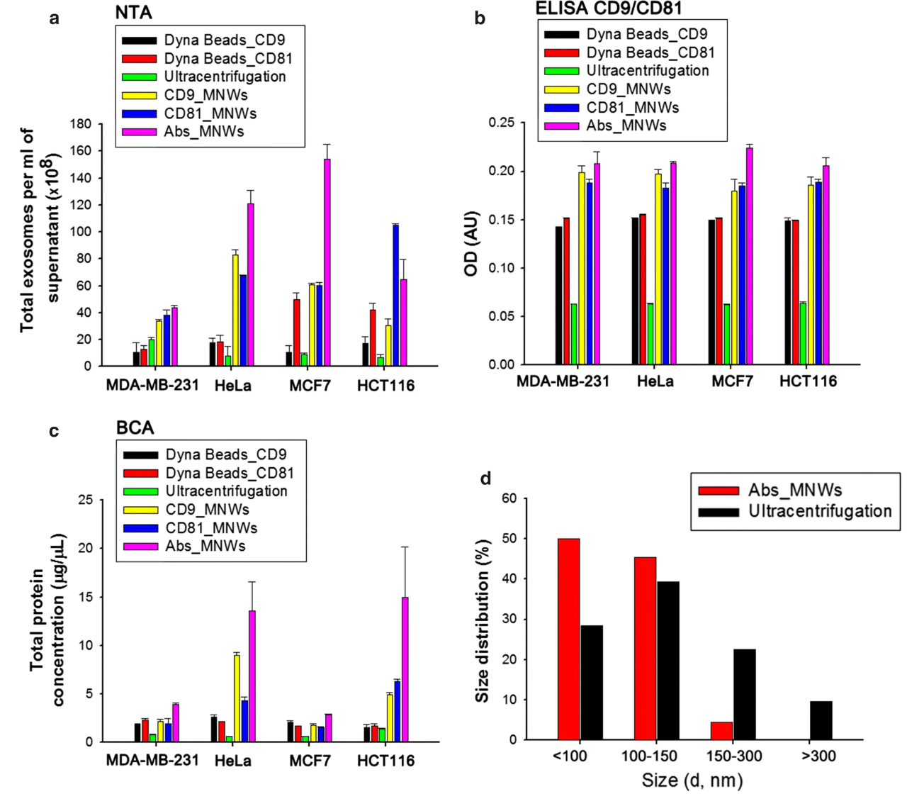
Lim, J. et al. J Nanobiotechnology. 2019.
Figure 3. Analysis of Exosomes Isolated from Cell Lines by Magnetic Beads and Magnetic Nanowires
Deliverables
1. High-resolution TEM images and particle size distribution heatmaps
2. Quantitative results of targeted molecules (e.g., miRNA levels, specific protein abundance)
3. Statistical analysis and visualized data charts (e.g., PCA analysis, differential molecule clustering)
Exosome research is transforming our understanding of cellular biology and disease mechanisms, yet its complexity demands precise experimental designs and highly reproducible technologies. Exosome Characterization Service in MtoZ Biolabs is committed to being your trusted research partner, helping you overcome technical challenges through standardized workflows, multidimensional analysis, and expert interpretation. Contact MtoZ Biolabs today to unlock the full potential of exosomes and drive your scientific discovery forward!
How to order?

