






Exosome and Extracellular Vesicle Proteomics Service for LC-MS/MS-Based Protein Analysis
Proteins detected in an extracellular vesicle preparation reflect both vesicle-associated material and components that co-isolate during sample processing. The resulting protein profile therefore depends not only on LC-MS/MS performance, but also on the biological source, extracellular vesicle (EV) preparation status, matrix background, and preservation history. These factors determine whether a detected protein can be interpreted as part of an EV-enriched preparation or only as a component of the analyzed sample.
LC-MS/MS-based exosome proteomics can support protein identification, relative quantification, and differential protein screening when the upstream sample and comparative design are technically coherent. The analytical route begins with the condition of the submitted material and proceeds through EV quality assessment, protein or peptide preparation, mass spectrometric measurement, and evidence-based interpretation.
Preparing EV Samples for LC-MS/MS
1. Starting Material and EV Preparation Status
(1) Source Materials and Pre-Isolated EVs
EV proteomics may begin with plasma, serum, urine, cerebrospinal fluid, cell culture-conditioned medium, pre-isolated EVs, EV protein lysates, or prepared peptides. Each starting point preserves a different amount of information about upstream processing. Source samples allow EV isolation and proteomic preparation to be planned as a connected procedure, whereas pre-isolated EVs depend on the separation method, buffer composition, storage history, and available characterization records. Protein lysates and peptides permit later analytical steps to proceed but provide less opportunity to assess vesicle integrity or correct losses introduced during earlier preparation.
(2) Purity, Integrity, and Matrix Background
EV enrichment does not imply complete separation from non-vesicular material. Plasma and serum preparations may retain abundant soluble proteins or lipoprotein-associated components, while conditioned medium may contain proteins derived from supplements, cell damage, or prolonged culture stress. Urine and cerebrospinal fluid present different concentration and matrix challenges. Freeze-thaw exposure, delayed processing, contamination, and unsuitable storage conditions may also alter vesicle integrity or increase background. These variables should be considered when comparing protein profiles across samples or interpreting apparently source-specific proteins.
2. Technical Quality Before Proteomics
(1) EV Characterization as Supporting Evidence
Particle-size and concentration measurements, electron microscopy, and EV marker protein assessment provide complementary evidence about an EV preparation. Nanoparticle tracking analysis describes particle distributions but does not determine the molecular identity of every measured particle. Transmission electron microscopy or cryo-electron microscopy supports morphological assessment, while western blotting for markers such as CD9, CD63, and CD81 contributes protein-level evidence. No single result establishes absolute purity or proves that every detected protein originated within or on an EV. Interpretation is stronger when multiple orthogonal measurements agree with the preparation method and sample source.
(2) Protein and Peptide Compatibility
Proteomic analysis requires efficient protein solubilization, controlled reduction and alkylation, reproducible enzymatic digestion, and removal of substances that interfere with chromatography or ionization. Residual detergents, salts, polymers, precipitation reagents, or highly abundant background proteins may suppress peptide signals and reduce effective sampling depth. Low-input EV preparations are especially sensitive to transfer loss and adsorption. Protein recovery, digestion efficiency, and peptide cleanup therefore influence both the number of identified proteins and the comparability of quantitative measurements.
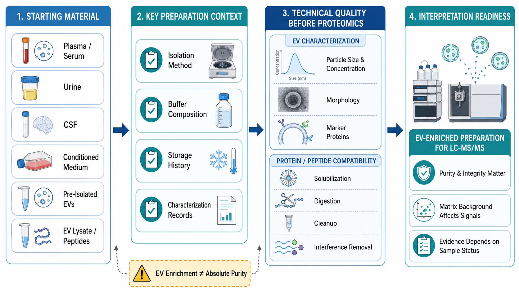
Figure 1. Sample-Dependent Entry Points for EV Proteomics
Selecting the Proteomics Measurement Strategy
1. LC-MS/MS Protein Identification
(1) Bottom-Up Identification and Protein Inference
In bottom-up proteomics, EV-associated proteins are digested into peptides before liquid chromatography and tandem mass spectrometry. Peptide-spectrum matches are evaluated against a sequence database, and accepted peptide evidence is assembled into protein groups. Shared peptides may support more than one protein sequence, so protein inference rules affect the final identification table. Identification confidence should be evaluated through peptide-level evidence, false discovery rate control, and consistency across replicates rather than through protein counts alone.
(2) What Protein Identification Establishes
Protein identification establishes that peptide evidence compatible with a protein or protein group was detected in the analyzed preparation. It does not by itself establish protein abundance, selective EV loading, vesicular localization, disease specificity, or biological function. A protein detected in an EV-enriched sample may represent vesicle cargo, a membrane-associated component, or co-isolated material. Claims about selective enrichment require an appropriate comparator, while claims about localization or function require independent evidence beyond discovery proteomics.
2. Quantitative Proteomics for Group Comparison
(1) Label-Free and Isobaric Labeling Designs
Label-Free quantification estimates relative peptide or protein abundance from signal intensities acquired separately for each sample. It accommodates flexible sample numbers but depends on consistent preparation, chromatographic performance, and batch control. Isobaric labeling with TMT or iTRAQ combines labeled samples for multiplexed analysis and supports direct relative comparison within the same multiplex. Channel allocation, reference design, sample balance, and ratio compression should be considered when interpreting labeled datasets. Neither strategy is universally preferable; the appropriate design depends on sample number, available input, group structure, and the required comparison framework.
(2) DIA Acquisition and Quantitative Consistency
Data-independent acquisition (DIA) is a mass spectrometric acquisition strategy rather than a labeling category. DIA repeatedly fragments broad precursor windows, producing systematic peptide sampling across runs and supporting consistent quantitative matrices. In routine EV proteomics, DIA is commonly implemented in a label-free format, although labeling strategy and acquisition mode remain conceptually separate design dimensions. The value of DIA depends on chromatographic stability, spectral interpretation, quality control, and a study design that distinguishes biological variation from technical variation.
From Quantitative Results to Differential Protein Candidates
1. Designing a Defensible Comparison
(1) Biological Groups, Replication, and Batch Structure
Differential protein analysis is only as interpretable as the comparison design. Biological groups should represent a defined experimental contrast, and biological replicates should capture variation among independent samples rather than repeated injections of the same preparation. Sample collection, EV isolation, digestion, labeling, and LC-MS/MS acquisition should be balanced across groups where possible. Confounding between biological condition and processing batch may create apparent differences that cannot be separated from technical effects.
(2) Normalization, Missing Values, and Statistical Criteria
Quantitative matrices require quality assessment before statistical testing. Normalization addresses systematic intensity differences but cannot correct severe preparation bias or unbalanced missingness. Missing values may arise from low abundance, stochastic sampling, interference, or processing failure, and the selected handling method should reflect the likely mechanism. Differential screening should consider effect size, variability, replicate consistency, multiple-testing control, and data completeness together. A fold-change threshold alone does not establish a robust candidate.
2. Interpreting Differential EV-Associated Proteins
(1) Functional Annotation and Candidate Prioritization
Gene Ontology annotation, pathway analysis, and protein-protein interaction analysis organize differential proteins into biological themes and relationships. These analyses depend on existing databases and the submitted protein list, so they generate interpretive hypotheses rather than direct experimental proof. Candidate prioritization can additionally consider quantitative consistency, effect size, peptide evidence, known cellular distribution, EV relevance, and the biological question. Prioritization should remain traceable to explicit criteria instead of treating statistical significance as equivalent to biological importance.
(2) Discovery Evidence and Follow-Up Validation
A differential EV-associated protein is a discovery-stage candidate, not a validated biomarker or confirmed mechanism. Targeted mass spectrometry such as PRM may assess selected peptides with higher measurement focus, while western blotting or ELISA may provide orthogonal protein-level evidence when suitable reagents and sample conditions are available. These approaches can confirm detectability or group-associated differences in an independent format, but broader biological or clinical claims require additional cohorts, controls, and functional experiments.
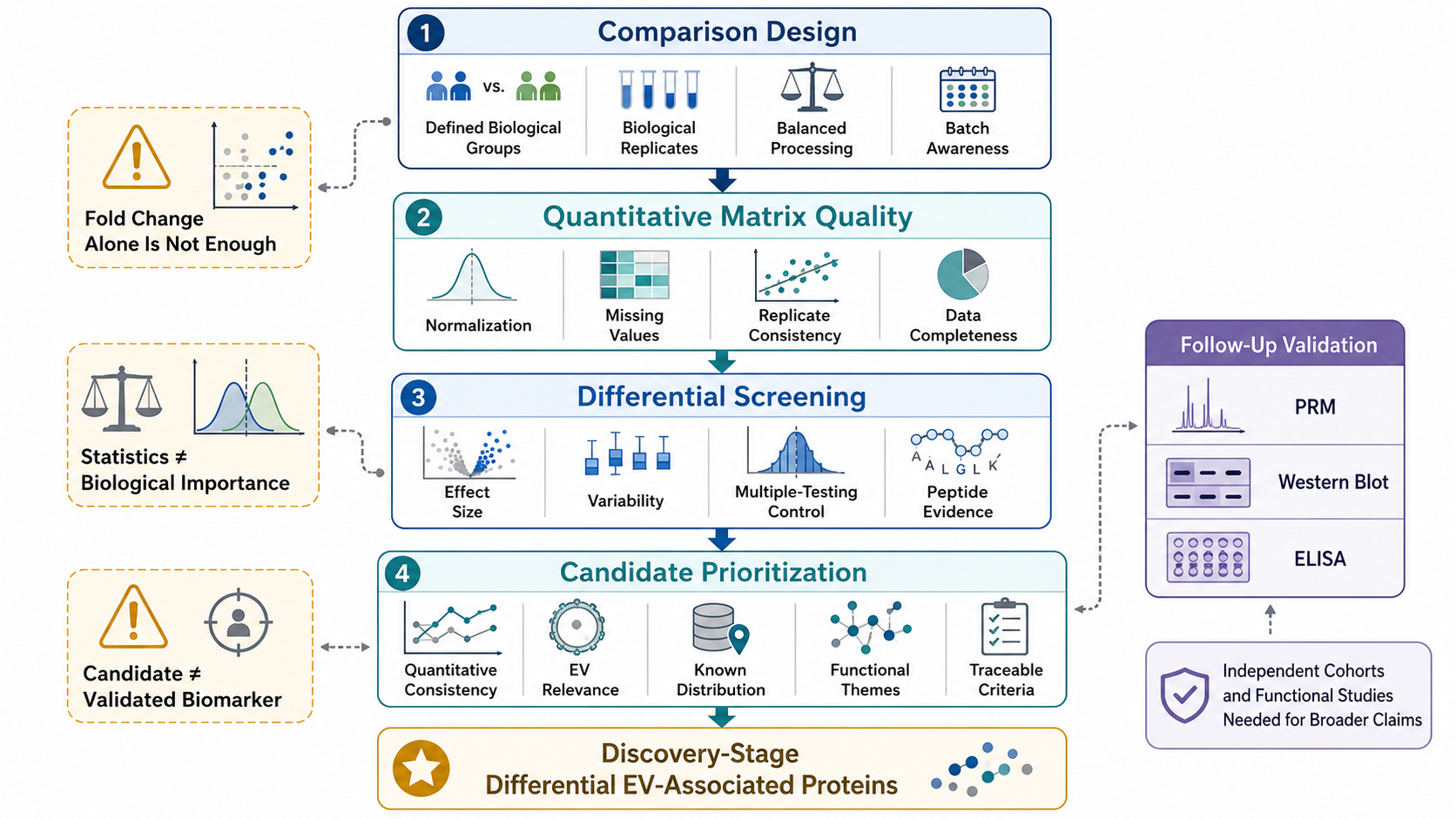
Figure 2. Evidence Framework for Differential EV Protein Candidates
Reliable exosome and extracellular vesicle proteomics depends on alignment among starting material, EV preparation quality, LC-MS/MS strategy, comparative design, and the intended evidence level. MtoZ Biolabs evaluates projects from source samples or pre-isolated EVs and supports LC-MS/MS protein identification, quantitative comparison, and differential protein analysis based on sample status, group design, and expected outputs. Submit your inquiry below for project evaluation.
MtoZ Biolabs, an integrated chromatography and mass spectrometry (MS) services provider.
Related Services
How to order?




