


Exosomal Surface Marker-based Exosome Characterization Service
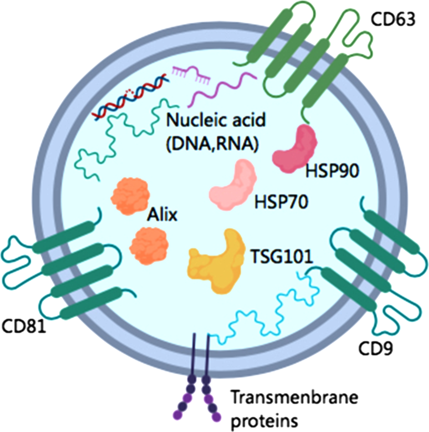
Exosomal Surface Marker-based Exosome Characterization Service focuses on analyzing exosomal surface markers to identify the origin, function, and disease relevance of exosomes. Exosomal surface markers are specific molecules present on the exosomal membrane, providing crucial information about exosome origin, type, and functionality. Different types of exosomes exhibit distinct surface markers, and by analyzing these markers, researchers can not only determine the cellular source of exosomes but also uncover their potential roles in physiological and pathological conditions. Common exosomal surface markers include, but are not limited to, CD9, CD63, and CD81, which are highly expressed on the exosomal membrane and play essential roles in exosome formation and function. MtoZ Biolabs offers the Exosomal Surface Marker-based Exosome Characterization Service enabling identification and quantification of exosomal surface markers. This service facilitates exosome classification, functional analysis, and biomarker discovery, supporting applications in early diagnosis, therapeutic monitoring, and disease research.
Wu J. Shen Z. Cancer Medicine. 2020.
Analysis Workflow
1. Exosome Isolation:
Exosomes are isolated using ultracentrifugation, density gradient centrifugation, or size exclusion chromatography (SEC).
2. Surface Marker Detection:
Based on research requirements, appropriate detection methods such as FACS (Fluorescence-Activated Cell Sorting), immunoaffinity enrichment, or ELISA are used to analyze exosomal surface markers.
3. Data Analysis and Reporting:
The detection results are analyzed, and a comprehensive report is generated, providing qualitative and quantitative information on exosomal surface markers. This information helps infer exosome origin, classification, and potential functions.
Applications
Disease Diagnosis: Exosomal surface markers serve as potential biomarkers for various diseases, including cancer, liver disease, and kidney disease, aiding in early detection and diagnosis.
Basic Research: Understanding the role of exosomes in intercellular communication, immune response, and pathogen transmission advances life science research.
Drug Development and Delivery Systems: As natural drug delivery carriers, exosomes can be optimized for efficient and targeted drug delivery based on their surface marker characteristics. This has significant potential in targeted therapies and vaccine development.
Service Advantages
1. Advanced Analysis Platform: MtoZ Biolabs established an advanced Exosomal Surface Marker-based Exosome Characterization Service platform, guaranteeing reliable, fast, and highly accurate analysis service.
2. One-Time-Charge: Our pricing is transparent, no hidden fees or additional costs.
3. High-Data-Quality: Deep data coverage with strict data quality control. AI-powered bioinformatics platform integrates all Exosomal Surface Marker-based Exosome Characterization Service data, providing clients with a comprehensive data report.
4. Multiplex Marker Analysis: Enables the simultaneous detection of multiple exosomal surface markers, facilitating a more comprehensive exosome analysis and enhancing the depth of research.
FAQ
Q. How Specific are Common Exosomal Surface Markers Such As CD9, CD63, and CD81?
CD9, CD63, and CD81 are the most commonly used exosomal markers, widely employed for identifying exosome presence and assessing exosome quality. These markers belong to the tetraspanin family and are ubiquitously expressed on exosomes derived from various cell types, making them useful for labeling the exosomal membrane. However, their specificity is relatively low, as these markers are not exclusive to exosomes and can also be found in other extracellular vesicles (EVs), such as microvesicles and endosomes. In order to improve specificity, multiple exosome markers are usually used in combination or cell type-specific markers (such as EpCAM, a marker unique to tumor cells) are used in combination with separation methods (such as ultracentrifugation, immunoenrichment, etc.) to confirm the source of exosomes.
Q. How are Different Surface Markers Related to Exosome Functions?
Exosomal surface markers are not only used for identifying exosome origin and classification but also play crucial roles in exosome function, including intercellular communication, immune regulation, and signal transduction. For example:
CD9, CD63, CD81: These classic exosomal membrane proteins are essential for exosome biogenesis, membrane fusion, trafficking, and endocytosis. Their expression levels are closely linked to exosome stability, intercellular communication, and cargo transport efficiency.
CD47: This surface marker helps exosomes evade immune clearance, acting as a “self-marker” that allows exosomes to circulate in the body for extended periods without being rapidly degraded.
EpCAM: As an epithelial cell surface marker, EpCAM is commonly found on epithelial-derived exosomes and plays a vital role in tumor exosome communication. It facilitates tumor metastasis and immune evasion by aiding in cellular targeting and exosomal interactions.
In summary, different surface markers are closely related to exosomal functions, influencing exosome formation, stability, targeting capabilities, and immune regulation.
Deliverables
1. Comprehensive Experimental Details
2. Materials, Instruments, and Methods
3. Total Ion Chromatogram & Quality Control Assessment (project-dependent)
4. Data Analysis, Preprocessing, and Estimation (project-dependent)
5. Bioinformatics Analysis
6. Raw Data Files
Case Study
This study conducted an in-depth proteomic analysis of the surface proteins of pancreatic ductal adenocarcinoma (PDAC) exosomes and identified specific biomarkers, including CLDN4, EPCAM, and CD151. Using these biomarkers, researchers developed a strategy to enrich cancer-derived exosomes from liquid biopsy samples. When analyzing blood samples from 103 PDAC patients, the detection rate of KRAS mutations in total exosomes was 44.1%. However, after enrichment using specific markers, the detection rate increased to 73.0%.
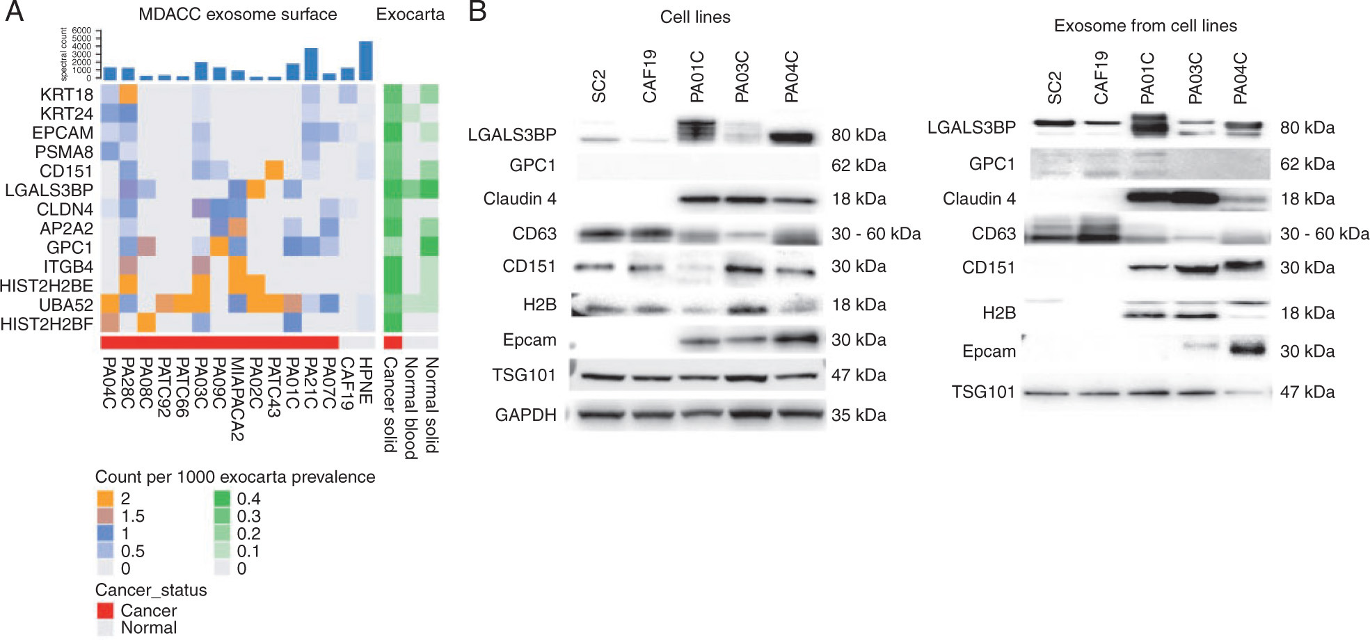
Castillo J. et al. Annals of Oncology. 2018.
How to order?

