


Cryo-TEM Imaging & Analysis
Cryogenic transmission electron microscopy (Cryo-TEM) is an advanced imaging technique that enables high-resolution visualization of biological macromolecules, nanomaterials, and complex soft matter under near-native conditions. By vitrifying aqueous samples in a thin layer of amorphous ice, Cryo-TEM preserves ultrastructural integrity without the need for chemical fixation or staining. This technique has become indispensable for structural biology, nanomedicine, pharmaceutical research, and materials science.
As a transformative tool, Cryo-TEM bridges the resolution gap between light microscopy and atomic-resolution techniques. It allows researchers to analyze morphology, particle size, internal structure, and heterogeneity with nanometer or even sub-nanometer precision. Moreover, Cryo-TEM is uniquely suited for examining dynamic and radiation-sensitive specimens, such as liposomes, exosomes, viruses, proteins, and polymeric assemblies.
Technical Principles
Cryo-TEM operates by freezing samples in vitreous (non-crystalline) ice at cryogenic temperatures, maintaining their native aqueous state. After rapid plunge-freezing, the specimen is inserted into a transmission electron microscope equipped with a cryo-holder. Imaging is performed under low-dose electron beams to minimize radiation damage, while ensuring structural preservation.
The electron beam passes through the thin frozen sample, generating contrast based on differences in electron density. Due to the absence of heavy metal stains, image contrast is inherently low, but high-resolution structural details can be reconstructed using techniques such as single-particle analysis or tomography. The ability to analyze samples in their hydrated and minimally disturbed form provides a significant advantage over conventional TEM.
Service at MtoZ Biolabs
MtoZ Biolabs offers a full-spectrum Cryo-TEM Imaging & Analysis service designed to support high-resolution structural visualization under native-like conditions. Our service platform integrates state-of-the-art cryo-electron microscopy instruments, automated sample vitrification systems, and advanced image acquisition software. Whether clients are working with protein complexes, exosomes, liposomes, soft matter, or nanomedicine formulations, our team delivers end-to-end project support—from sample preparation and cryo-fixation to image processing and 3D reconstruction.
We accept a wide range of sample types, and our protocols are tailored to the specific physical and biochemical properties of each project. With robust data quality control, customizable imaging strategies, and comprehensive bioimage analysis capabilities, we ensure reproducible and publication-ready outcomes.
Analysis Workflow
1. Sample Preparation and Vitrification
Samples are applied onto grids and blotted to form a thin film. The grids are rapidly vitrified by plunge-freezing into liquid ethane using a state-of-the-art vitrification system.
2. Cryo-Transfer and Imaging
The vitrified grids are transferred into a cryo-TEM instrument via a cryo-holder to maintain low temperatures. Imaging is performed using a high-performance cryo-TEM system equipped with direct electron detectors and automated stage control.
3. Image Acquisition and Quality Control
Low-dose images are captured to balance contrast and sample preservation. The imaging parameters (e.g., defocus, electron dose) are carefully optimized for each sample.
4. Data Processing and Analysis
We offer downstream processing services, including particle picking, 2D class averaging, 3D reconstruction, and tomographic analysis depending on the project goal.
5. Data Delivery and Consultation
Clients receive raw and processed image files, accompanied by detailed analysis reports. Optional one-on-one consultations help interpret structural insights and guide further experiments.
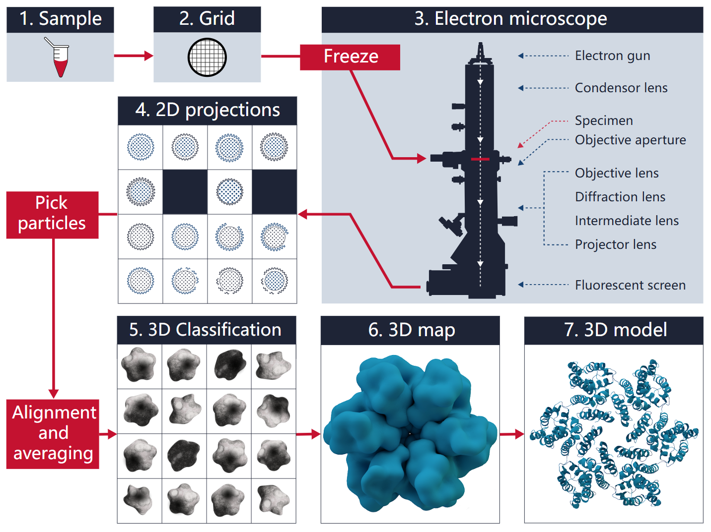
Figure 1. Workflow for Cryo-TEM Imaging & Analysis
Service Advantages
☑️Native-State Visualization: Imaging under cryogenic conditions preserves the sample’s hydrated morphology without chemical fixation or staining, maintaining structural fidelity.
☑️Custom Sample Preparation: Tailored vitrification protocols utilizing hydrophilic EM grids and precisely controlled environmental conditions to support a wide range of biomolecular and nanomaterial samples.
☑️Flexible, Confidential Service Model: We support both exploratory and hypothesis-driven projects, offering scalable analysis packages under strict data confidentiality protocols.
☑️Rapid Turnaround and Consultation: Efficient scheduling and timely delivery of raw and processed datasets, along with optional project consultation to guide next steps.
Applications
1. Structural Biology
Cryo-TEM allows visualization of biomolecules such as proteins, nucleic acids, and large assemblies (e.g., membrane proteins, ATP synthase) in their native conformations. It supports high-resolution single-particle analysis and structural reconstruction, critical for elucidating mechanisms of biological function, protein–protein interactions, and ligand binding.
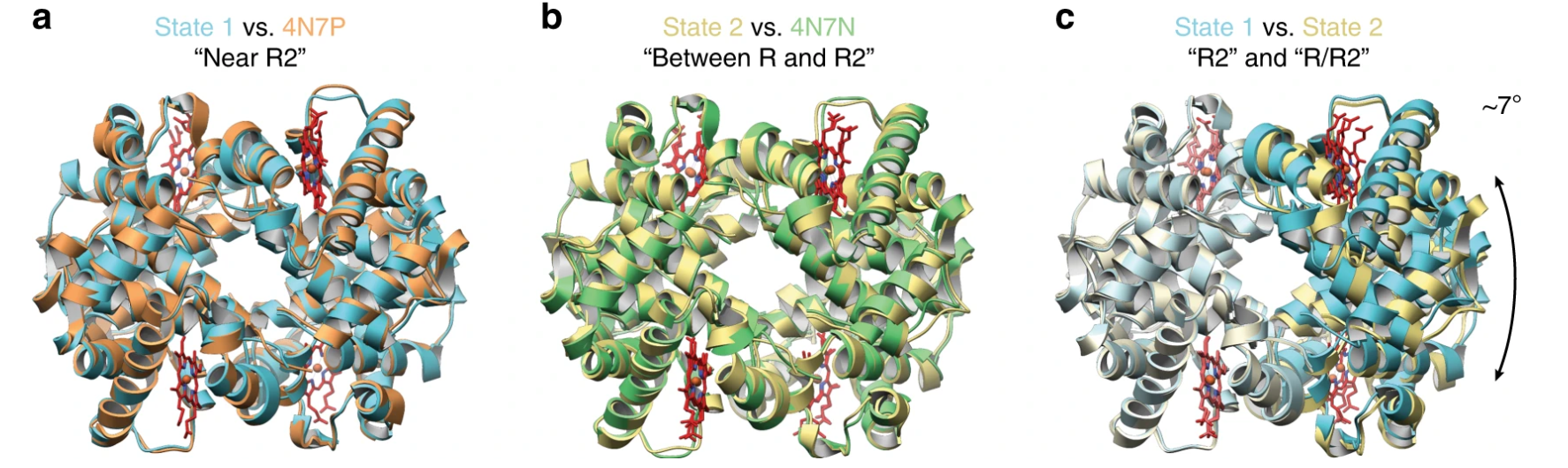
Figure 2. Multiple Conformational States of human methemoglobin (metHb) Determined using Single-Particle Cryo-TEM
2. Extracellular Vesicle Characterization
Cryo-TEM provides detailed morphology, size distribution, and cargo content insights for extracellular vesicles (EVs), including exosomes and microvesicles. It enables researchers to confirm vesicle purity, analyze structural heterogeneity, and assess loading efficiency in drug delivery or diagnostic applications.
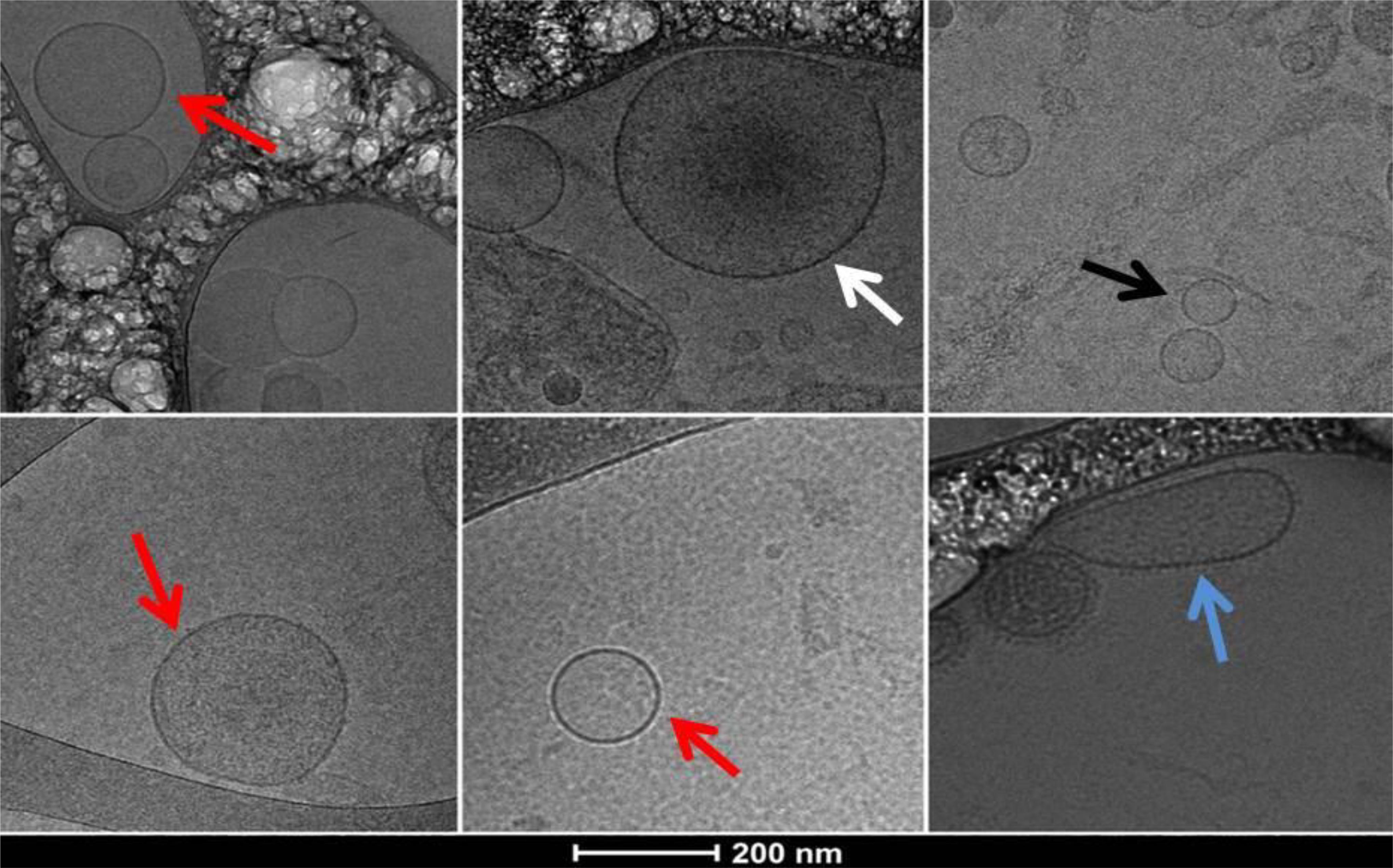
Figure 3. Cryo-TEM Images of Extracellular Vesicles Obtained from Platelet-Poor Plasma Pellet Sample of a Patient with Hypersplenism Showing a Heterogeneous Population of EVs
3. Subcellular Imaging
Cryo-TEM can reconstruct 3D cellular architectures, organelles, and cytoskeletal networks at nanometer resolution. This expands its use in cellular biology research.
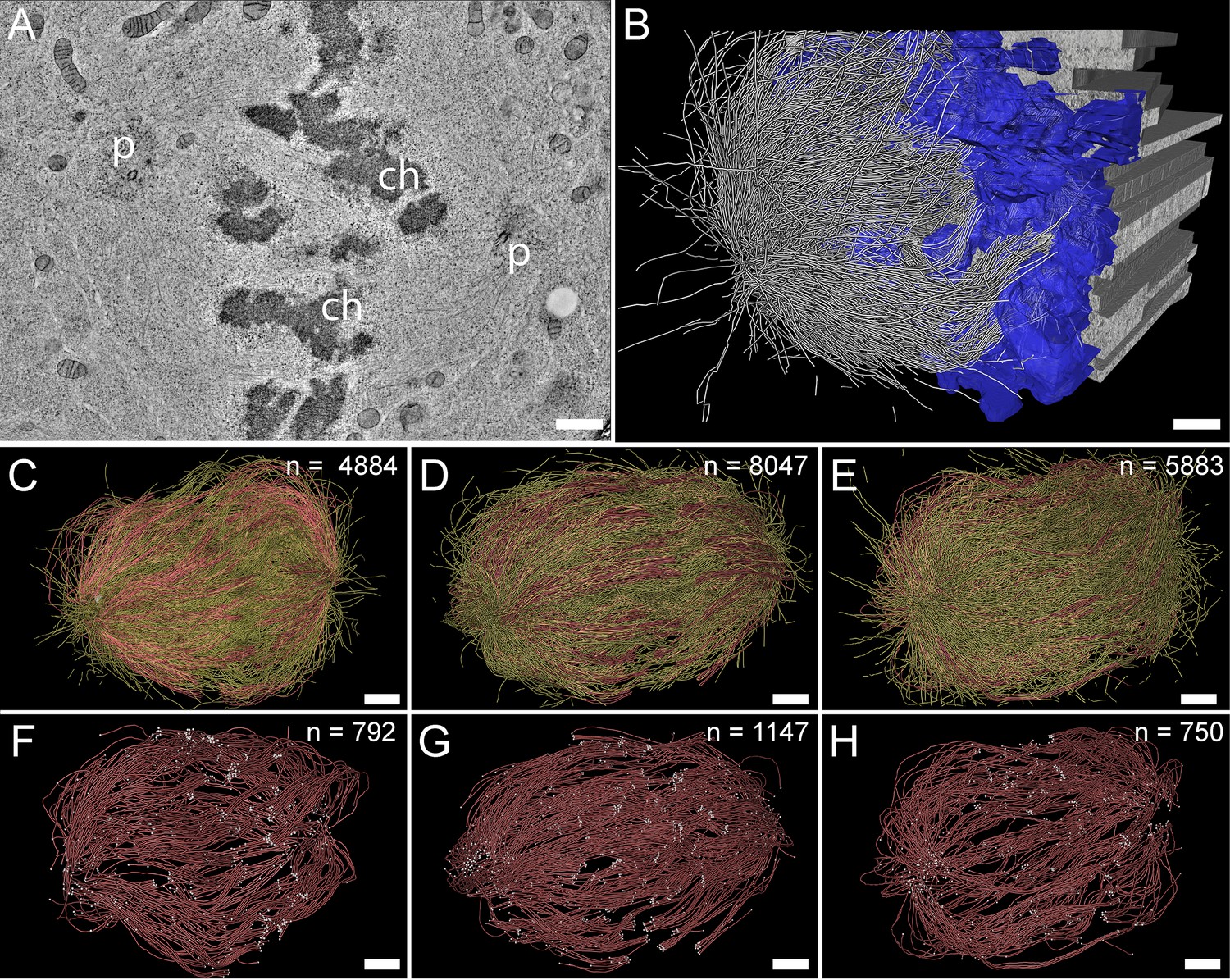
Kiewisz, R. et al. Elife. 2022.
Figure 4. Three-Dimensional Reconstruction of Metaphase Spindles by Large-Scale Electron Tomography
4. Structural Virology
The technique is pivotal for investigating viral morphology and dynamics, including intact virions, replication intermediates, and viral-host complexes. Cryo-TEM enables direct imaging of viral capsids, surface proteins and envelopes, informing vaccine targets and antiviral strategies.
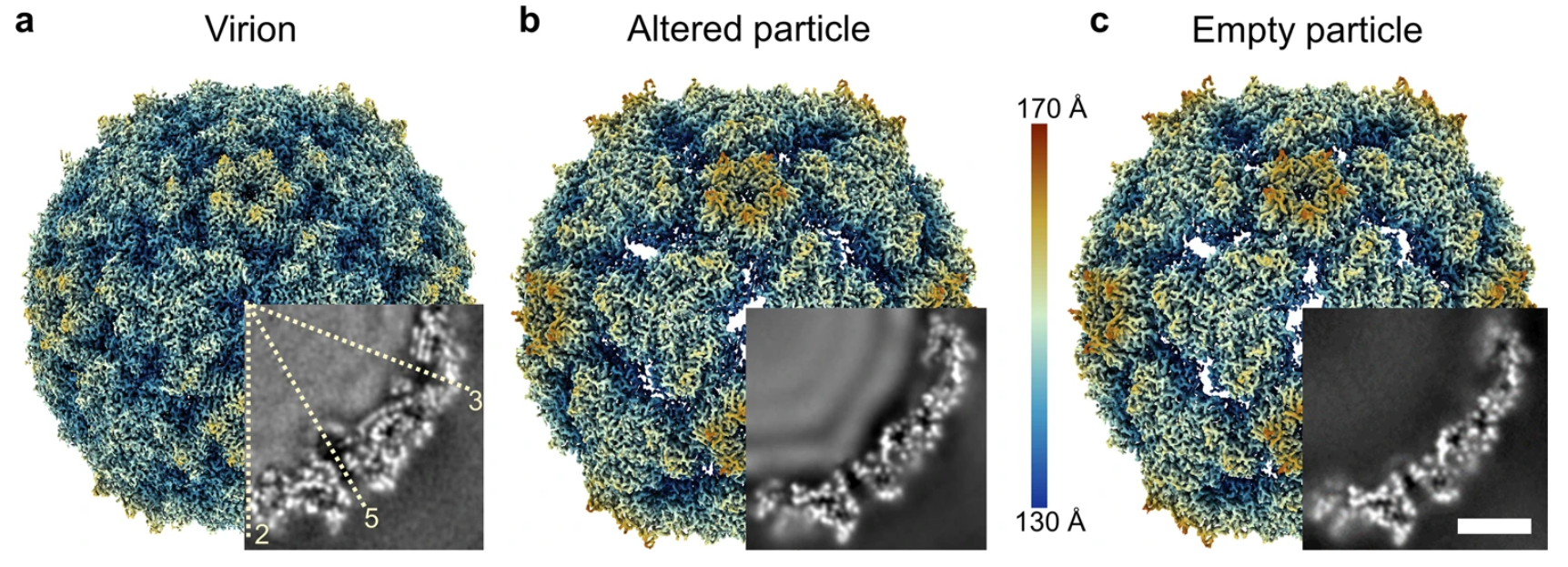
Büttner, C. R. et al. Commun Biol. 2022.
Figure 5. Cryo-Electron Density Map of Coxsackievirus A6 (CV-A6) Virion (a), Altered Particle (b), and Empty Particle
5. Drug Discovery
Cryo-TEM enables direct visualization of drug–target complexes, supporting structure-based design and lead optimization. It helps reveal binding modes, conformational changes, and target accessibility critical to early drug discovery.
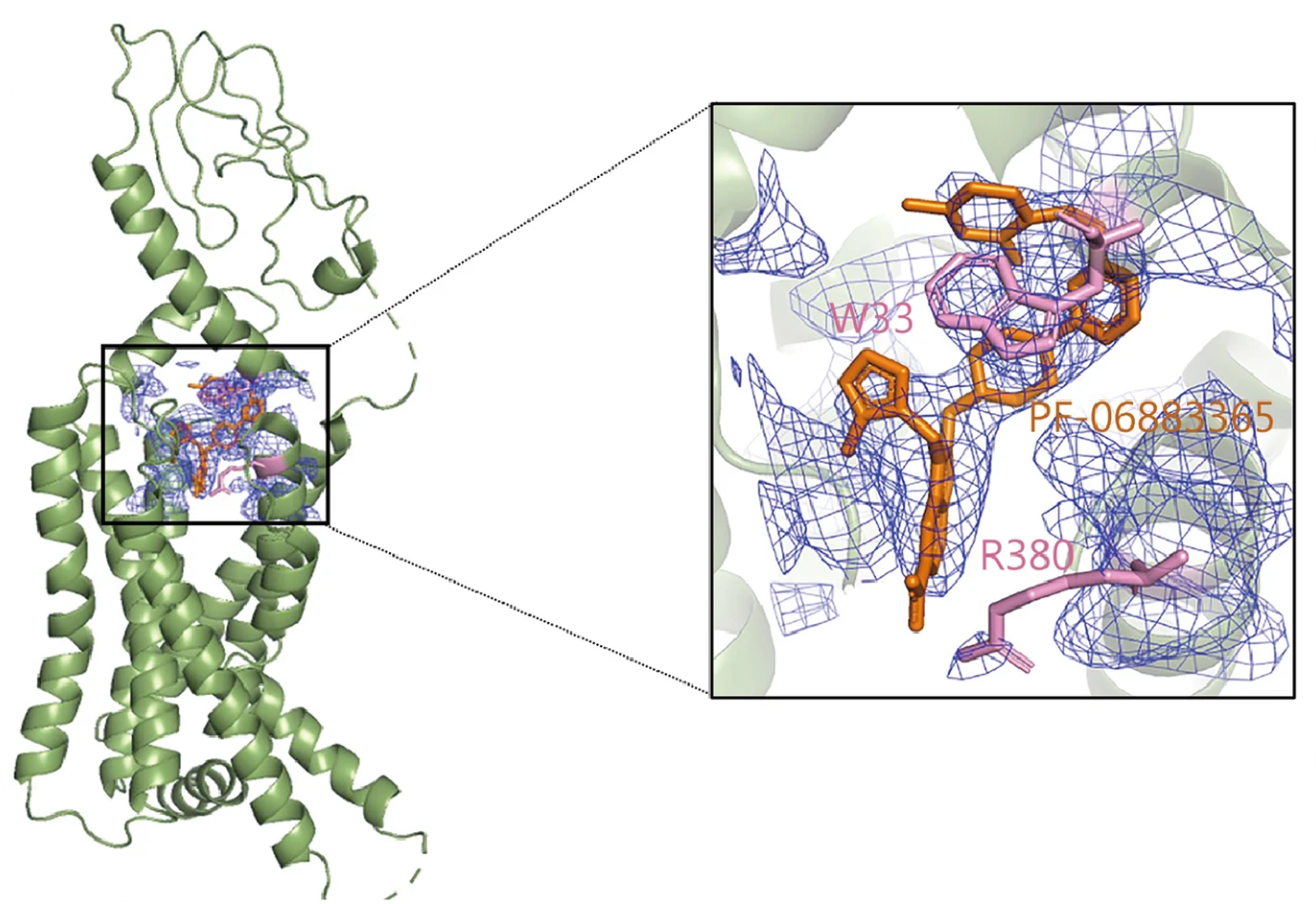
Zhu, K. F. et al. Mil Med Res. 2023.
Figure 6. Cartoon Display of Cryo-TEM Structure of Glucagon-Like Peptide-1 Receptor (GLP-1R) Bound with Small Molecule PF-06883365
FAQ
Q1: What types of samples are suitable for Cryo-TEM Imaging & Analysis service?
Cryo-TEM is ideal for hydrated, nanoscale specimens such as proteins, protein complexes, viruses, liposomes, exosomes, lipid nanoparticles, polymeric micelles, hydrogels, and other soft materials. It is particularly effective for fragile or structure-sensitive samples that require imaging in a near-native state.
Q2: How should I prepare and ship my samples?
Samples should be shipped on dry ice or in liquid nitrogen dry shippers to maintain cryogenic or frozen conditions. For detailed submission instructions, please contact us—we’ll provide guidelines tailored to your sample type.
Q3: Can the Cryo-TEM Imaging & Analysis service be used for quality control or formulation studies?
Yes. Cryo-TEM is increasingly used in pharmaceutical and nanomedicine applications for evaluating particle size distribution, aggregation states, vesicle morphology, and stability under various formulation conditions.
How to order?

