


Cryo-EM for Biological Tissues Structure Characterization Service
- Muscle tissues
- Liver tissues
- Kidney tissues
- Tumor tissues
- Vascular tissues
- Other specialized tissues (e.g., immune system, cardiac tissues)
Cryo-EM for Biological Tissues Structure Characterization Service enables high-resolution analysis of biological tissues under near-physiological conditions using cryo-electron microscopy (Cryo-EM). Unlike conventional electron microscopy, which often compromises sample integrity through dehydration-induced shrinkage and aggregation, Cryo-EM preserves native hydrated structures via rapid vitrification without chemical fixatives or heavy metal staining. Recent technological advances have significantly enhanced Cryo-EM resolution, establishing it as a critical tool for visualizing cells, tissues, and functional macromolecular assemblies.
Within the Cryo-EM framework, cryo-electron tomography (Cryo-ET) plays a pivotal role by enabling 3D reconstruction of tissues, cells, and organelles without the need for crystallization. By integrating high-pressure freezing, cryo-ultramicrotomy, and Cryo-FIB-SEM techniques, Cryo-ET captures dynamic biological processes and intricate spatial organization in their native states.
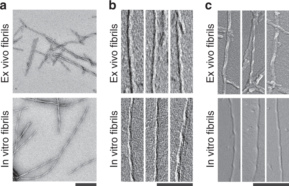
Kollmer, M. et al. Nat. Commun. 2019.
Figure 1. Fibrillar Structures in Alzheimer's Disease Brain Tissue Revealed by Cryo-EM
MtoZ Biolabs offers high-quality Biological Tissues Structure Characterization Service that supports fundamental research, disease mechanism elucidation and innovative drug discovery by leveraging a state-of-the-art Cryo-EM platform and an experienced expert team.
Services at MtoZ Biolabs
MtoZ Biolabs offers Cryo-EM for Biological Tissues Structure Characterization Service to deliver tailored ultrastructural analysis across a wide range of tissues, including:
We tailor sample preparation and imaging strategies based on tissue characteristics, delivering end-to-end solutions covering sample processing, imaging, data analysis, and 3D reconstruction.
Analysis Workflow
To ensure the highest quality outcomes for Biological Tissues Structure Characterization Service based on Cryo-EM, MtoZ Biolabs adheres to a meticulously standardized workflow:
1. Sample Preprocessing and Vitrification
Rapid freezing to preserve the natural ultrastructure.
2. Cryo-Sectioning and FIB Milling
Producing optimal imaging thickness through cryo-ultramicrotomy or Cryo-FIB.
3. High-Resolution Data Acquisition
Capturing images with cutting-edge direct electron detectors.
4. Data Preprocessing and 2D Classification
Particle selection, alignment, and denoising to optimize 3D reconstruction inputs.
5. 3D Reconstruction and Analysis
Applying Single Particle Analysis (SPA) or Cryo-ET to build accurate 3D structures.
6. Structural Annotation and Delivery
Providing 3D density maps, local resolution evaluation, and preliminary modeling outputs.
Why Choose MtoZ Biolabs?
Choosing MtoZ Biolabs for Cryo-EM for Biological Tissues Structure Characterization Service grants you access to:
✅ State-of-the-Art Technology: Equipped with top-tier Cryo-EM microscopes and next-generation direct detectors for achieving sub-nanometer resolution.
✅ Native-State Imaging: Samples are imaged in their true hydrated condition without the artifacts induced by chemical fixation or staining.
✅ Broad Tissue Compatibility: Adaptable for a wide variety of tissue types and complexities, ensuring broad research applicability.
✅ Minimal Sample Requirements: Enabling high-quality analyses from limited or precious tissue samples.
✅ Expert End-to-End Support: Our experienced Cryo-EM team manages every step from sample preparation to 3D structure modeling, ensuring efficient, high-quality delivery.
Applications
The Cryo-EM for Biological Tissues Structure Characterization Service supports a wide range of research and application areas, including:
· Tissue Ultrastructure Elucidation: Revealing subcellular architecture in liver, neural, and tumor tissues.
· Disease Mechanism Investigation: Analyzing ultrastructural changes in tumors, neurodegenerative disorders, and metabolic diseases.
· New Drug Development Assistance: Characterizing target localization and tissue microenvironment features to inform small molecule, antibody, and cell therapy research.
· Cell Communication and Exosome Research: Visualizing extracellular vesicle distribution and function at high resolution within tissue microenvironments.
Case Study
Case 1: Cryo-EM Reveals Ultrastructural Changes in Huntington’s Disease Neural Tissues
In a study on Huntington’s disease (HD), researchers used Cryo-ET to analyze the ultrastructure of neurons derived from HD patient iPSCs and primary neurons from BACHD mice. Cryo-EM imaging revealed prominent subcellular abnormalities in HD models, including sheet-like aggregates enclosed by double membranes, distorted mitochondrial cristae, and enlarged mitochondrial RNA granules.
By integrating AI-driven image analysis with mitochondrial proteomics, the study further uncovered the molecular basis of early mitochondrial dysfunction in HD pathogenesis. These results demonstrate the strength of Cryo-EM in capturing disease-associated ultrastructural changes at the tissue level, providing critical insights for early diagnosis and targeted therapy development in neurodegenerative diseases.
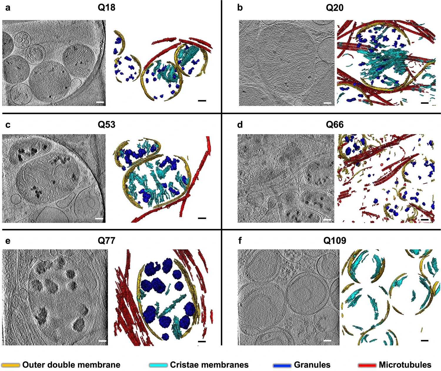
Wu, G. H. et al. Nat. Commun. 2023.
MtoZ Biolabs offers Cryo-EM for Biological Tissues Structure Characterization Service to support high-resolution analysis of subcellular features in complex tissues and cellular models, driving advances in life science and disease mechanism research.
FAQ
Q1: What are the essential sample requirements for Biological Tissues Structure Characterization Service based on Cryo-EM?
Tissues must maintain proper hydration and should be sectioned to a thickness of 100–300 nm to avoid freezing artifacts. MtoZ Biolabs offers comprehensive sample preparation optimization to ensure imaging quality.
Q2: Can Cryo-EM analyze large or highly complex tissue samples?
Yes. Although Cryo-EM imposes thickness limitations, techniques like cryo-ultramicrotomy and Cryo-FIB milling enable localized refinement, allowing high-resolution reconstruction of large or complex tissues. MtoZ Biolabs supports detailed characterization across various tissue types.
Q3: How does Cryo-EM differ from conventional TEM for tissue analysis?
Unlike traditional TEM, Cryo-EM preserves the tissue’s natural hydration and ultrastructure without chemical fixation or staining, avoiding artifacts associated with dehydration. MtoZ Biolabs' advanced Cryo-EM platform ensures more accurate and authentic biological tissue representation.
How to order?

