


Visual Proteomics Service
Visual Proteomics Service integrates advanced high-resolution microscopy with cutting-edge mass spectrometry, enabling precise analysis of protein localization, interactions, and functions within their native cellular environment. Visual Proteomics bridges the gap between molecular details and cellular architecture by integrating cryo-electron microscopy (Cryo-EM) or advanced fluorescence microscopy with mass spectrometry-based protein identification. By mapping proteins within their spatial and functional environments, visual proteomics provides a holistic perspective that traditional proteomics approaches often miss.
Deep visual proteomics (DVP) extends the capabilities of traditional visual proteomics by integrating artificial intelligence-driven image analysis, single-cell or single-nucleus laser microdissection, and ultra-high-sensitivity mass spectrometry. Deep visual proteomics preserves spatial context while linking protein abundance to complex cellular phenotypes. Deep visual proteomics enables researchers to classify distinct cell states based on proteomic profiles and detect spatially resolved proteome changes in tissues, such as those associated with cancer progression or microenvironmental heterogeneity. This capability makes deep visual proteomics particularly valuable for studying tumor microenvironments, cellular heterogeneity, and disease progression.
Service at MtoZ Biolabs
MtoZ Biolabs offers a comprehensive Visual Proteomics Service that seamlessly integrates state-of-the-art imaging platforms with advanced mass spectrometry systems. Our Visual Proteomics Service includes the innovative deep visual proteomics approach, which integrates AI-driven image analysis, single-cell or single-nucleus laser microdissection, and ultra-high-sensitivity mass spectrometry. With a team of experienced scientists and an optimized workflow, we provide precise, reliable, and customizable solutions tailored to meet diverse research needs. Researchers can leverage our Visual Proteomics Service to obtain actionable insights that foster innovation in structural biology, systems biology, and translational medicine. Free project evaluation, welcome to learn more details.
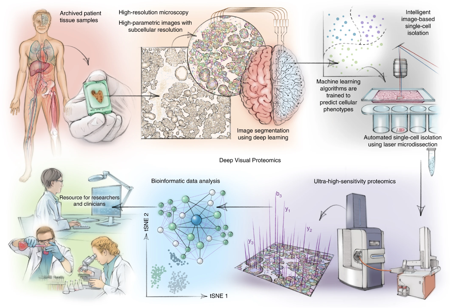
Figure 1. DVP Concept and Workflow
Service Advantages
1. Advanced Analysis Platform: MtoZ Biolabs established an advanced Visual Proteomics Service platform, guaranteeing reliable, fast, and highly accurate analysis service.
2. One-Time-Charge: Our pricing is transparent, no hidden fees or additional costs.
3. High-Data-Quality: Deep data coverage with strict data quality control. AI-powered bioinformatics platform integrates all Visual Proteomics Service data, providing clients with a comprehensive data report.
4. Customizable Service: Personalized service to meet various research needs.
5. Fast Turnaround: The process from sample handling to report generation is efficient, shortening the analysis cycle.
Applications
1. Structural Biology
Visual Proteomics Service enables detailed mapping of protein structures and complexes, providing insights into molecular architecture and assembly mechanisms.
2. Systems Biology
By integrating spatial and proteomic data, researchers can uncover protein interactions, signaling pathways, and dynamic cellular processes in their native contexts.
3. Cancer Research
Deep visual proteomics identifies spatial proteomic changes within tumor microenvironments, aiding in the discovery of biomarkers and therapeutic targets for precision oncology.
4. Neuroscience
The service facilitates the study of protein networks in neurons and synapses, revealing mechanisms underlying neurodegenerative diseases and brain function.
5. Drug Development
Visual Proteomics Service accelerates drug discovery by identifying drug-protein interactions and evaluating drug effects on protein networks in cellular and subcellular environments.
6. Infectious Disease Research
Researchers can utilize visual proteomics to study host-pathogen interactions, revealing protein localization and dynamics critical for understanding disease mechanisms.
Case Study
This study introduces single-cell Deep Visual Proteomics, integrating imaging, laser microdissection, and mass spectrometry to map spatially resolved proteomes in tissues with single-cell precision.
Rosenberger, F. A. et al. Nat Methods. 2023.
FAQ
Q: How does deep visual proteomics help in understanding disease progression, such as cancer or neurodegenerative disorders?
In cancer research, deep visual proteomics reveals spatially resolved proteomic changes within the tumor microenvironment, uncovering pathways such as immune evasion, metabolic reprogramming, and cell migration during tumor progression. It allows precise mapping of protein networks across different cell states, aiding in the identification of biomarkers and therapeutic targets.
In neurodegenerative disorders, deep visual proteomics characterizes protein misfolding, aggregation, and their spatial distribution in affected brain regions. By linking proteomic data with cellular phenotypes, it uncovers molecular mechanisms driving disease progression, such as disrupted synaptic signaling or protein trafficking. The integration of spatial and proteomic insights provided by deep visual proteomics enables a comprehensive understanding of disease dynamics, accelerating the development of targeted therapies and precision medicine strategies.
MtoZ Biolabs, an integrated chromatography and mass spectrometry (MS) services provider.
Related Services
Cryo-EM De Novo Structure Analysis Service
Cryo-EM Protein-Small Molecule Complex Structure Characterization Service
How to order?

