


Transmission Electron Microscope (TEM) based Exosome Characterization Service
Transmission electron microscope (TEM) based exosome characterization is an advanced technique that utilizes transmission electron microscopy (TEM) to achieve high-resolution imaging and structural analysis of exosomes. Exosomes are nanoscale vesicles secreted by cells, widely involved in intercellular communication, disease progression, and drug delivery. TEM employs an electron beam to penetrate the sample, directly revealing the vesicular structure, bilayer membrane characteristics, size distribution, and morphological integrity of exosomes. It provides ultra-high-resolution images far beyond the capabilities of optical microscopy and is considered the "gold standard" for confirming the presence and quality of exosomes.
Transmission electron microscope (TEM) based exosome characterization service is extensively applied in exosome-related basic research, drug delivery system development, engineered exosome validation, and quality control of exosome products in fields such as cancer, neurodegenerative diseases, and metabolic disorders. Through TEM, researchers can confirm the structural features of exosomes, assess isolation purity, and examine the morphology of vesicles after modification or cargo loading. This provides solid morphological evidence to support the application of exosomes in scientific research and clinical translation.
Yang, R J. et al. Nature Protocols, 2023.
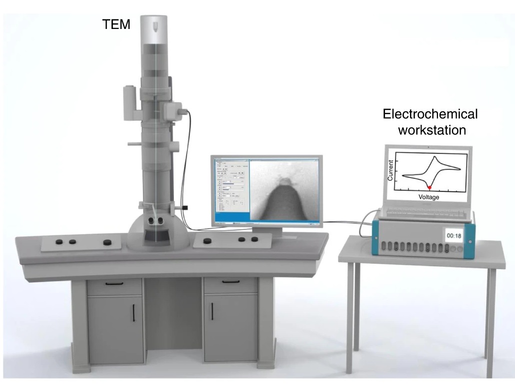
Figure 1. Setup for in Situ TEM Observation.
Services at MtoZ Biolabs
Based on high-resolution Transmission Electron Microscopy (TEM), the transmission electron microscope (TEM) based exosome characterization service provided by MtoZ Biolabs offers comprehensive morphological and structural characterization of exosome samples. Through TEM imaging, this service allows direct visualization of exosome size, membrane structure, and integrity, delivering clear vesicle images to confirm exosome presence, purity, and morphology. The service includes sample fixation, embedding, ultrathin sectioning, staining, and imaging, and ultimately provides high-quality TEM images along with a detailed image analysis report.
Service Advantages
1. Ultra-High-Resolution Imaging
By utilizing advanced transmission electron microscopy, we provide nanoscale high-resolution images that allow for clear observation of the characteristic vesicular structure, bilayer membrane, and internal morphology of exosomes, offering an intuitive visualization of their true architecture.
2. One-Time-Charge
Our pricing is transparent, no hidden fees or additional costs.
3. Standardized Sample Processing Workflow
We employ professionally standardized protocols for exosome sample preparation and negative staining to prevent damage or aggregation during analysis, ensuring high-quality imaging and stable, reproducible data.
4. Flexible Customizable Service
Tailored to the client’s research goals and project requirements, we offer personalized exosome characterization solutions, including structural evaluation of specially engineered exosomes (e.g., drug delivery vesicles), enhancing the specificity and effectiveness of the service.
Applications
1. Morphological Confirmation of Exosomes
By high-resolution TEM imaging, the vesicle structure and bilayer membrane of exosomes are visually displayed to confirm their typical morphology, providing morphological verification for newly isolated exosome samples or exosomes from different sources.
2. Structural Validation of Engineered Exosomes
The transmission electron microscope (TEM) based exosome characterization service can be used to visualize the structure of exosomes after engineering modifications, such as loading with drugs, RNA, proteins, or other functional molecules. This service confirms whether the vesicle modification has been successfully achieved, making it highly suitable for monitoring exosome integrity during the development of drug delivery systems, gene therapy vectors, and other therapeutic applications.
3. Disease-Associated Exosome Studies
In cancer, neurodegenerative diseases, cardiovascular diseases, and other research areas, TEM is used to observe morphological changes of exosomes secreted under disease conditions, revealing the relationship between exosome structure and function during disease progression.
4. Development of Novel Exosome-Based Carriers
For the development of exosomes as natural nano-delivery systems, the transmission electron microscope (TEM) based exosome characterization service can be applied to examine the surface structure and membrane fusion state of newly engineered vesicles. This service supports the validation of innovative technologies such as targeted drug delivery and vaccine development by providing detailed structural insights into engineered exosome carriers.
Case Study
1. A Method for Extraction of Exosomes from Breast Tumour Cells and Characterisation by Transmission Electron Microscopy
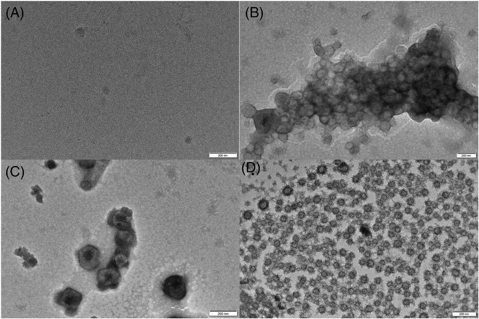
This study aims to establish a method for isolating exosomes from breast tumor cells and to characterize their morphological features using Transmission Electron Microscopy (TEM). The subjects of the study are exosomes secreted by breast tumor cells, which are extracted and purified through a combination of ultracentrifugation and membrane filtration techniques. Subsequently, TEM is employed to visualize the extracted exosomes and analyze their size, morphology, and integrity. The results demonstrate that the successfully isolated exosomes exhibit the typical cup-shaped morphology, with diameters ranging from approximately 30 to 150 nm, and possess intact vesicular membrane structures consistent with classical exosome characteristics. The samples also show high purity without noticeable contaminants. The study concludes that this method can stably and efficiently isolate high-quality exosomes from breast tumor cells and achieve effective characterization through TEM, thereby providing reliable technical support for subsequent functional studies and biomarker discovery related to exosomes.
Liu, Z A. et al. Journal of Microscopy, 2023.
Figure 2. TEM of Exosomes Stained with Phosphotungstic Acid.
FAQ
Q1: What Specific Information about Exosomes Can TEM Characterization Provide?
A1: TEM can visually display the overall morphology, size distribution, membrane structure, and bilayer membrane features of exosomes. It confirms whether exosomes possess intact vesicular structures and helps identify the presence of impurities or other particles.
Q2: Can TEM Characterization Provide Data on Exosome Size Distribution?
A2: Yes, TEM can offer statistical analysis by observing a large number of exosome images, generating size distribution graphs and average diameter data, which help assess the uniformity and purity of exosome samples.
How to order?

