



Quantitative proteomics techniques are the practical LC-MS/MS routes that turn digested peptides into relative protein matrices, or into peptide amounts when a matched stable isotope-labeled internal standard (SIS) is present (Bantscheff et al. 2012). The review below compares the technique classes used in project work: unlabeled discovery, isobaric multiplexing, and targeted confirmation. It is not a disease-application catalog and not a bulk dye-assay list.
Technique choice still separates labeling chemistry from acquisition mode. Unlabeled digests can be acquired as data-dependent acquisition (DDA) or data-independent acquisition (DIA). Isobaric tandem mass tag (TMT) work is typically acquired as DDA to read reporter ions. Closed peptide lists are acquired as parallel reaction monitoring (PRM) or multiple reaction monitoring (MRM) / selected reaction monitoring (SRM). In MtoZ Biolabs offerings, unlabeled discovery is commonly run as DDA label-free or DIA, multiplexed isobaric work is typically evaluated as TMT, and named proteins are typically followed by PRM, with MRM when a scheduled triple-quadrupole panel already exists.
Technique Classes Used in Quantitative Proteomics
Discovery techniques keep an open protein inventory. Unlabeled DDA label-free quantification (LFQ) extracts MS1 peak areas after identification (Cox et al. 2014). Unlabeled DIA extracts fragment chromatograms from scheduled isolation windows (Gillet et al. 2012). Isobaric TMT labels digests, mixes them, and reads relative channel abundance from reporter ions (Thompson et al. 2003). Metabolic labeling such as SILAC belongs in the labeled-discovery class when cells can be grown on heavy amino acids.
Confirmation techniques start after names freeze. PRM and MRM remeasure scheduled peptides from extracted fragment signals (Lange et al. 2008; Peterson et al. 2012). SIS-supported targeted assays convert light-to-heavy ratios into peptide amounts (Gerber et al. 2003). Relative targeted peak areas without SIS remain relative.
Quantitative proteomics techniques differ by which digests can be mixed, which precursors are isolated, and which claim the extracted signal can support.
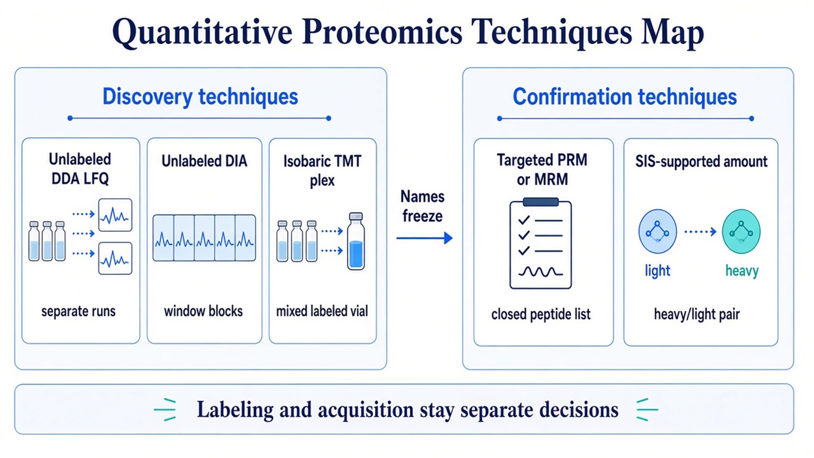
Figure 1. Discovery techniques build open relative matrices; confirmation techniques remeasure a closed peptide list, with SIS required only for amount claims.
Researchers can refer to What Are the Quantitative Methods for Proteomics? for the underlying definition of quantitative signals and claims.
How the Main Techniques Compare in Practice
Technique |
Quantitative signal |
Sample handling |
Typical claim |
Main constraint |
|---|---|---|---|---|
Unlabeled DDA LFQ |
MS1 peak area |
Separate injections |
Relative discovery |
Missingness and run-order drift |
Unlabeled DIA |
Fragment chromatograms |
Separate injections |
Relative discovery |
Window scheme and extraction QC |
TMT isobaric plex |
Reporter ions |
Label, mix, one LC path |
Relative multiplex |
Freezable set; possible ratio compression |
SILAC-style metabolic label |
Light/heavy precursors |
Mix after culture labeling |
Relative in cells |
Not a default for tissue or plasma |
PRM or MRM |
Targeted fragment peaks |
Closed inclusion list |
Relative confirmation |
Off-list proteins stay unseen |
Targeted plus SIS |
Light/heavy targeted peaks |
Closed list with calibrant |
Peptide amount |
Unique peptide and assigned SIS stock |
A comparison table does not replace a methods record. Acquisition settings, false discovery rate (FDR) thresholds, unique-peptide rules, and missing-value or impurity corrections still have to be declared with the chosen technique.
Researchers who need the three-axis selection logic after this comparison can refer to Protein Quantification Method.
Shared Workflow Steps That Change by Technique
Every quantitative technique still moves through matched extraction, digestion, LC-MS/MS, identification or targeted extraction, and a declared contrast. What changes is where samples diverge.
Unlabeled techniques keep each digest on its own injection. Alignment and normalization carry the cross-run burden. TMT adds channel labeling and pooling before acquisition, so load matching and amine chemistry become critical before mixing. Targeted techniques insert peptide selection and method scheduling before the cohort is acquired. SIS, when used, adds spike timing and calibration before amount reporting.
Technique success depends on that order more than on a biomarker keyword. Unequal input, confounded run order, incomplete labeling, or an oversized targeted panel can break the claim after the instrument run looks complete.
Parameters and Limits That Decide Technique Fit
Unlabeled techniques need interleaved group order, stable chromatography, and a missing-value rule that matches abundance-dependent dropout (Lazar et al. 2016). DIA adds library or library-free extraction settings. TMT needs a channel map, labeling completeness, and a plan for co-isolation if fold changes look smaller than expected. Targeted techniques need unique peptides, duty-cycle fit, and qualifier rules. Amount techniques need an assigned SIS concentration.
Limits follow the same map. Unlabeled matrices do not invent unidentified proteins. TMT ratios are relative inside a plex and do not accept late unlabeled files as extra channels. Targeted techniques do not survey an open proteome. Ordinary discovery intensities are not ng per mL. Pathway enrichment is optional after QC. It is not a substitute for a failed technique choice.
Instrument brand names, kit channel counts, and multi-omics storytelling are implementation or interpretation layers. They are not the technique definition.
How to Choose a Technique for the Next Project
The useful test is whether the protein list is open, whether samples can be mixed, and whether the unit is a ratio or an amount.
Project need |
Technique that often fits |
Recheck before committing |
|---|---|---|
Open list, samples may still be added |
Unlabeled DDA LFQ or unlabeled DIA |
Run order identical to treatment |
Open list, full set can be mixed now |
TMT relative plex |
Later harvests without a bridge |
Relative comparison in labeled cells |
SILAC-style metabolic labeling |
Tissue or plasma without culture labeling |
Named peptides to remeasure |
PRM or MRM |
Panel longer than one duty cycle |
Mass or molar unit for named peptides |
Targeted assay plus matched SIS |
Relative peaks read as concentration |
Only total protein load matching |
Bulk dye or A280 assay |
Not a proteome technique |
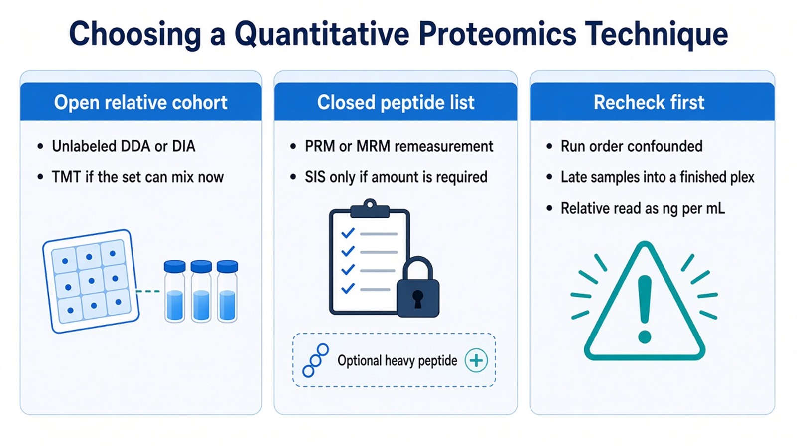
Figure 2. Choose a discovery or confirmation technique from list status and claim unit; confounded design and amount-without-SIS claims need repair first.
Unlabeled discovery more often fits open enrollment. TMT more often fits a freezable multi-group set. Targeted confirmation more often fits after names freeze. Exceptions exist when a tiny validated panel never needed discovery, or when a short closed cohort must share one injection to limit run-order drift.
Researchers who already have sample type, group design, and a requested claim can also review the MtoZ Biolabs Quantitative Proteomics Service for project-specific technique planning. For closed-list trade-offs after discovery, see Advantages and Disadvantages of Targeted Proteomics.
Frequently Asked Questions
1. Is label-free quantification a different technique from DIA?
Label-free names the chemistry. DIA names the acquisition schedule. Unlabeled digests are often acquired as DIA, and the two decisions remain separate in a technique statement.
2. Does absolute quantification equal PRM or MRM?
No. PRM and MRM are targeted acquisition techniques. Absolute amount requires a matched SIS and a declared calibration on top of that targeted schedule.
3. Should every discovery project finish with targeted confirmation?
No. Targeted confirmation fits when names can freeze and a unique peptide is available. Keep discovery while the inventory is still open or when the remaining question is still untargeted screening depth.
4. Can TMT and unlabeled files be reviewed as one technique matrix?
Not without an explicit bridge design. They use different quantitative objects and missingness patterns. Mixing them in one statistical model without that bridge creates contrasts the data cannot defend.
5. Where do spectral counting techniques sit in this review?
Spectral counting can rank detectability inside discovery data. For group fold-change statistics, intensity-based unlabeled, reporter-ion, or targeted traces are the stronger quantitative objects.
MtoZ Biolabs can provide quantitative proteomics techniques across unlabeled DDA or DIA discovery, TMT relative plexes, and targeted PRM or MRM follow-up when sample type, study design, and the intended claim are available.
If further evaluation of discovery versus confirmation routing, relative versus SIS-supported amount, or whether a bulk load assay is sufficient is needed, project details can be submitted for a feasibility review.
Reference
- M. Bantscheff, S. Lemeer, M.M. Savitski, B. Kuster (2012). Quantitative mass spectrometry in proteomics: critical review update from 2007 to the present. Anal. Bioanal. Chem., 404, 939-965. https://doi.org/10.1007/s00216-012-6203-4
- J. Cox, M.Y. Hein, C.A. Luber, I. Paron, M. Mann (2014). Accurate proteome-wide label-free quantification by delayed normalization and maximal peptide ratio extraction, termed MaxLFQ. Mol. Cell. Proteomics, 13, 2513-2526. https://doi.org/10.1074/mcp.M113.031031
- S.A. Gerber, J. Rush, O. Stemman, M.W. Kirschner, S.P. Gygi (2003). Absolute quantification of proteins and phosphoproteins from cell lysates by tandem MS. Proc. Natl. Acad. Sci. U.S.A., 100, 6940-6945. https://doi.org/10.1073/pnas.0832254100
- L.C. Gillet, P. Navarro, S. Tate, H. Rost, N. Selevsek, L. Reiter, R. Bonner, R. Aebersold (2012). Targeted data extraction of the MS/MS spectra generated by data-independent acquisition: a new concept for consistent and accurate proteome analysis. Mol. Cell. Proteomics, 11, O111.016717. https://doi.org/10.1074/mcp.O111.016717
- V. Lange, P. Picotti, B. Domon, R. Aebersold (2008). Selected reaction monitoring for quantitative proteomics: a tutorial. Mol. Syst. Biol., 4, 222. https://doi.org/10.1038/msb.2008.61
- C. Lazar, L. Gatto, M. Ferro, C. Bruley, T. Burger (2016). Accounting for the multiple natures of missing values in label-free quantitative proteomics data sets to compare imputation strategies. J. Proteome Res., 15, 1116-1125. https://doi.org/10.1021/acs.jproteome.5b00981
- A.C. Peterson, J.D. Russell, D.J. Bailey, M.S. Westphall, J.J. Coon (2012). Parallel reaction monitoring for high resolution and high mass accuracy quantitative, targeted proteomics. Mol. Cell. Proteomics, 11, 1475-1488. https://doi.org/10.1074/mcp.O111.013698
- A. Thompson, J. Schafer, K. Kuhn, S. Kienle, J. Schwarz, G. Schmidt, T. Neumann, R. Johnstone, A.K. Mohammed, C. Hamon (2003). Tandem mass tags: a novel quantification strategy for comparative analysis of complex protein mixtures by MS/MS. Anal. Chem., 75, 1895-1904. https://doi.org/10.1021/ac0262560
