



Mass spectrometry (MS) protein quantification estimates how protein abundance differs across samples from ion signals recorded after proteins are usually digested into peptides. The measurement is not a colorimetric total-protein assay such as bicinchoninic acid (BCA) or Lowry. A protein-level value is inferred from identified peptide features, so the valid claim is relative change of the same protein, or an amount only when a matched standard and a calibration model are part of the design. Unlabeled discovery, isobaric tagging, metabolic labeling, and targeted monitoring are separate routes to that claim. They are not interchangeable names for one instrument method.
Researchers who already have defined samples, comparison groups, or a specific project objective can also review the MtoZ Biolabs Quantitative Proteomics Service for project-specific feasibility and analysis planning.
What Question Mass Spectrometry Protein Quantification Can Answer
The first decision is the scientific claim, not the instrument brand. MS can support a relative comparison, an amount estimate, or an identity check. Mixing those three claims is the usual source of over-read results.
Relative change, amount, and identity are different endpoints
Relative quantification asks whether the same protein is higher or lower across groups after a comparable workflow. Absolute or copy-number claims need a matched isotope standard and a calibration model that matches the claimed level, peptide or protein. Intact-mass measurement confirms molecular weight and major proteoforms. It does not, by itself, report a proteome-wide abundance matrix.
Colorimetric assays remain useful for estimating total protein load before digestion. They do not identify which proteins changed. Enzyme-linked immunosorbent assay (ELISA) and Western blot quantify selected proteins when antibodies and linearity are adequate. They do not replace an untargeted or multiplexed MS comparison.
Protein values are usually inferred from peptides
Bottom-up proteomics digests proteins, most often with trypsin, and treats selected peptides as surrogates of the parent protein (Bantscheff et al. 2007). Peptide quantification is the operational measurement behind most protein quantification tables. Shared peptides, missed cleavage, variable modifications, and unequal ionization mean that peptide intensity is not a direct copy-number ranking across different proteins.
Intact-protein or top-down quantification is a different experiment. It is useful when the analyte must remain the intact chain. It is not the default discovery route for a complex tissue or biofluid digest.
When Mass Spectrometry Quantification Fits a Study Design
MS quantification fits when the study needs protein-resolved change across a defined contrast, and when samples can enter a digestion and LC-MS/MS plan that stays comparable. It is a poor substitute for a quick total-protein reading, and it is a poor discovery method if the only need is one antibody-defined target with an already validated immunoassay.
Clinical tissues, cultured cells, and biofluids can all be quantified when extraction, contaminants, and a searchable protein database are compatible with the chosen route. Metabolic labeling such as stable isotope labeling by amino acids in cell culture (SILAC) requires a system that incorporates heavy amino acids. Chemical tags require peptides that can be labeled after digestion. Unlabeled discovery requires independent injections under one chromatographic regime.
Group definitions, the intended contrast, and whether later samples may still enroll should be fixed before the first digest is queued. A later change in column, instrument, or labeling set can create a new analytical block that ordinary processing does not stitch together.
How Peptide Signals Become Protein Values
Proteins are extracted, digested, and separated by liquid chromatography. The mass spectrometer records precursor ions and fragment spectra. Peptide identities come from fragment evidence searched against a protein database. Quantitative values come from peak area, reporter ions, or scheduled transitions, depending on the route. Software then summarizes peptide evidence into protein-level values.
A larger signal means more ion current was recorded for that feature in that run or that channel, subject to digestion recovery, loading, ionization, interference, and peak quality. Protein inference can change the reported number when peptides are shared, filtered, or missing.
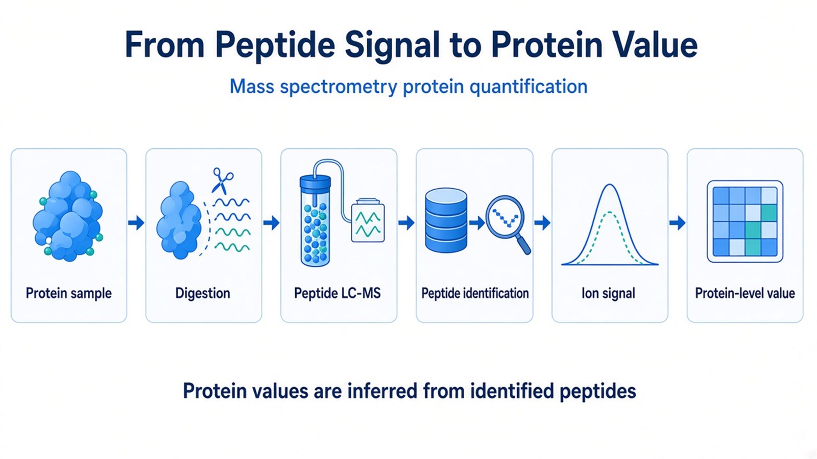
Figure 1. Most MS protein quantification infers protein-level values from identified peptide ion signals after digestion, separation, and identification.
A protein-level MS value is usually inferred from peptide ion signals, not from a colorimetric total-protein reading.
Which Quantification Route Matches the Design
Labeling, acquisition, and assay scope are separate layers. Data-dependent acquisition (DDA) and data-independent acquisition (DIA) name how precursors are fragmented. They do not decide whether a label was used. In MtoZ relative-discovery offerings, DIA is typically run on unlabeled digests, while tandem mass tag (TMT) workflows are typically acquired in DDA to read reporter ions.
Study need |
Route that often fits |
Main quantitative signal |
Reassess when |
|---|---|---|---|
Relative discovery, independent injections |
Unlabeled LFQ |
MS1 peak area or DIA fragment chromatograms |
Near-complete values or same-run multiplex are required |
Relative comparison inside one pooled run |
Isobaric labeling such as TMT |
Reporter ions |
Samples cannot be labeled, or the cohort exceeds a coherent multiplex plan |
Metabolic comparison in a labelable system |
SILAC |
Light-to-heavy peptide pairs |
The organism or buffer cannot incorporate heavy amino acids |
Predefined peptides, amount or confirmation |
Targeted MS such as MRM or PRM |
Scheduled transitions |
The protein list is still open |
Ion-current comparison without identification is not a protein quantification result. Spectral counting can support rough ranking in older DDA files, but protein-level fold changes for a biological claim should use intensity or reporter-ion values.
Unlabeled discovery has its own alignment, missing-value, and run-order constraints.
For studies focused primarily on that unlabeled route, What Is Label-Free Quantification in Proteomics? provides a more detailed explanation of MS1 peak-area LFQ, spectral counting, and unlabeled DIA as a separate acquisition choice.
When sample number, multiplexing, or acquisition strategy makes the choice less straightforward, researchers can refer to Comparing LFQ and Isobaric Labeling Strategies in Proteomics Quantification for a more detailed strategy-selection framework.
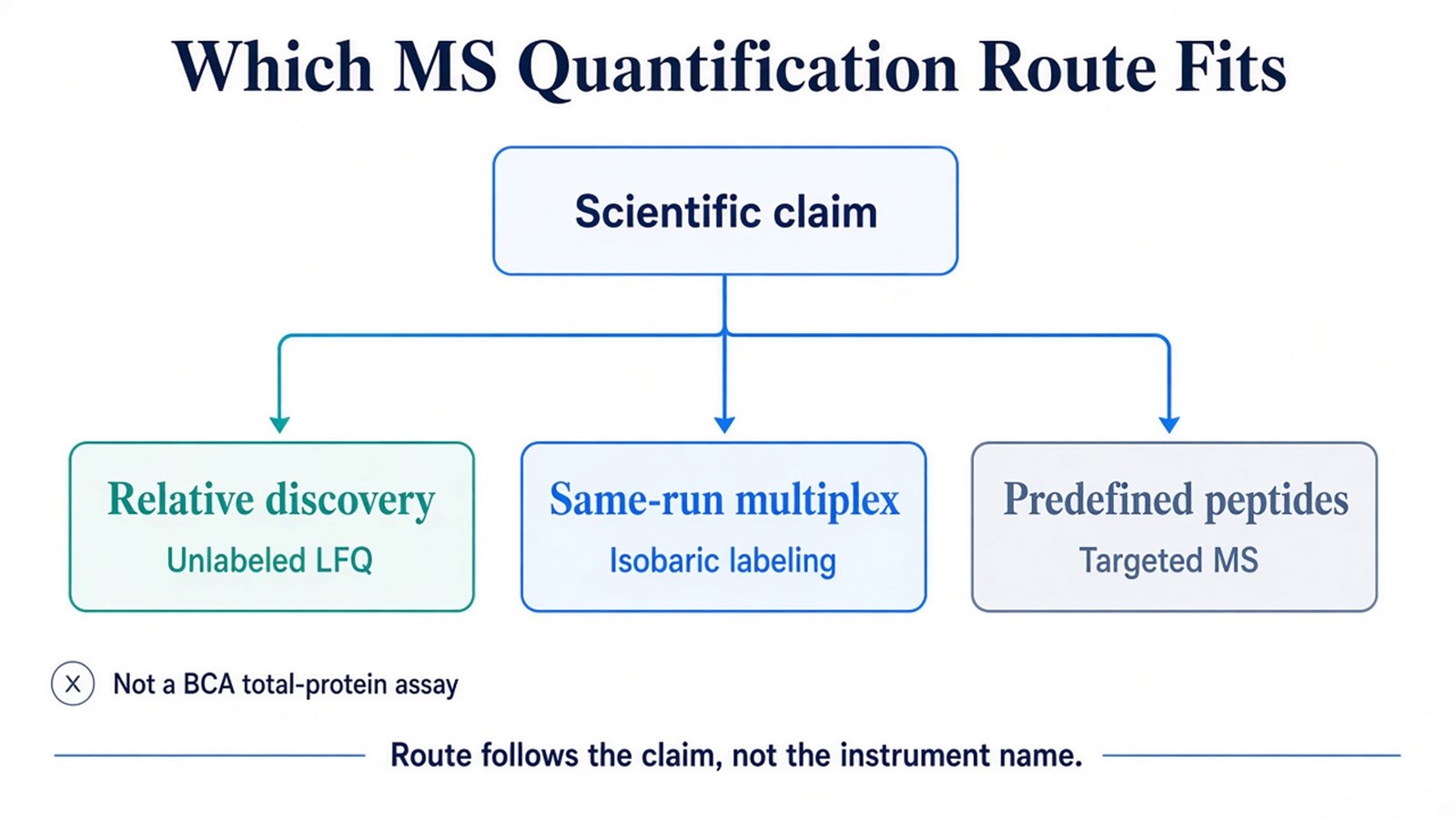
Figure 2. The usable MS route follows the claim: relative discovery, same-run multiplex, or a predefined peptide amount.
Conditions That Make a Quantitative Comparison Valid
Search queries about conditions are often looking for experimental requirements, not a list of laboratory skills. The comparison is only as strong as the steps that make independently handled samples, or pooled labeled channels, actually comparable.
Extraction and digestion must stay matched across the contrast. Detergents, salts, and polymers that survive cleanup can suppress ionization or damage chromatography. Incomplete digestion creates missed-cleavage peptides that do not represent the parent protein evenly. Loading differences can look like biological change if they are not normalized or, in labeled work, if channels are not balanced.
Chromatography has to remain stable enough for the chosen metric. Unlabeled peak-area comparison depends on aligned retention time. Isobaric comparison depends on co-isolated peptides that can compress reporter ratios. Targeted comparison depends on the analyte staying inside its scheduled window.
If the claim is amount, a matched internal standard must be added at a point that matches that claim. A heavy peptide spiked after digestion cannot correct digestion loss that already occurred. Quality-control injections can monitor drift. They cannot rescue a run order in which all controls were acquired before all treated samples, or a labeling set that mixed digestion batches with biological groups.
The checks above are planning guides. They are not acceptance cutoffs, and they do not convert a candidate protein list into a diagnostic result.
How to Read a Protein Quantification Result
An MS quantification table supports the comparison that the design actually measured. After identification, quantification, and protein summarization, a higher protein value can support a relative statement about that protein across groups in this study. Peptide support, missingness, channel interference, and the stated normalization belong next to that statement.
Ordinary discovery intensities should not rank protein A against protein B by copy number, transfer a ratio from one column or instrument to another study as a calibrated quantity, or stand in for a clinical cutoff. A large fold change on one poorly shared peptide is weaker evidence than a smaller change supported by several unique peptides with complete observations.
PTM-aware quantification is a related but narrower experiment. A phosphorylation or glycosylation site needs a modified peptide as the analyte. Global protein abundance and site occupancy should not be treated as the same number.
Relative MS quantification supports same-protein comparison inside one coherent study.
Frequently Asked Questions
1. Is mass spectrometry protein quantification the same as a BCA or Lowry assay?
No. BCA and Lowry estimate total protein load. MS identifies peptides and infers protein-level change from those ion signals. A colorimetric reading can size a digest. It cannot report which proteins changed.
2. Why do protein quantification reports start from peptides?
Most discovery workflows digest proteins before LC-MS/MS. The recorded features are peptide ions. Protein values are summarized from those peptides. Shared or modified peptides can change the protein number even when the underlying spectra look intense.
3. Can MS report absolute protein concentration without standards?
Not from ordinary unlabeled or isobaric discovery intensities. Amount or copy-number claims need a matched standard and a calibration model. Targeted assays such as multiple reaction monitoring (MRM) can support that claim when the standard and the transition are validated in the study matrix.
4. What sample conditions matter most before an MS quantification run?
Matched extraction and digestion, cleanup compatible with LC-MS, a defined contrast, and a plan that keeps labeling sets or unlabeled chromatography under one analytical regime. If later samples may still enroll, that enrollment has to be part of the first sequence plan.
Once the comparison design, unlabeled or labeled sample set, and expected protein-level readout are defined, the project can be evaluated as a quantitative proteomics study. Researchers can review the MtoZ Biolabs Quantitative Proteomics Service for project-specific sample evaluation, feasibility assessment, and workflow planning.
