



The principle of co-immunoprecipitation (Co-IP) is three binding events in one capture. An antibody binds a bait antigen. Protein A or Protein G on beads binds the Fc of that antibody. Proteins that remain attached to the bait through lysis and wash stay on the collected resin and can be eluted for Western blot or mass spectrometry. Co-IP is not a synonym for immunoprecipitation (IP). IP uses the first two events to recover one nominated chain. Co-IP uses the same two events under conditions meant to keep a neighborhood. The pellet after Co-IP is an association that survived the written buffer. It is not, by itself, a direct contact, a network map, or a native-cell snapshot.
A lysate contains the bait, proteins that were bound to it, proteins that bind the antibody scaffold, and proteins that bind the resin. The principle explains why a partner can appear. It also explains why a bead binder can appear. Specificity of the antibody–antigen pair is necessary. It is not sufficient.
Researchers who already have an IP-grade antibody, a nominated pair, or a complex they want to inspect by this principle can also review the MtoZ Biolabs Co-Immunoprecipitation Protein Interaction Analysis Service for project-specific feasibility and analysis planning.
The Three Binding Events
The antibody is chosen against one bait. In the lysate it forms an antigen–antibody complex if the epitope is accessible and the protein is not already denatured past recognition. Protein A/G beads, covalent or non-covalent, bind the Fc and allow magnetic or centrifugal collection. Partners are not bound by the antibody unless they share an epitope. They ride if a non-covalent contact with the bait, or a bridge through another subunit, survives detergent, salt, and wash time.
Calling this “highly specific isolation” collapses those three events into one. The antibody can be specific for the bait and still collect Fc binders, Protein A/G binders, and abundant proteins that stick to agarose or magnetic surfaces. For how IP and Co-IP judge different pellets after the same chemistry, Differences Between Immunoprecipitation and Co-Immunoprecipitation provides a complementary pellet-rule discussion.
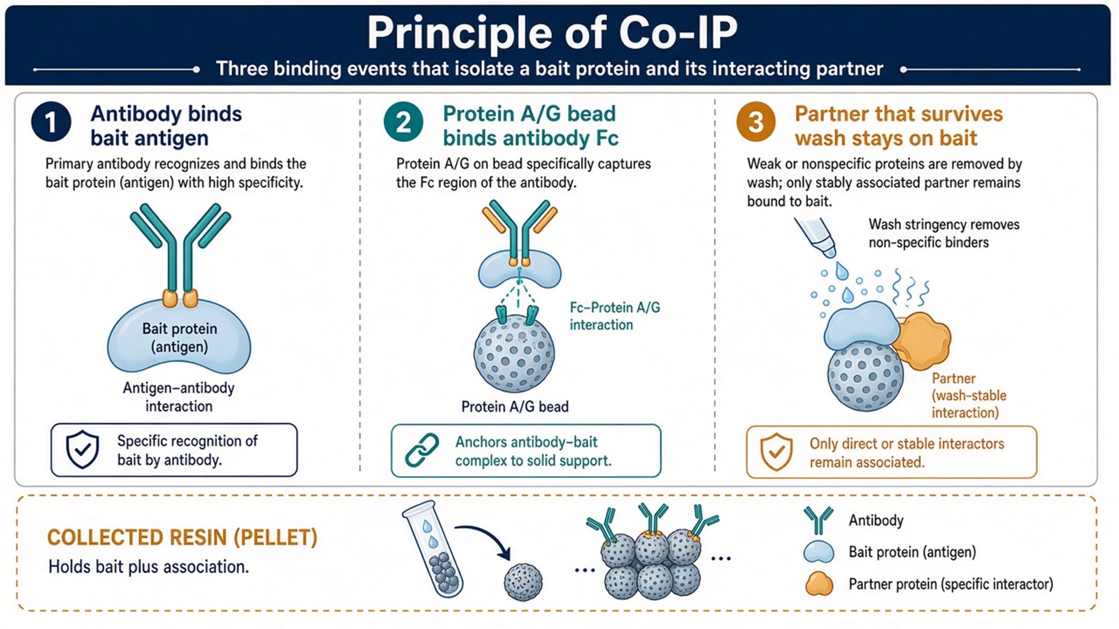
Figure 1. Event 1 is antigen–antibody. Event 2 is Fc–Protein A/G. Event 3 is a wash-stable association with the bait. Only the first two are designed. The third is inferred.
Name the three events before the first blot. A band in the eluate does not say which event produced it.
Capture of Protein-Protein Interactions
While the antibody occupies the bait, proteins that were in a complex with that bait may remain attached. The collected material can therefore contain the bait, direct contacts, bridged subunits, and proteins that never touched the bait. Elution with acid, peptide competition, or a denaturing buffer releases that mixture from the beads. Release does not sort the mixture.
A stable complex in the living cell can still fall apart in NP-40. A weak or transient contact can still survive if local concentration on the bead is high. The principle is physical co-recovery, not a Kd. Indirect members of a complex are expected, not a contamination class by themselves. Bead and antibody background are a different class. They are not solved by calling the method native.
Subsequent Analysis and Identification
The same principle supports two readouts. Western blotting tests a nominated prey on the same blot as the bait. It answers a binary presence question under the stated wash. It does not open the rest of the pellet. Mass spectrometry identifies peptide-supported protein groups in the eluate, including proteins that were not nominated. It does not identify every component of the physiological complex, and it does not construct a network from one bait IP. For the wet-lab order that follows this principle into an LC-MS/MS file, Process of IP Mass Spectrometry provides a complementary step list. For how that file is ranked, How to Analyze IP Mass Spectrometry Results? provides a complementary analysis order.
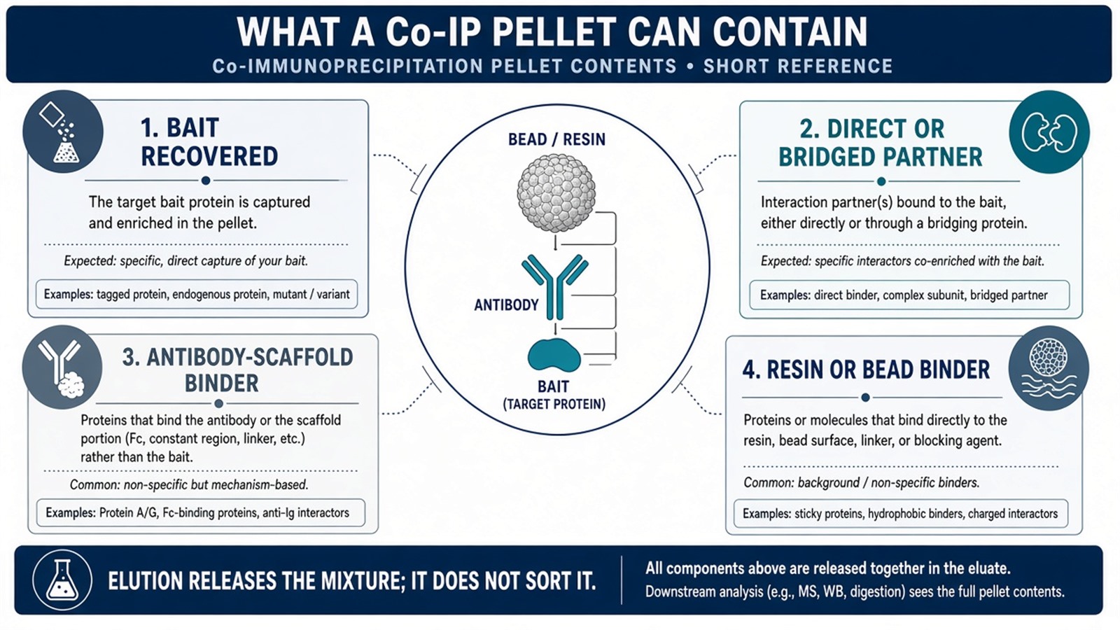
Figure 2. Co-recovery is the principle. Direct contact, a bridged subunit, Fc binding, and bead binding can all occupy the same eluate.
Advantages and Challenges of the Technique
Co-IP can be run on endogenous proteins when an IP-grade antibody exists, and it does not require a genetic tag. That is an experimental convenience. It is not proof that the buffer matches the living cell. Lysis opens membranes, dilutes local concentrations, and replaces cytoplasm with detergent. “Near-physiological” is accurate only as a contrast to fully denaturing purification, not as a claim that native complexes are intact.
Sensitivity follows bait occupancy, antibody affinity, and detection method. Co-IP is not automatically the method of choice for low-abundance partners. A scarce prey can be missed when occupancy is low, even if the principle is correctly applied. For that occupancy problem, Co-IP for Low-Expression Protein Interactions provides a complementary discussion.
The main challenges sit inside the principle. Antibody specificity and affinity decide whether the bait is collected. Non-specific binding to Fc or resin produces false partners. Interaction stability depends on buffer, salt, time, and temperature. Those limits are not later software problems. They are properties of the three events.
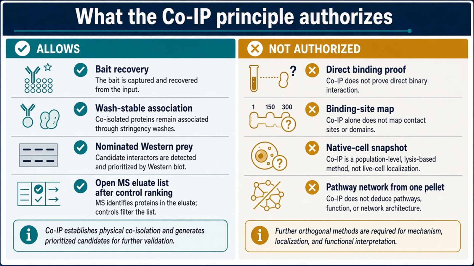
Figure 3. What the principle supports: bait recovery and a control-ranked association. What it does not: a binding residue, an absolute copy number, or a pathway map from one pellet.
What the Principle Does Not Authorize
A successful bait blot shows that events 1 and 2 worked. It does not show that a prey band is a direct interactor.
An open MS list from the eluate is a capture inventory. It is not a reconstructed interactome.
Reciprocal Co-IP strengthens a nominated pair. It still reports co-recovery, not a contact residue. For why Co-IP-MS does not map binding sites, Can Co-IP Followed by Mass Spectrometry Determine the Binding Sites Between Two Proteins? provides a complementary limit discussion.
Experimental Limits
The principle reduces the mixture to what the antibody and resin can hold. It does not remove background. A matched IgG or isotype, and a beads-only or empty-tag arm, are how the third event is interpreted. Diagnostic language sits outside ordinary research reporting unless a separately validated assay framework is in place.
Project Planning and Service Scope
A usable Co-IP plan names the bait epitope, the Protein A/G resin, the lysis class intended to keep the complex, the matched control, and whether the next readout is a nominated Western or an open MS table. MtoZ Biolabs scopes endogenous or tagged Co-IP, and Co-IP-MS, on that written principle. Interpretability still follows the three events, not the instrument name.
Once the principle is specified, the project can be a pair confirmation or an open partner discovery. Researchers who need an LC-MS/MS reading of a Co-IP pellet can review the MtoZ Biolabs Co-IP-MS Protein Interaction Analysis Service for project-specific sample evaluation, feasibility assessment, and workflow planning.
Frequently Asked Questions
1. What is the principle of co-immunoprecipitation?
An antibody binds the bait. Protein A/G beads bind the antibody Fc. Proteins that remain with the bait through lysis and wash are collected and eluted. The readout inspects that pellet.
2. Is Co-IP the same as IP?
No. Both use antibody and Protein A/G. IP asks whether the bait was recovered. Co-IP asks which other proteins were recovered with it.
3. Does a co-precipitated protein bind the bait directly?
Not necessarily. Bridged complex members, antibody-scaffold binders, and resin binders can occupy the same pellet.
4. Does Co-IP preserve native protein state?
It is less denaturing than SDS purification when a mild buffer is used. Lysis still dilutes and deterges the cell. Native-cell language needs a method that never opened the cell.
5. Can Western blot and mass spectrometry use the same principle?
Yes. Both read the eluted pellet. Western tests a nominated prey. MS opens the protein-group list. Neither readout changes the three binding events.
Reference
- Phizicky E.M., Fields S. (1995). Protein-protein interactions: methods for detection and analysis. Microbiol. Rev., 59, 94-123. https://doi.org/10.1128/mr.59.1.94-123.1995
- Gingras A.C., Gstaiger M., Raught B., Aebersold R. (2007). Analysis of protein complexes using mass spectrometry. Nat. Rev. Mol. Cell Biol., 8, 645-654. https://doi.org/10.1038/nrm2208
- ten Have S., Boulon S., Ahmad Y., Lamond A.I. (2011). Mass spectrometry-based immuno-precipitation proteomics: the choice of method matters. Proteomics, 11, 1153-1159. https://doi.org/10.1002/pmic.201000548
- Dunham W.H., Mullin M., Gingras A.C. (2012). Affinity-purification coupled to mass spectrometry: basic principles and strategies. Proteomics, 12, 1576-1590. https://doi.org/10.1002/pmic.201100523
- Lin J.S., Lai E.M. (2017). Protein-protein interactions: co-immunoprecipitation. Methods Mol. Biol., 1615, 211-219. https://doi.org/10.1007/978-1-4939-7033-9_17
