


Phosphorylation Site Identification Services
- Targeted Drug Development: Validate kinase inhibitor specificity and assess off-target effects.
- Disease Mechanism Studies: Investigate phosphorylation-driven mechanisms in cancer and neurodegenerative diseases.
- Signaling Pathway Analysis: Map phosphorylation cascades in immune response, metabolic regulation, or apoptosis.
- Biomarker Discovery: Identify phosphorylation signatures associated with disease progression or treatment response.
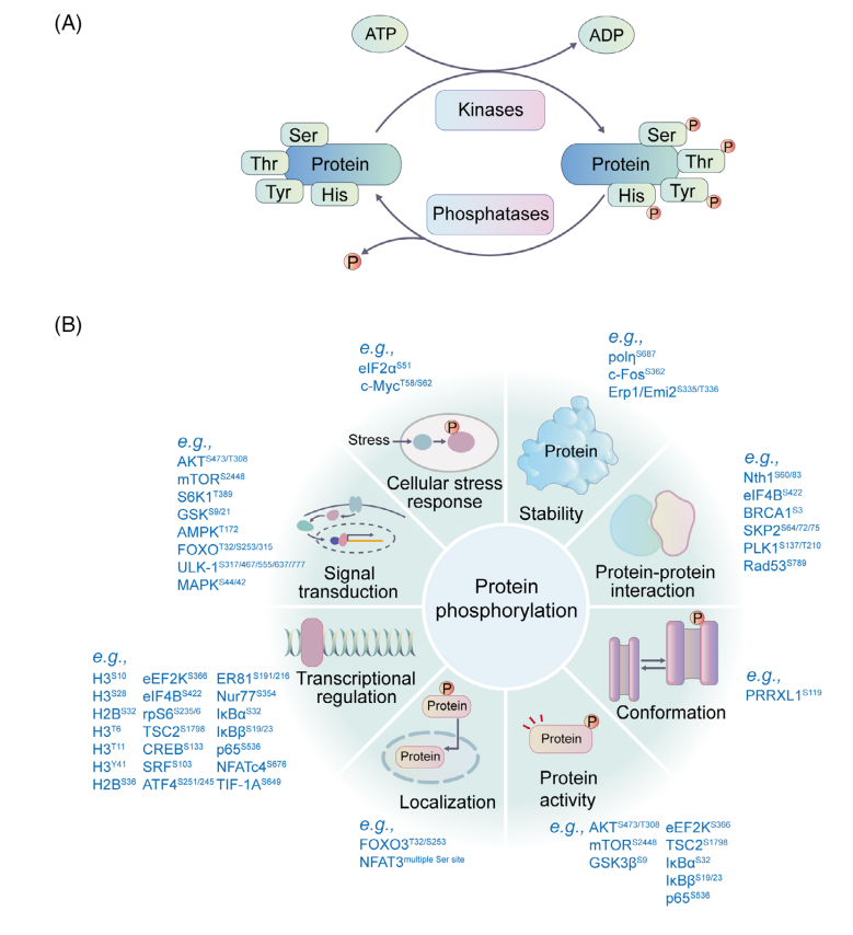
Phosphorylation is one of the most extensively studied post-translational modifications (PTMs), driving essential biological processes such as cell growth, proliferation, and survival. Mapping phosphorylation sites is crucial for understanding signaling mechanisms, disease targets, and drug development. MtoZ Biolabs utilizes high-resolution, high-mass-accuracy mass spectrometry combined with advanced bioinformatics tools to provide rapid, high-sensitivity, and high-throughput Phosphorylation Site Identification Services, covering analyses from single proteins to whole-cell levels.
Zhong, Q. et al. MedComm (2020). 2023.
Figure 1. The Phosphorylation Process and the Potential Functions of Phosphorylation
Services at MtoZ Biolabs
1. Target Protein Phosphorylation Site Identification
MtoZ Biolabs utilizes a high-resolution LC-MS/MS platform combined with phosphopeptide enrichment strategies to achieve precise localization and quantitative analysis of phosphorylation modifications on specific target proteins. This service identifies phosphorylation sites on serine, threonine, and tyrosine residues, helping researchers uncover regulatory mechanisms involved in protein signal transduction and functional modulation.
2. Phosphoproteomic Site Identification
MtoZ Biolabs employs IMAC or TiO₂ enrichment coupled with high-throughput mass spectrometry to achieve systematic identification and dynamic comparison of phosphorylation sites across the entire proteome. This service provides a comprehensive phosphorylation landscape under different conditions, supporting studies of cellular signaling pathways and molecular regulatory mechanisms.
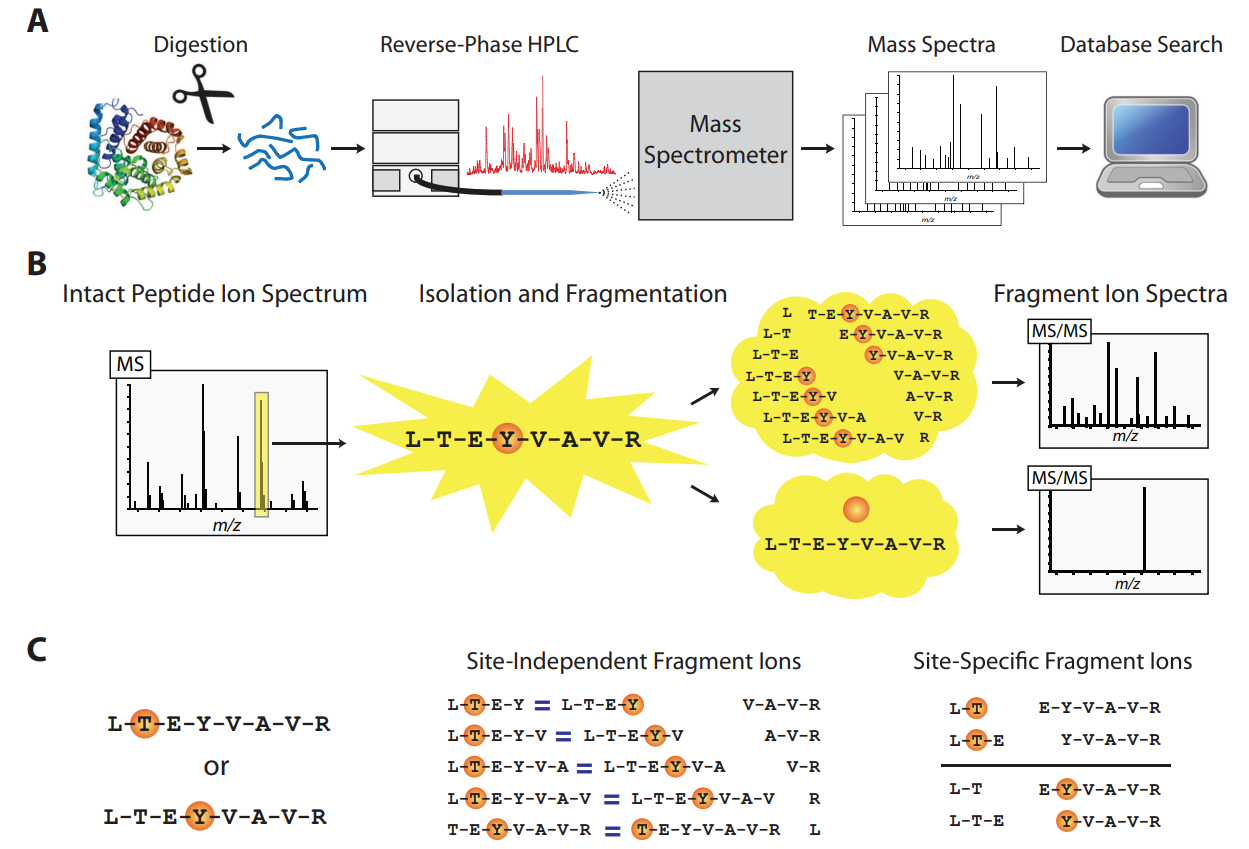
Analysis Workflow
1. Sample Preparation: Target proteins are purified via immunoprecipitation (IP), gel electrophoresis, or precipitation.
2. Enzymatic Digestion: Proteins are digested into peptides suitable for mass spectrometry analysis using trypsin/Lys-C.
3. Phosphopeptide Enrichment: Selective enrichment of phosphorylated peptides is performed using immobilized metal affinity chromatography (IMAC) or titanium dioxide (TiO₂) technology to enhance detection sensitivity.
4. LC-MS/MS Analysis: Phosphorylation sites on serine (S), threonine (T), and tyrosine (Y) residues are precisely mapped using high-resolution mass spectrometry (e.g., Orbitrap) coupled with nano-liquid chromatography.
Dephoure, N. et al. Mol Biol Cell. 2013.
Figre 2. The Workflow of Phosphorylation Site Identification
Service Advantages
✅ Ultrasensitive Enrichment Technology: Optimized IMAC/TiO₂ workflows to capture low-abundance phosphorylation signals, enhancing detection depth.
✅ High-Precision Localization: Advanced mass spectrometry platforms such as Orbitrap Fusion Lumos, combined with proprietary algorithms, achieve >95% site localization confidence.
✅ Adaptability to Complex Samples: SCX or SDS-PAGE fractionation ensures data reliability for high-complexity samples.
✅ Multidimensional Insights: Includes dynamic phosphorylation quantification (Label-free/TMT), kinase-substrate network prediction, and KEGG pathway annotation to uncover biological significance.
Case Study
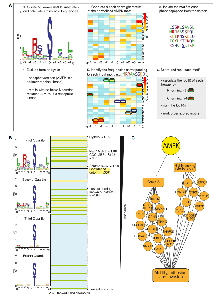
1. Identification of AMPK Phosphorylation Sites Reveals a Network of Proteins Involved in Cell Invasion and Facilitates Large-Scale Substrate Prediction
AMPK is a central regulator of cellular energy sensing and is increasingly recognized for its role in non-metabolic processes. This study employed a chemical genetics screen combined with a peptide capture strategy to identify direct AMPK phosphorylation sites in whole cells. Analysis revealed that AMPK substrates include numerous proteins involved in cell motility, adhesion, and invasion. Notably, AMPK phosphorylation of the RHOA guanine nucleotide exchange factor NET1A inhibits extracellular matrix degradation. Additionally, the study established an AMPK motif matrix and developed a pipeline for predicting AMPK substrates, providing valuable insights for future research. Phosphorylation Site Identification Services enables the identification of phosphorylation sites on proteins, facilitating the analysis of kinase-substrate interactions. By integrating advanced phosphoproteomics approaches, it provides high-confidence phosphorylation site information, supporting the investigation of key regulatory mechanisms in signaling pathways, including those involved in cell migration and invasion.
Schaffer, BE. et al. Cell Metab. 2015.
Figure 3. Use of the AMPK Phosphorylation Motif to Rank Phosphorylation Sites Identified at Low Frequency in the Screen
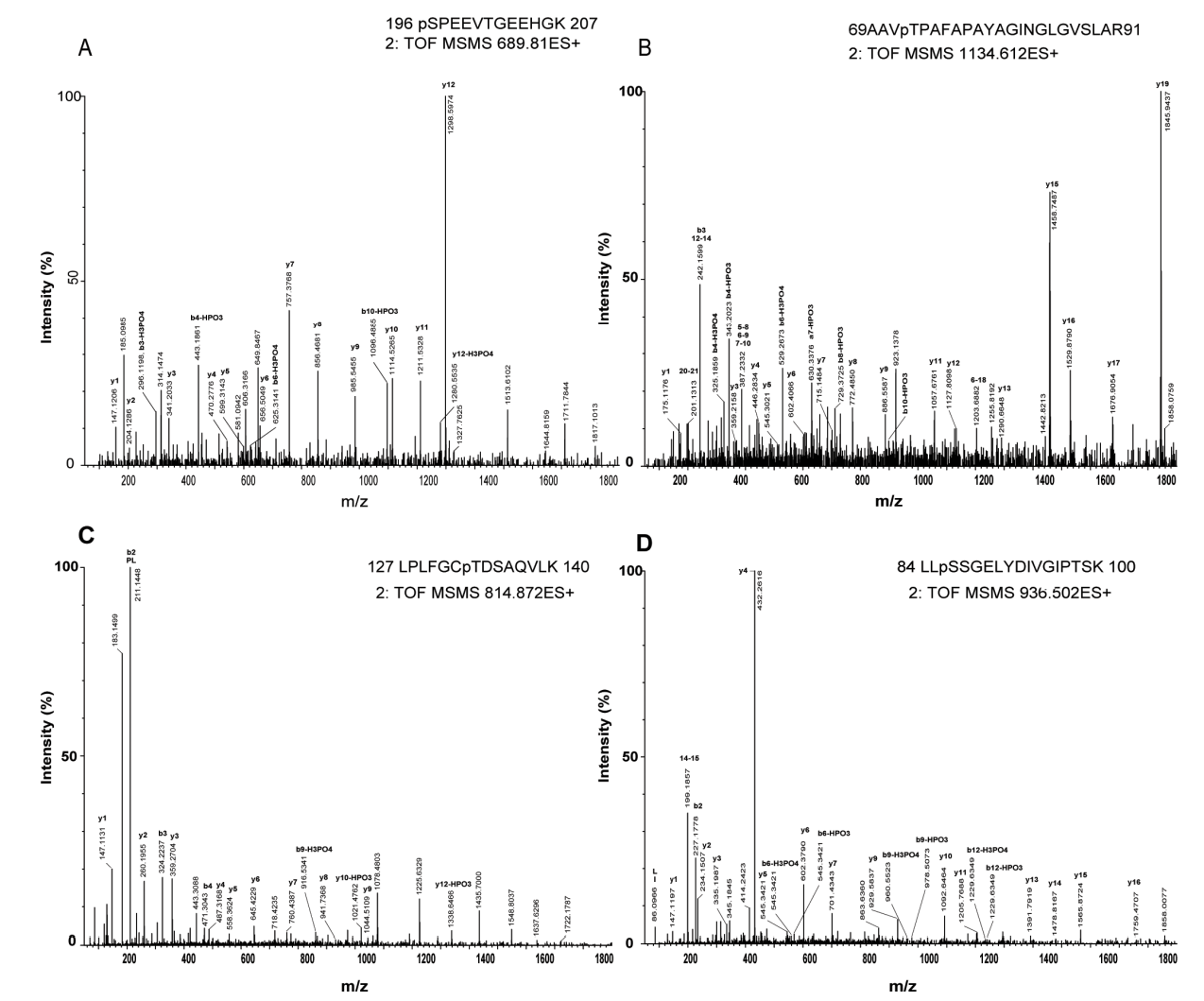
2. Enrichment and Analysis of Intact Phosphoproteins in Arabidopsis Seedlings
This study employed protein extraction under non-denaturing conditions combined with immobilized metal-ion affinity chromatography (IMAC) to enrich intact phosphoproteins, followed by identification using two-dimensional gel electrophoresis (2-DE) and liquid chromatography-tandem mass spectrometry (LC-MS/MS). A total of 144 phosphorylated peptides and sites were identified, among which 18 phosphopeptides and 8 phosphorylation sites were recorded in the PhosPhAt 4.0 and P3DB databases. The 82 identified phosphoproteins were mainly involved in carbohydrate metabolism, photosynthesis/respiration, and oxidative stress response. Compared to peptide-level enrichment methods, intact phosphoprotein enrichment enhanced the detection of phosphorylated threonine and tyrosine residues. Differences in phosphorylation patterns between seedlings and mature leaves suggest that this approach can be applied to study the dynamic changes in protein phosphorylation during plant development. Phosphorylation Site Identification Services utilizes high-resolution mass spectrometry combined with phosphoprotein/phosphopeptide enrichment strategies to identify phosphorylation sites in biological samples. By optimizing enrichment and analysis methods, it enhances the coverage of different phosphorylated residues, making it suitable for studying dynamic changes in protein phosphorylation and its role in biological processes.
Aryal, UK. et al. PLoS One. 2015.
Figur 4. Identification of Phosphorylation Sites Using Tandem Mass Spectrometry (MS/MS)
Applications
Deliverables
1. Comprehensive Experimental Details
2. Materials, Instruments, and Methods
3. Detailed Reports on Fragment Ion Spectra, Site Localization Probability (pRSL), and Peptide Sequences
MtoZ Biolabs, an integrated chromatography and mass spectrometry (MS) services provider.
Related Services
Quantitative Phosphoproteomics Service
Post-Translational Modifications Proteomics Service
Purified Protein Post-Translational Modification Sites Identification Service
How to order?

