



A Far-Western blot asks whether a labeled bait stays on a membrane-bound prey after wash. The mechanism is residual probe on a solid support, not solution equilibrium (Wu et al. 2007). SDS-PAGE unfolds most prey, so the probe can bind only if a linear motif remains or if the blot is renatured. Prefer this path when a purified bait exists and the claim is overlay retention. Reassess when renaturation is unnamed for a fold-dependent pair, the label occupies the interface, or a band is written as native binding, proven direct contact, or a Kd.
Researchers who already have a purified bait, a prey blot, and a written overlay claim can also review the MtoZ Biolabs Far-Western Blot Analysis Service for project-specific feasibility. What that residual mark is allowed to mean is covered in Principle of Far-Western Blot in Protein-Protein Interaction Analysis.
Why the Probe Can Stay After Wash
The Far-Western mechanism starts after the prey is immobilized. Electrophoresis and transfer put proteins on a membrane at a size window. A labeled bait is then offered in solution. Molecules that find an accessible surface can remain through wash. Molecules that do not are removed. The recorded band is leftover probe at that window. It is not a concentration of free bait and free prey at equilibrium.
That is why the assay is not a Western blot. A Western antibody recognizes an epitope on the prey. Far-Western uses the bait as the probe. The chemistry can work without a prey antibody. It cannot work if the bait no longer presents the contact surface after labeling. Biotin, an epitope tag, or an enzyme fused onto the interface can blank a real pair.
Do not write the mechanism as native binding under near-physiological conditions. The prey has usually been through SDS and heat. Do not write the same mechanism as proof of a direct contact. Residual probe can mark a linear motif, a restored fold, or a non-native surface created by denaturation.
The mechanism is leftover labeled bait on a membrane-bound prey. It does not certify a binary protein-protein contact in solution.
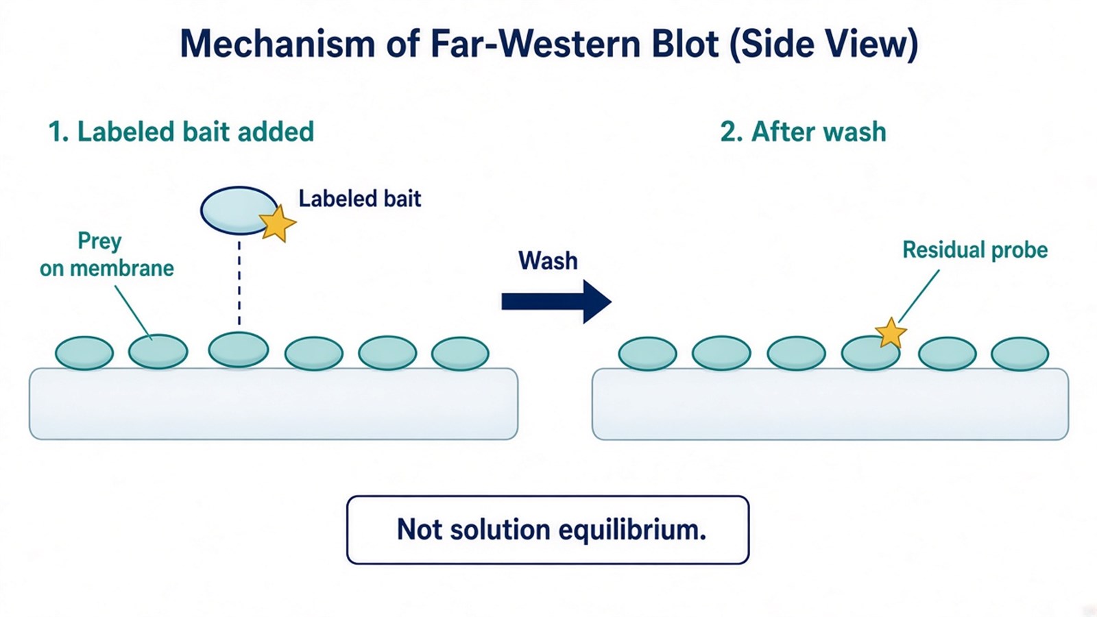
Figure 1. The band reports residual probe. Solution Kd is a different measurement.
Renaturation Is Part of the Binding Mechanism
SDS-PAGE unfolds most prey before transfer. The mechanism of later binding therefore depends on what survives on the membrane. A linear motif can remain readable on a denatured blot. A folded pocket cannot, unless the blot is renatured. Renaturation is not a decorative wash. It is the step that decides which class of contact the probe can still see.
Skipped renaturation has two failure modes. The intended pair can disappear because the pocket never returned. A new, non-native surface can appear and stick to the probe. Either outcome can look like a clean mechanism if controls are missing. Name whether the blot was left denatured or taken through a written renaturation series before the probe is added. How that choice sits in the run order is covered in Workflow of Far-Western Blot for Detecting Protein-Protein Interactions.
Probe activity belongs to the same mechanism. If labeling occupies the interface, the blot is blank for a chemical reason. If the prey never reached a visible load, the blot is blank for a transfer reason. Neither blank proves the pair is absent in solution.
The mechanism is a poor first choice when no purified bait exists, when the pair is known to need a fold the blot cannot restore, or when the next sentence is endogenous co-recovery. Capture after wash belongs on Co-IP or a pull-down, compared in What Are the Main Differences Between GST-Pull Down and Co-IP?.
Renaturation decides which contacts the probe can still form. It does not return a cellular fold by default.
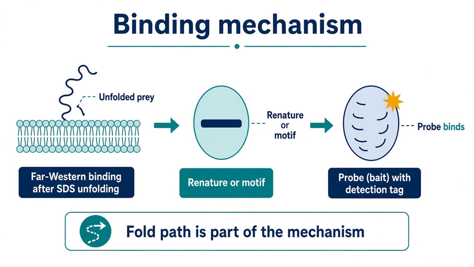
Figure 2. Unfolded prey, then the named path, then probe. The order is the binding mechanism.
Signal Specificity and the Validation Boundary
A residual-probe mechanism is only as specific as the arms that travel with it. Probe-only or no-prey shows label that sticks to empty membrane. A blocked or mutated bait shows whether the mark still appears after the intended surface is broken. A no-renaturation arm, when fold is required, shows whether the signal depends on refolding. Unmatched probe concentration or unmatched exposure makes the contrast unreadable.
Intensity can compare the same prey across matched lanes. Intensity does not become a dissociation constant. Grayscale mixes load, transfer, renaturation, probe activity, and exposure. Those variables do not rearrange into Kd, and they do not become gene regulation or a drug target.
|
Mechanism check |
What a pass looks like |
What the chemistry cannot say |
|---|---|---|
|
Residual probe |
Labeled bait stays after wash at a size window |
Solution equilibrium or a Western epitope |
|
Membrane renaturation |
Named denature or refold path matched to the pair |
Native cellular binding |
|
Signal specificity |
Probe-only and a non-binder on the same recipe |
Every dark lane is a partner |
|
Result boundary |
Candidate overlay retention under those arms |
Proven direct binding or a Kd |
A practical path is:
- Write the overlay sentence the residual mark must support.
- Name whether the pair needs fold and whether renaturation is planned.
- Confirm prey load and that labeling left the contact surface usable.
- Run probe-only and a non-binder with the same recipe.
- Send a passing pair to an orthogonal pull-down or Co-IP.
Do not write Far-Western blot as a more physiological confirmation than Co-IP because the probe is a protein. Do not write a successful band as a completed interactome. When a listed advantage depends on this mechanism holding, the trade-off is reviewed in Far-Western Blot: Advantages and Disadvantages.
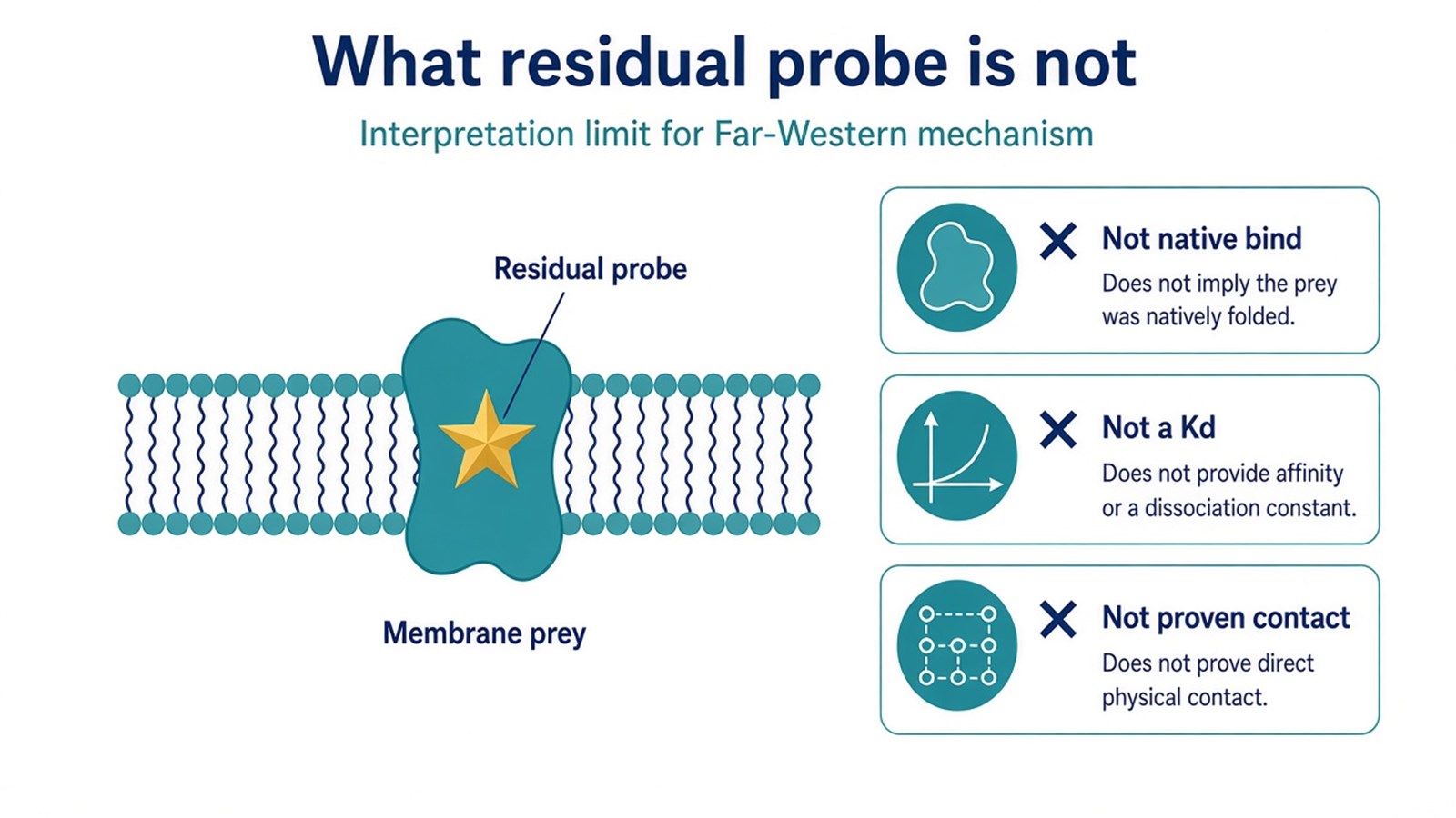
Figure 3. The mechanism stops at residual probe versus probe-only. A chemiluminescent band is not a completed interaction analysis.
Once the bait, prey load, renaturation path, and negative arms are named, the project can be scoped as a Far-Western overlay test. Researchers can review the MtoZ Biolabs Far-Western Blot Analysis Service for sample evaluation.
Frequently Asked Questions
1. What is the mechanism of Far-Western blot?
A labeled bait is offered to membrane-bound prey. Residual probe after wash is the signal. That is overlay retention, not solution binding.
2. Why does SDS-PAGE change the mechanism?
It unfolds most prey. The probe can then see a linear motif, a renatured fold, or a non-native surface. Name which path you used.
3. Does residual probe prove two proteins bind directly?
No. Leftover label on a membrane is not a binary contact in solution. Direct binding needs an orthogonal pair assay.
4. Can the detection signal be read as Kd?
No. Intensity mixes load, transfer, renaturation, probe activity, and exposure. It can compare the same prey across matched lanes.
5. Is Far-Western blot a native or physiological method?
Not after SDS-PAGE. Call it native only if a non-denaturing separation was used and that choice is written. Even then, the prey is still on a membrane.
Reference
- Y. Wu, Q. Li, X.Z. Chen (2007). Detecting protein-protein interactions by Far western blotting. Nat. Protoc., 2, 3278-3284. https://doi.org/10.1038/nprot.2007.459
