



Primer design for site-directed mutagenesis in a protein-interaction project encodes a written codon change. It does not choose a biologically meaningful residue by itself, and a sequenced mutant does not prove that two proteins bind. The original complementary-pair description remains one valid architecture. It is not the only one, and it does not match the later sentence that sends a PCR product into a cloning vector.
Two polymerase chain reaction (PCR) routes are in common use. Whole-plasmid mutagenesis uses a complementary forward and reverse pair, each carrying the mismatch, then DpnI to cut the methylated parental plasmid. Overlap-extension uses two fragment primers that meet at the mutation, then a stitching reaction before the product is cloned. Length, melting temperature (Tm), and flanking sequence follow that architecture. A generic 20–30 nucleotide PCR primer is often too short for a complementary mutagenic pair.
Researchers who already have a wild-type and mutant pair and need a wash-stable recovery readout can also review the MtoZ Biolabs Protein-Protein Interaction Analysis Service for project-specific feasibility and analysis planning.
Choose the Architecture Before the Oligo
The original six-step list stays useful if step 2 is split. Complementary primers that match except at the mutation site belong to whole-plasmid PCR. They are not the primers used when the next bench step is to clone a linear PCR product. Mixing those two recipes is the usual reason a first-round mutagenesis fails after the oligos look correct on paper.
Substitution, insertion, and deletion are not one primer problem. A single-codon substitution sits in the middle of both complementary oligos, with matched flanks. A short insertion or deletion still uses that layout if the bulge remains small. A longer indel is usually designed as overlap-extension or inverse PCR, not as a 25-mer complementary pair.
Purity and working concentration are order and PCR notes. They are not design parameters. HPLC- or PAGE-purified long oligos can reduce truncated species. They do not correct a Tm that was calculated on the wrong architecture.
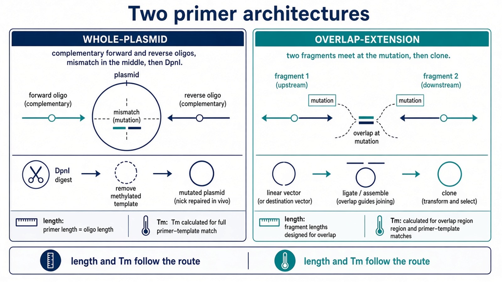
Figure 1. Left: complementary oligos and DpnI. Right: two fragments that meet at the mutation, then cloning. Length and Tm follow the route, not a generic 20–30 nucleotide rule.
Name the PCR architecture, the codon, and the later interaction assay before the oligos are ordered. A complementary pair is not a cloning primer.
The Mutation Site in a Protein-Interaction Project
Site selection is an interaction decision that the primer only writes into DNA. Common choices are an interface alanine, a charge reversal, or a residue nominated by a structure or a cross-link. The oligo does not know which of those is justified. A residue picked because it is easy to mutate is still a valid DNA change. It is a weak interaction hypothesis.
Silent restriction sites can be added beside the mutation to speed colony screens. They do not replace Sanger or NGS confirmation of the open reading frame. Host codon preference should be used for the new amino acid. A rare codon can drop expression and later look like a lost interaction.
If the mutant will be a tagged bait in a panel, empty-tag and wild-type arms still have to be written first. For those project locks, Planning an Affinity Purification Mass Spectrometry Project provides a complementary planning page.
Length, Tm, and Secondary Structure
The original length, Tm, and hairpin rules remain, with architecture-specific bounds. Complementary mutagenic pairs are commonly 25–45 nucleotides, with the mismatch near the center and at least eight to ten matched bases on each side. Tm is calculated for the full oligo, including the mismatch, and the two primers should be close to each other. Whole-plasmid protocols often target a high Tm, near 78 °C in the original kit language. That number is a protocol setting, not a proof of efficient annealing on every template.
Overlap-extension primers follow ordinary PCR Tm matching at the outer ends. The overlapping inner ends must also share enough identity to stitch. Online calculators help only when the same salt and mismatch rules are used for both oligos.
Secondary structure still matters. Self-hairpins and primer-dimers compete with template annealing, especially when the mutation raises local GC. Moving the mismatch by a few bases, or lengthening one flank, is a design fix. Lowering primer concentration is a PCR fix and should not be used to rescue a pair that folds on itself.
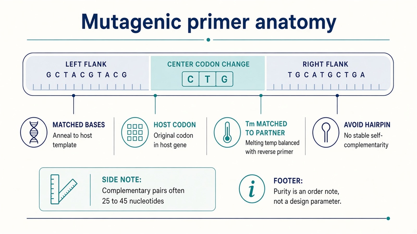
Figure 2. The codon change sits in the middle. Flanks hold the oligo on the template. Tm is matched for the chosen architecture. Host codon preference is a design choice, not a later rescue.
After the Oligos Arrive
Whole-plasmid PCR is followed by DpnI digestion of the parental methylated template, transformation, and sequencing across the mutation and the nearby open reading frame. Overlap-extension products are cloned into the intended vector, then sequenced the same way. The original cloning sentence belongs only to the second route.
Sequence confirmation ends the DNA claim. It does not end the interaction claim. Expression, solubility, and localization can change with a single substitution. A mutant that disappears from a Co-IP pellet may be unfolded, mislocalized, or poorly expressed. Wild-type and mutant must be compared under matched capture. For that recovery readout, How to Combine Co-IP with Mass Spectrometry for Protein Interaction Analysis? provides a complementary capture page.
Experimental Limits
Primer design reduces a project to one architecture, one codon, and one later assay. Diagnostic interface mapping, loss-of-binding as proof, and therapeutic-mutation language sit outside ordinary research reporting unless a separately validated binding assay is already named. A clean chromatogram does not replace that assay.
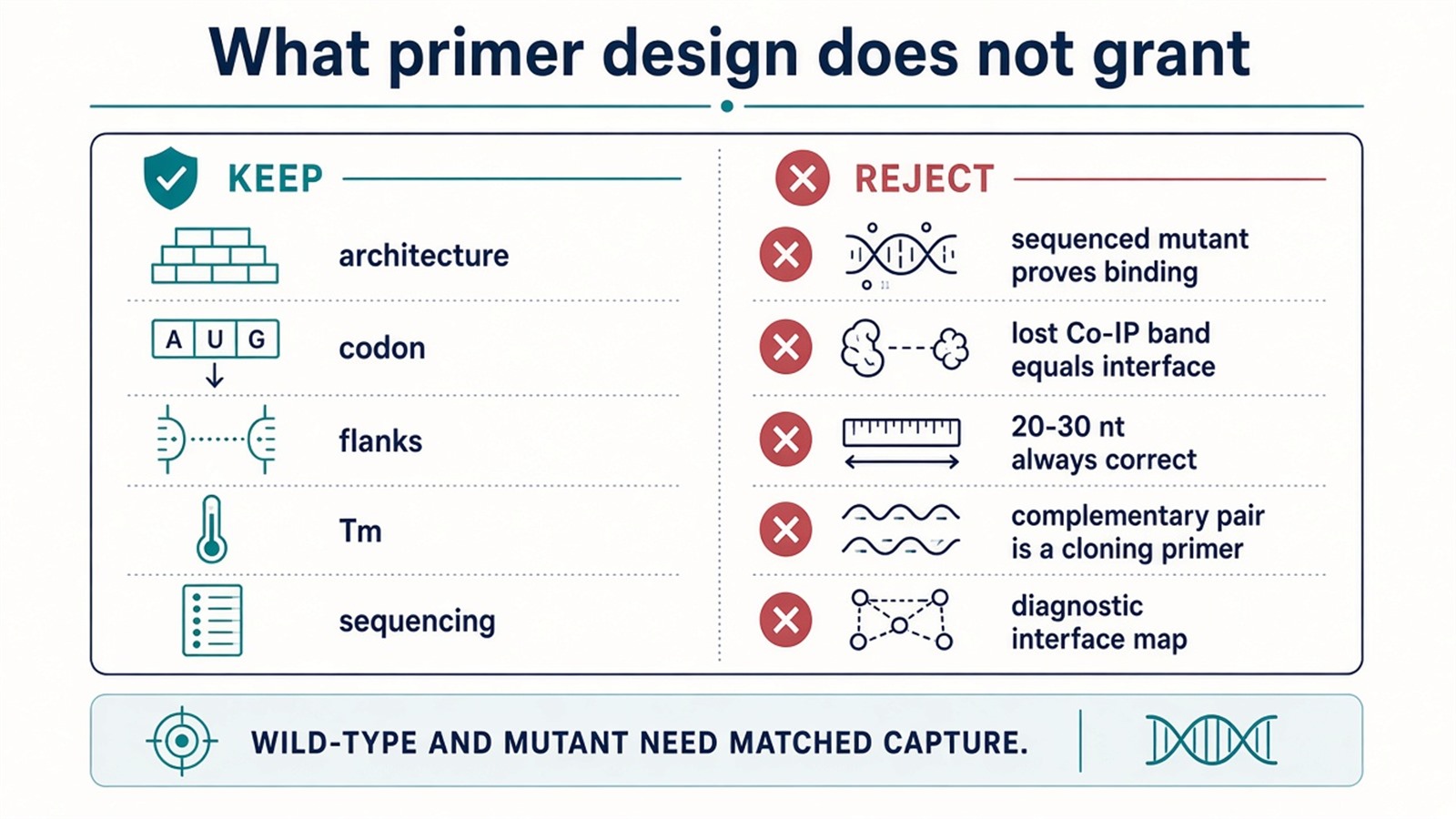
Figure 3. Keep: architecture, codon, flanks, Tm, and sequencing. Not granted: interaction proof, interface mapping, or a lost Co-IP band as unfolding-free evidence.
Project Planning and Service Scope
A usable request names the intended amino-acid change, the PCR architecture, the expression host, and the orthogonal recovery or binding assay that will compare wild-type and mutant. MtoZ Biolabs scopes protein-interaction MS on Orbitrap-class LC-MS/MS after the constructs exist. Interpretability still follows those arms, not a mutagenic-primer heading.
Once the next sentence is a wild-type versus mutant recovery check, this page should hand off. Researchers who need that pellet can review the MtoZ Biolabs Co-IP MS Protein Interaction Analysis Service for project-specific sample evaluation, feasibility assessment, and workflow planning.
Frequently Asked Questions
1. Must mutagenic primers be complementary to each other?
Only in whole-plasmid mutagenesis. Overlap-extension uses two fragment primers that meet at the mutation, then a cloning step.
2. Is 20–30 nucleotides the correct primer length?
That range fits many ordinary PCR primers. Complementary mutagenic pairs are commonly longer, often 25–45 nucleotides, with the mismatch in the center.
3. Does a sequenced point mutant prove a protein interaction?
No. The sequence confirms the codon change. Interaction evidence still needs a matched wild-type versus mutant assay.
4. Can primer design pick the residue to mutate?
No. The oligo writes a codon that the project already chose. Interface, alanine-scan, and charge-reversal choices sit upstream of design.
5. Is cloning the next step after every mutagenesis PCR?
No. Whole-plasmid products are usually DpnI-treated and transformed. Cloning belongs to overlap-extension or a later subcloning step.
Reference
- Ho S.N., Hunt H.D., Horton R.M., Pullen J.K., Pease L.R. (1989). Site-directed mutagenesis by overlap extension using the polymerase chain reaction. Gene, 77, 51-59. https://doi.org/10.1016/0378-1119(89)90358-2
- Zheng L., Baumann U., Reymond J.L. (2004). An efficient one-step site-directed and site-saturation mutagenesis protocol. Nucleic Acids Res., 32, e115. https://doi.org/10.1093/nar/gnh110
- Liu H., Naismith J.H. (2008). An efficient one-step site-directed deletion, insertion, single and multiple-site plasmid mutagenesis protocol. BMC Biotechnol., 8, 91. https://doi.org/10.1186/1472-6750-8-91
- Edelheit O., Hanukoglu A., Hanukoglu I. (2009). Simple and efficient site-directed mutagenesis using two single-primer reactions in parallel to generate mutants for protein structure-function studies. BMC Biotechnol., 9, 61. https://doi.org/10.1186/1472-6750-9-61
- Sambrook J., Russell D.W. (2001). Molecular Cloning: A Laboratory Manual. 3rd ed. Cold Spring Harbor Laboratory Press.
