



Proximity labeling marks proteins that sit near a bait fusion during a defined labeling window in living cells. After streptavidin enrichment, LC-MS/MS reports biotinylated neighbors. The list is a spatial snapshot, not a binding table. A neighbor can share a compartment, pass through a nanometer-scale radius, or sit on a shared scaffold without contacting the bait. Enzyme class changes the time window and the background chemistry. Negative controls and filtering decide which names stay ranked. Bait fusion placement and labeling time belong in the same plan as the empty-enzyme control. Orthogonal recovery is still required if the next sentence must name a protein-protein interaction.
Researchers who already have a bait fusion, a localization check, and a written proximity claim can review the MtoZ Biolabs Proximity Labeling Mass Spectrometry service for feasibility. Method-level choice among enrichment and pair routes is covered in Measuring Protein-Protein Interactions.
Proximity Labeling Evidence Is Spatial, Not Binding
A proximity labeling experiment asks who was close enough to receive a reactive biotin intermediate while the bait fusion was in place. The detection step then identifies proteins that arrived on streptavidin after lysis. Those two jobs must stay separate. Fusion placement, labeling time, and the claim sentence stay with the research plan. Enrichment and search start after labeling has already happened, or after labeled cells are shipped with matched controls.
Do not read a long accession list as proof that the bait bound those proteins. Roux and colleagues designed a promiscuous biotin ligase fusion to recover proximal proteins in mammalian cells, and the same paper treats proximity and interaction as related but not identical claims (Roux et al. 2012). A later Co-IP can test whether a named pair still co-recovers from a physiological lysate. Antibody follow-up is scoped through the IP-Mass Spectrometry Service.
In-vitro retention on a recombinant bait is a different sentence again. If the next experiment must show that a purified pair stays on resin after a defined wash, use the Fusion Protein Interaction Analysis Service | Pull-Down and MS path rather than stretch a biotin list.
Ligase and Peroxidase Mechanisms Change the Interpretable Window
Two chemistries dominate current live-cell designs. A biotin ligase fusion activates biotin and transfers biotin-AMP to nearby lysines over minutes to hours. A peroxidase fusion, supplied with biotin-phenol and a short hydrogen peroxide pulse, generates a phenoxyl radical that labels nearby tyrosine-rich surfaces in seconds.
Literature enzyme families map onto those two chemistries. The BioID class is a slower ligase window, often run with biotin for many hours. The TurboID class is a faster ligase window that can finish in about ten minutes when biotin is added (Branon et al. 2018). The APEX class is the peroxidase pulse used for mitochondrial and other compartment maps on a one-minute scale (Rhee et al. 2013). Service intake still uses the Proximity Labeling Mass Spectrometry name. Enzyme family is a design choice, not a separate catalog line.
The window changes what a name is allowed to mean. An overnight ligase run reports who lived near the fusion for a long biotin period. A ten-minute ligase run can follow a shorter treatment. A one-minute peroxidase pulse can follow an acute stimulus. None of those windows convert proximity into contact. Peroxidase labeling also adds peroxide stress and phenol chemistry that a ligase run does not share. Ligase labeling can biotinylate distant abundant proteins if the fusion is overexpressed or mislocalized.
Write the enzyme class, fusion termini, localization evidence, biotin or phenol dose, and exact labeling time with the shipment. A fusion that fails a localization check is not yet a proximity bait.
The comparison below keeps ligase and peroxidase evidence on separate tracks before ranking.
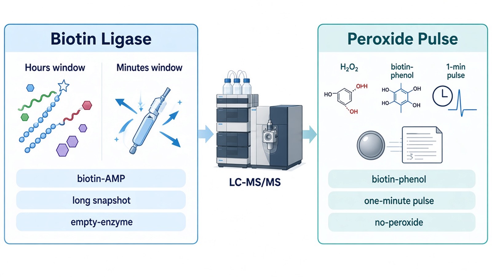
Figure 1. Biotin Ligase vs Peroxidase Labeling Windows.
Negative Controls That Must Travel With the Bait Fusion
Empty-enzyme controls are part of the experiment, not an optional bioinformatics filter. The same ligase or peroxidase without the bait, or with only a localization signal, must be processed through the same biotin or phenol step, lysis, streptavidin capture, and LC-MS/MS method. A no-biotin or no-peroxide arm measures substrate-independent recovery. Parental cells without the fusion measure endogenous biotinylation and streptavidin binders.
Do not replace those tubes with a published contaminant list. Recurrent-contaminant catalogs help interpret leftovers after experimental subtraction (Mellacheruvu et al. 2013). They do not measure enzyme-only biotinylation in the current wash.
If the claim is a stimulus-dependent neighbor, add an unstimulated bait-fusion arm. If the claim is a compartment-restricted map, add a fusion that is expressed but excluded from that compartment when such a construct exists. Relative intensity can be compared across bait and empty-enzyme tubes. Proteins equal in both tubes stay annotated. They should not be drawn as edges.
Background Filtering Versus Orthogonal Validation
Filtering ranks names. It does not finish a protein-protein interaction claim. Typical filters compare bait versus empty-enzyme intensity, drop proteins that appear in no-substrate arms, and flag keratins, streptavidin binders, and the enzyme itself. After filtering, the usable set is the bait-enriched subset, often still large.
A ranked neighbor is a candidate for a later recovery test. Reciprocal Co-IP asks whether the pair can be recovered from lysate after labeling is no longer the readout. Pull-down asks whether a defined pair is retained on an immobilized bait. Affinity of a prepared pair is an SPR question, not a default identification output. Mixed enrichment-MS projects that already have a named pair can be reviewed under the MS-Based Protein-Protein Interaction Analysis Service.
|
Planning item |
Stays with the research plan |
Enters the detection service |
|---|---|---|
|
Enzyme chemistry |
Ligase versus peroxidase window |
Labeled cells or streptavidin product |
|
Negative controls |
Empty-enzyme, no-substrate, parental |
Parallel tubes for search and intensity comparison |
|
Claim sentence |
Neighbor map, stimulus snapshot, or compartment list |
Identification table scoped to that sentence |
|
Follow-up on a named pair |
Reciprocal Co-IP, pull-down, or SPR |
Not completed by the biotin list |
|
Direct tissue labeling |
FFPE or frozen tissue without a fusion |
Not accepted as proximity intake |
The table is a handoff rule, not a ranking of enzyme brands.
Method Selection, Sample Handoff, and Follow-Up
Proximity labeling fits after a bait can be fused to a labeling enzyme and still reach the intended compartment. It fits weak or short-lived associations that washing may remove. It fits membrane or poorly soluble baits when lysis would break the complex before capture. It fits poorly as a first assay when no fusion exists, when the fusion mislocalizes and cannot be checked, or when the publication sentence is direct binding.
It also fits poorly when the only available material is FFPE or frozen tissue without a prior labeling step. Those matrices are not a substitute for a live-cell fusion. Co-IP from tissue remains the first route when an IP-grade antibody exists and the question is co-recovery after lysis.
Ship labeled cell pellets or already enriched streptavidin products. Planning amounts for cells are at least 1x10^7 cells per sample for transient or stable expression. Labeled samples should travel with the matched negative-control set. Liquid products should keep SDS below 0.1 percent. Pellets and solutions travel frozen on dry ice after a snap freeze. Do not ship Western-negative labeling reactions, severely degraded pellets, or high-salt, high-detergent lysates that were never cleaned.
State species and the protein database if a custom FASTA will be used. Orbitrap-class high-resolution LC-MS/MS is the usual acquisition platform. Search should use UniProt or a client FASTA in MaxQuant or an equivalent disclosed workflow.
Intake is faster when the spatial claim, enzyme window, and control set are mapped before shipment. The diagram below links fusion design, controls, and sample format to a proximity request.
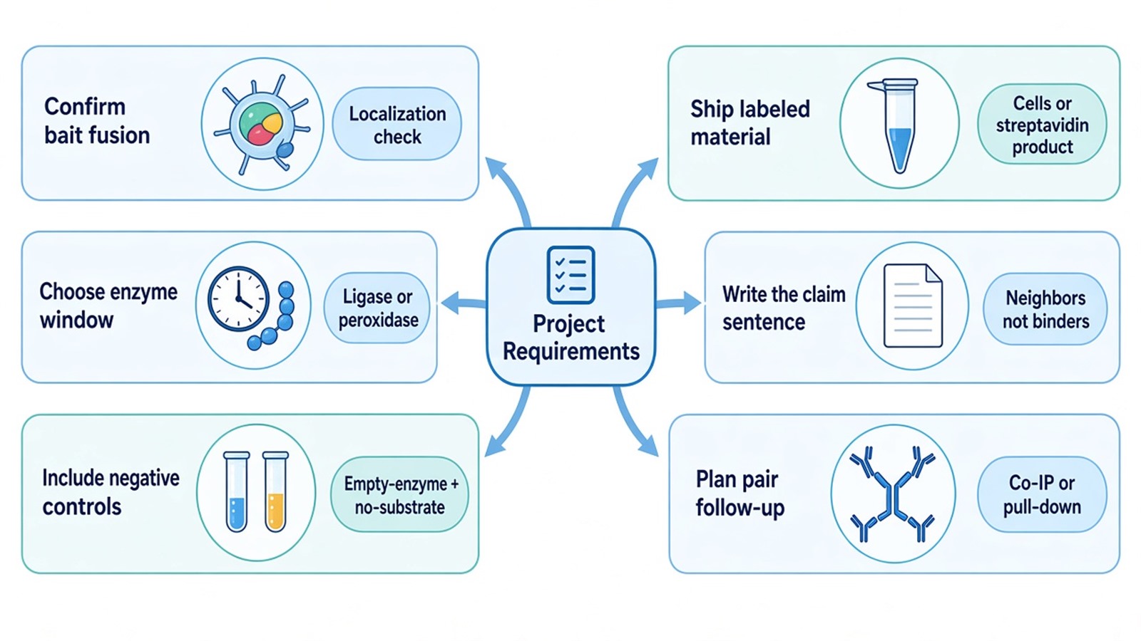
Figure 2. Matching Project Requirements With Proximity Labeling Intake.
Deliverables typically include identification tables, bait versus empty-enzyme intensity comparisons, and a short methods note on digest and search settings. Deliverables do not include a claim of direct cellular binding, a guaranteed partner count, or a finished reciprocal Co-IP. To start intake, send the fusion map, enzyme window, control plan, and intended claim to info@MtoZ-Biolabs.com or use the site inquiry form so the detection path can be confirmed before shipment.
Frequently Asked Questions
1. Can a proximity labeling list replace reciprocal Co-IP for a named pair?
No. The list reports biotinylated neighbors during the labeling window. Reciprocal Co-IP tests whether the pair can still be recovered from lysate. The two sentences are different.
2. Is an empty-enzyme tube enough if biotin was added to every culture?
No. Empty-enzyme measures fusion-independent biotinylation. A no-biotin or no-peroxide arm measures substrate-independent recovery on streptavidin. Both arms are required before names are ranked.
3. How many cells are typically planned for a labeled pellet?
Plan at least 1x10^7 cells per sample for transient or stable expression, and ship the matched control pellets in the same batch. Feasibility still depends on fusion expression and residual detergent.
4. Can FFPE or frozen tissue be submitted for direct live-cell labeling?
No. Direct labeling on FFPE or frozen tissue without a prior live-cell fusion is outside this intake. Tissue Co-IP can be reviewed when an IP-grade antibody and a co-recovery claim already exist.
5. Does a one-minute peroxidase pulse prove a stimulus-dependent complex?
No. The pulse reports who was near the fusion during that minute. An unstimulated bait-fusion arm and a later recovery test are still required if the sentence must name a complex.
Researchers can keep the spatial claim separate from later pair tests, then confirm fusion design, controls, and sample format through the MtoZ Biolabs Proximity Labeling Mass Spectrometry service.
Reference
- K.J. Roux, D.I. Kim, M. Raida, B. Burke (2012). A promiscuous biotin ligase fusion protein identifies proximal and interacting proteins in mammalian cells. J. Cell Biol., 196, 801-810. https://doi.org/10.1083/jcb.201112098
- T.C. Branon, J.A. Bosch, A.D. Sanchez, N.D. Udeshi, T. Svinkina, S.A. Carr, J.L. Feldman, N. Perrimon, A.Y. Ting (2018). Efficient proximity labeling in living cells and organisms with TurboID. Nat. Biotechnol., 36, 880-887. https://doi.org/10.1038/nbt.4201
- H.W. Rhee, P. Zou, N.D. Udeshi, J.D. Martell, V.K. Mootha, S.A. Carr, A.Y. Ting (2013). Proteomic mapping of mitochondria in living cells via spatially restricted enzymatic tagging. Science, 339, 1328-1331. https://doi.org/10.1126/science.1230593
- D. Mellacheruvu, Z. Wright, A.L. Couzens, J.P. Lambert, N.A. St-Denis, T. Li, Y.V. Miteva, S. Hauri, S.E. Sardiu, T.Y. Low, et al. (2013). The CRAPome: a contaminant repository for affinity purification-mass spectrometry data. Nat. Methods, 10, 730-736. https://doi.org/10.1038/nmeth.2557
