



BiFC Protein Interaction
Protein-protein interaction analysis asks which proteins remain together after a defined capture or complementation test and matched controls. BiFC answers that question for a named pair inside a cell. Two proteins are fused to complementary fluorescent-protein fragments. Fluorescence appears only if those fragments reconstitute. The path fits an expression system that can accept both fusions and the claim is candidate proximity of that pair, not binary direct binding or functional regulation (Hu et al. 2002). Prefer this path when a named pair must be scored in intact cells. Reassess when a fragment-only or non-interacting control is missing, expression is unmatched, or fluorescence is written as proven contact.
Researchers who already have a named pair, fusion status, and a written interaction claim can also review the MtoZ Biolabs Protein-Protein Interaction Analysis for orthogonal capture after a BiFC shortlist.
Which Experimental System Can Support BiFC
BiFC is a named-pair protein-protein interaction assay. Each partner carries one non-fluorescent fragment of a split fluorescent protein. If the partners come close enough, the fragments can reconstitute and emit light. That is a proximity readout in the cell that expressed the fusions. It is not an open partner discovery method, and it is not a more complete interactome than Co-IP-MS.
The system is readable when three things are named. First, both proteins can be expressed as fusions in the same cell. Second, fragment orientation and a fluorescent-protein split are chosen before any image is scored. Third, the cell can be imaged or sorted without treating autofluorescence as a pair. How a BiFC signal is later read is covered in BiFC Analysis. Another binary in-cell pair test is compared in Yeast 2 Hybrid Analysis.
The system is a poor first choice when tagging changes localization or function, when the claim is an unknown-partner list, or when the claim is reconstituted binding of a purified domain. Open-partner ranking after antibody capture belongs on IP-MS or Co-IP-MS, reviewed under the IP-Mass Spectrometry Service.
BiFC reports candidate proximity of a named pair after fragment reconstitution. It does not certify a binary protein-protein contact.
Negative Controls Before Any Fluorescent Pair Is Kept
Negative controls are the denominators of BiFC. They are not optional after one cell looks bright.
Each fragment expressed alone shows self-assembly and autofluorescence. A non-interacting pair that uses the same fragments, the same promoters, and the same localization signals shows complementation that does not depend on the intended interface. An interface mutant, when the pair is already named, tests whether fluorescence still appears after the contact surface is broken. Unmatched expression of one fusion is not a negative control. A brighter promoter on the bait only makes reconstitution unreadable.
Localization must be matched. A nuclear bait plus a cytosolic prey can still fluoresce if overexpression forces both into one compartment. Fragment-only and non-interacting arms must travel the same transfection, fixation or live-imaging recipe, and exposure settings.
System check |
What a pass looks like |
What the analysis cannot say |
|---|---|---|
Experimental system |
Named pair, both fusions expressed, chosen split |
An untagged endogenous interactome |
Negative control |
Fragment-only and a non-interacting pair on the same recipe |
Every bright cell is a true pair |
Candidate screening |
Fluorescence ranked versus those arms |
Intensity as binding affinity |
Result boundary |
Candidate proximity under those arms |
Direct contact or gene regulation |
A practical path is:
- Write the proximity sentence the image or sort must support.
- Express both fusions and confirm they reach the intended compartment.
- Run fragment-only and a non-interacting pair with the same recipe.
- Keep only signals that pass that contrast.
- Send the shortlist to an orthogonal Co-IP, pull-down, or pair assay.
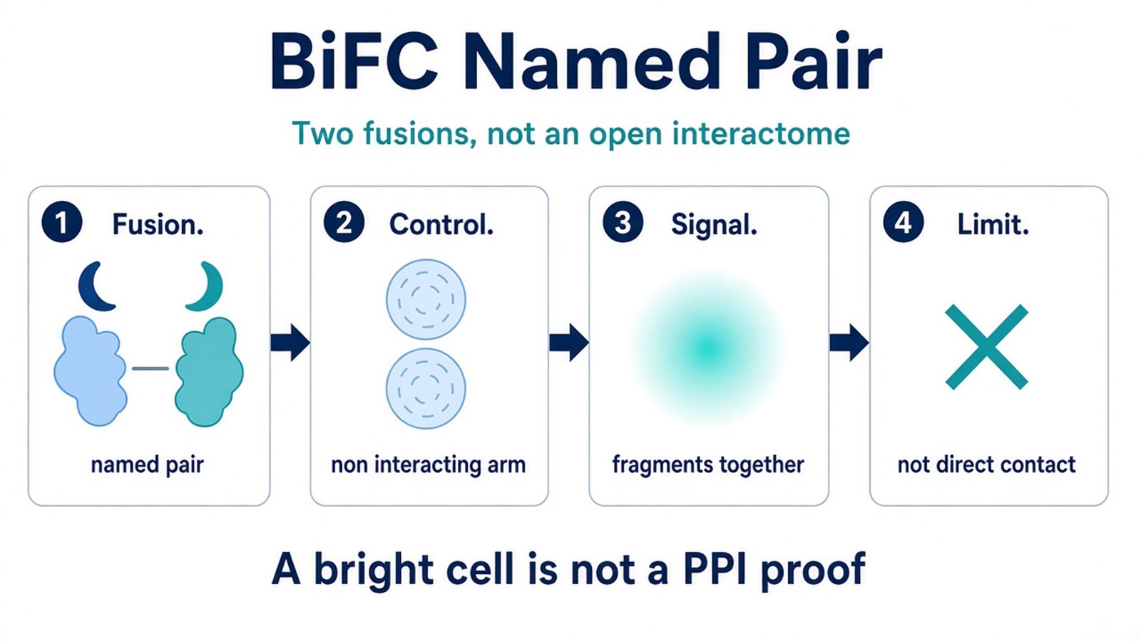
Figure 1. BiFC starts after both fusions and a non-interacting arm are named. A bright cell is not a completed interaction result.
Screening Candidate Interacting Pairs
Candidate screening starts after the intended pair is brighter than fragment-only and the non-interacting arm. If those controls are as bright as the test pair, stop calling the pair an interaction.
Fluorescence means the fragments reconstituted. It does not mean the two proteins touch, and it does not mean they stay together. Reconstitution is usually irreversible, so later darkness cannot be read as dissociation. Intensity can compare the same pair across matched arms. Intensity does not rank two different pairs as tighter binders.
Do not write a BiFC-positive pair as an open interactome. Do not write a nuclear signal as proof that the pair regulates transcription. Do not write a multi-color BiFC panel as a completed network. Do not write CRISPR tagging as a required rescue of an overexpression artifact.
Candidate screening ranks a named pair versus matched negative arms. It does not certify a binary protein-protein contact or an off-rate.
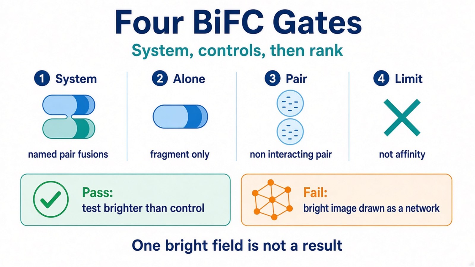
Figure 2. Screening is a control outcome. Extra exposure time does not replace a non-interacting pair.
Result Boundaries and Common Biases
The common bias is writing BiFC as high-sensitivity protein interaction analysis because fluorescence appeared without an added dye. Another bias is skipping the non-interacting arm because one image looks clean. Overexpression can force complementation. Self-assembly of split fragments can create background. The reconstituted reporter can trap a pair that would have separated.
Do not write a successful fluorescent cell as proof of direct binding. Do not write BiFC as real-time monitoring of association and dissociation. Do not write a subcellular pattern as functional regulation. Prefer BiFC when a named pair must be scored in intact cells and both fusions are acceptable. Prefer Y2H when the next sentence is a binary test in yeast. Prefer Co-IP or IP-MS when the next sentence is endogenous co-recovery or unknown partners.
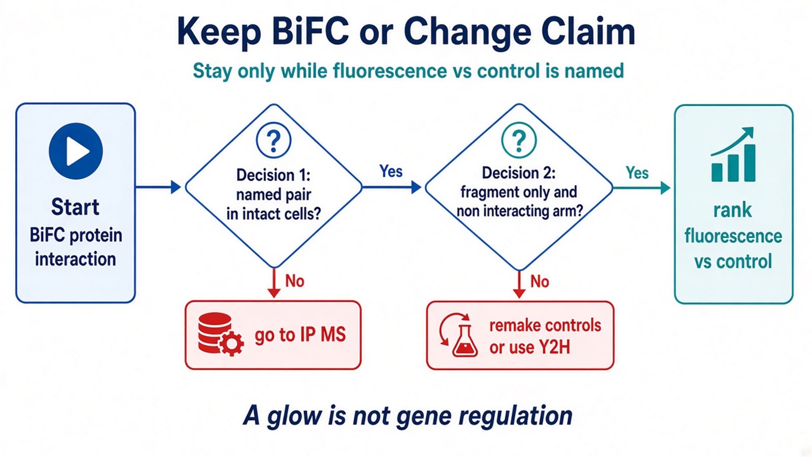
Figure 3. Keep BiFC only while fluorescence versus a non-interacting arm can be named. A confocal image is not a completed interaction analysis.
Once the pair, fusion status, split class, and negative arms are defined, the project can be scoped as a BiFC proximity test. Researchers can review the MtoZ Biolabs Protein-Protein Interaction Analysis for sample evaluation and orthogonal capture planning.
Frequently Asked Questions
1. Does BiFC prove two proteins bind directly?
No. It reports fragment reconstitution after the fusions come close. Direct contact needs an orthogonal pair assay.
2. Can I skip the non-interacting control if the image is bright?
No. Fragment-only and a non-interacting pair must see the same recipe. One bright field is not a BiFC result.
3. Can BiFC watch a pair come apart?
No. Reconstitution is usually irreversible. Later loss of signal is not an off-rate.
4. Is BiFC the same as Co-IP or IP-MS?
No. BiFC scores a named pair by fluorescence. Co-IP and IP-MS recover proteins after capture. Depth in a Co-IP-style eluate is typically hundreds to about a thousand proteins, depending on the sample.
5. When should this assay move to Co-IP or IP-MS?
Move when the next written sentence is endogenous co-recovery or unknown partners. Keep the BiFC shortlist as the input, not as the proof.
Reference
- C.D. Hu, Y. Chinenov, T.K. Kerppola (2002). Visualization of interactions among bZIP and Rel family proteins in living cells using bimolecular fluorescence complementation. Mol. Cell, 9, 789-798. https://doi.org/10.1016/S1097-2765(02)00492-6
