



TMT and iTRAQ applications in proteomics research are closed-multiplex isobaric designs that compare reporter-ion ratios across defined groups inside one pooled LC-MS/MS run (Thompson et al. 2003; Ross et al. 2004). Typical uses include frozen case-control or treatment contrasts, multi-arm drug or stimulus panels, short time courses collected before mixing, signaling response studies, and comparative proteomics across tissue states when amine labeling fits. Applications deliver candidate relative protein changes inside a plex map, not clinical cutoffs, drug approval claims, or absolute concentrations without separate calibrated assays. Prefer TMT or iTRAQ when every arm can enter one kit and the scientific claim is same-run relative change; reassess label-free LFQ or DIA when enrollment stays open or labeling fails.
Researchers who already have defined samples, comparison groups, or a specific project objective can also review the MtoZ Biolabs TMT Quantitative Proteomics Analysis Service and Quantitative Proteomics Service for project-specific isobaric application feasibility and analysis planning.
Shared Application Logic for TMT and iTRAQ
Tandem mass tag (TMT) and isobaric tags for relative and absolute quantitation (iTRAQ) are both amine-reactive isobaric chemistries. Samples are digested, tagged, pooled, and quantified from low-mass reporter ions after fragmentation. MS1 shows one precursor per labeled peptide; channels are read at MS2 or, for some TMT workflows, at SPS-MS3 when cleaner reporters are required.
Application logic is therefore the same class: a contrast that can be frozen before mixing, a channel map with reference or bridge channels when needed, and a relative claim about the same protein across those channels. Kit size and reporter layout differ between TMT and iTRAQ products. Plex planning must match the actual reagent, not a generic isobaric label.
Researchers can refer to Mass Spectrometry TMT Technology for MS1-isobaric readout detail and to TMT-Based Proteomics for labeling study design.
Isobaric applications compare channels inside a closed mix, not MS1 peak areas across independent unlabeled runs.
Research Applications That Often Match a Closed Plex
Disease and comparative proteomics candidate lists
Case versus control tissue, disease model versus wild type, or two defined tissue states can use TMT or iTRAQ when sample number fits the kit and peptides can be labeled at primary amines. Differential proteins remain candidates for follow-up. They are not diagnostic markers, prognostic scores, or companion diagnostics without targeted validation.
Drug, stimulus, and signaling response panels
Treated versus control cells, a small compound panel, hormone or stress stimuli, or genotype contrasts can sit in one plex when channels are randomized and the full set is available before pooling. A protein that changes after treatment supports a relative response readout inside that experiment. It is not proof of target engagement, mechanism of action, or clinical efficacy.
Short time-course or dose series frozen before labeling
A bounded on-treatment series or dose ladder fits isobaric multiplexing when every time point or dose is collected before the labeling reaction. Open clinical enrollment that will continue after mixing needs a new plex, an explicit bridge design, or an unlabeled route instead.
Systems-level contrasts with predefined arms
Multi-condition screens that compare several arms under one experimental record can use higher-plex TMT kits when chemistry and instrument time match the panel. Global pathway or network figures interpret the differential list. They do not repair incomplete labeling, ratio compression, or a confounded channel map.
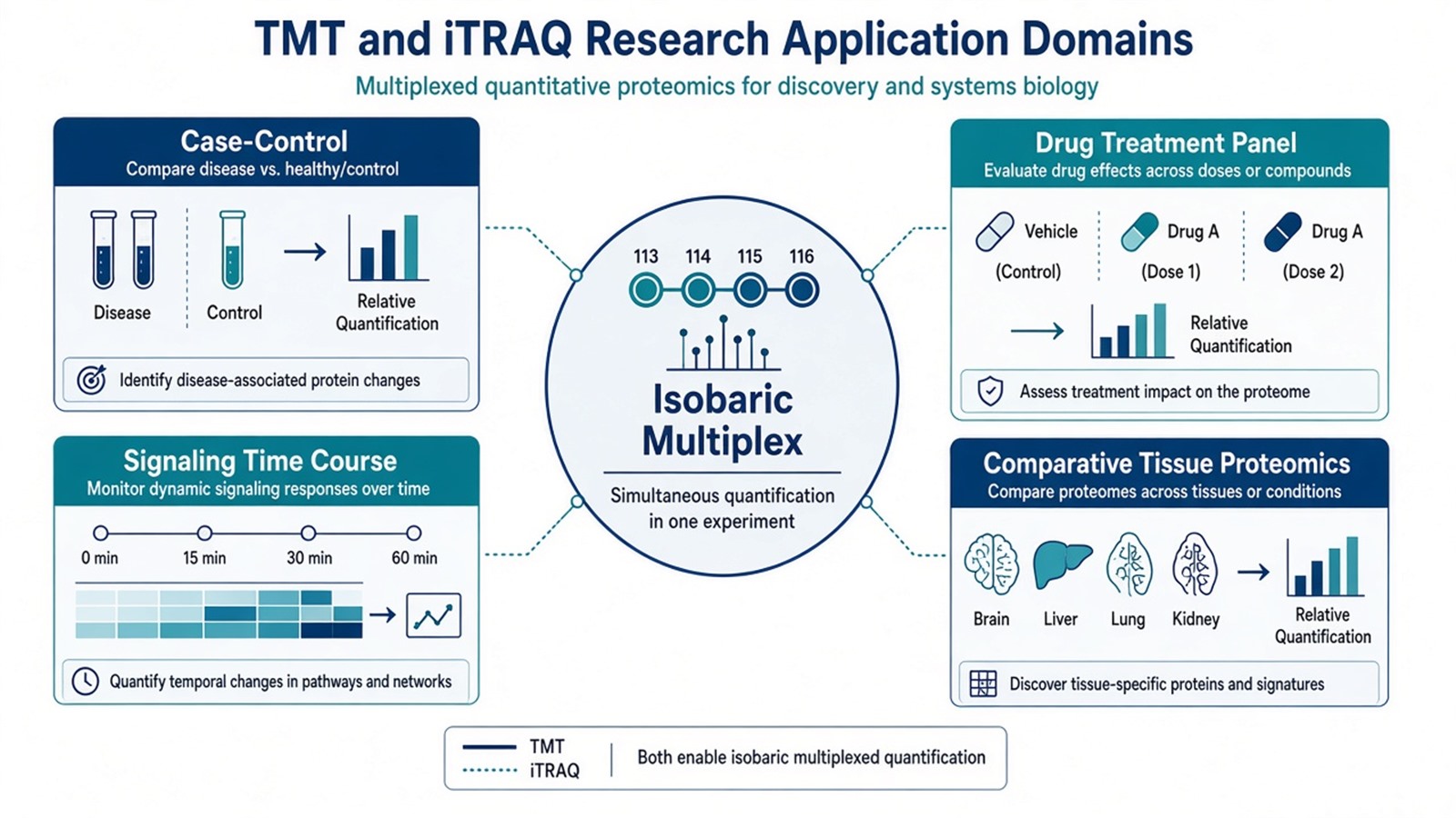
Figure 1. TMT and iTRAQ applications are defined by a frozen multiplex contrast, not by a disease keyword alone.
For biomarker-oriented framing, see Application of TMT Quantitative Proteomics in Biomarker Discovery. For isotopic labeling overview, see Quantitative Analysis of Proteomics Using Isotopic Labeling Methods.
Judgment Criteria: TMT, iTRAQ, or a Different Route
Intended research use |
TMT or iTRAQ often fits |
Reassess |
|---|---|---|
Frozen multi-arm contrast in one mix |
Closed plex with channel QC |
Late samples after pooling |
Same-run relative ratios across channels |
MS2 or SPS-MS3 isobaric readout |
Absolute amount without standards |
Open cohort with ongoing enrollment |
Not as one extending plex |
Label-free LFQ or unlabeled DIA |
Short confirmed peptide list |
Not primary isobaric discovery |
PRM or MRM with optional SIS |
Labeling incompatible matrix |
Chemistry fails at amines |
Unlabeled discovery or targeted panel |
Ratio compression from co-isolated peptides can shrink true fold changes at MS2 (Ting et al. 2011). Fractionation, narrower isolation where possible, or SPS-MS3 on supported TMT workflows can reduce mixing. Those steps change reporter purity; they do not convert discovery ratios into validated clinical cutoffs.
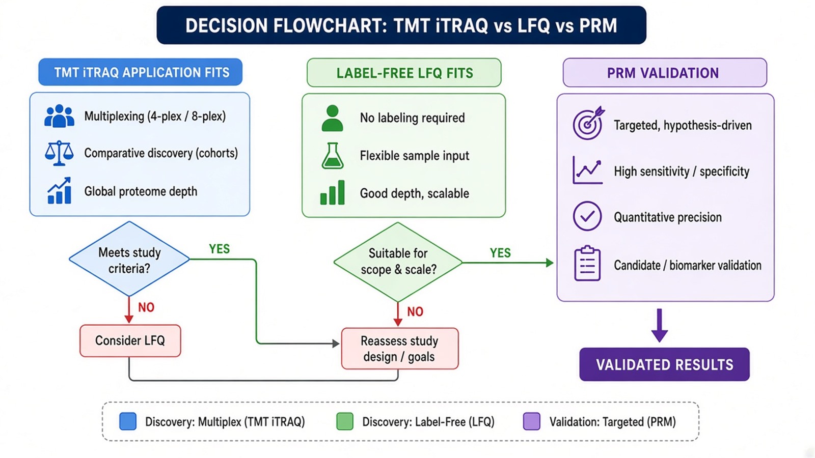
Figure 2. Application choice follows whether the contrast can close into one isobaric mix and what claim the table must support.
For LFQ versus isobaric strategy, see Comparing LFQ and Isobaric Labeling Strategies in Proteomics Quantification and TMT vs Label Free Quantitative Proteomics. For advantage and disadvantage lists, see Advantages and Disadvantages of TMT-Based Quantitative Proteomics Analysis.
Limits and Application Boundaries
TMT and iTRAQ inherit isobaric limits. Incomplete labeling, wrong channel assignment, post-mix sample additions, and MS2 ratio compression can distort apparent biology. Enrichment analysis shows overrepresentation within the quantified background; it does not prove pathway activity. Personalized medicine, early diagnosis, and regulatory biomarker claims sit outside ordinary research isobaric reporting unless separately validated assays exist.
Typical defensible applications when design and QC align:
- Preclinical disease versus control proteome contrasts with planned orthogonal confirmation.
- Closed drug or genetic perturbation panels with randomized channel placement.
- Short treatment time courses or dose ladders collected before labeling.
- Cell signaling or stress-response studies with matched prep across arms.
- Cross-tissue or cross-genotype comparisons that fit one or bridged plex series.
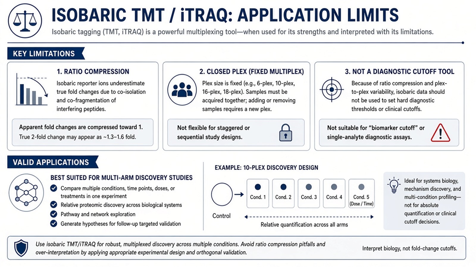
Figure 3. Isobaric applications support relative discovery matrices; clinical and absolute claims need separate assay designs.
For compression mechanism, see How Does Ratio Compression Arise in TMT-Based Quantitative Proteomics and Why Is It Limited to Relative Quantification?. For validation handoff, see How to Plan a Targeted Proteomics Validation Study and PRM-MS Mass Spectrometry.
Moving From Isobaric Applications to Follow-Up Assays
A TMT or iTRAQ differential list is an input to follow-up, not a final biomarker or drug-target certificate. Report peptide support, missing reporters, plex map, normalization, and scan level with every candidate. Orthogonal measurement on a short list, often parallel reaction monitoring (PRM), Western blot, or enzyme-linked immunosorbent assay (ELISA), tests whether the same protein still changes in a new aliquot.
Once the application contrast, channel map, and expected relative readout are defined, the project can be evaluated as a TMT or iTRAQ research study. Researchers can review the MtoZ Biolabs TMT Quantitative Proteomics Analysis Service for sample evaluation, feasibility assessment, and workflow planning.
Frequently Asked Questions
1. Are TMT and iTRAQ applications interchangeable in study design?
They share isobaric pooled-multiplex logic but differ in kit chemistry, channel count, and reporter details. Plex planning must match the reagent in hand, not a generic isobaric name.
2. Can TMT or iTRAQ alone validate a disease biomarker?
No. Discovery plexes nominate candidates with relative channel change. Diagnostic or prognostic validation needs locked assays and separate evidence.
3. When is label-free quantification a better application match?
When samples must stay as independent injections, enrollment continues after the first batch, or amine labeling is incompatible with the matrix or study timeline.
4. Does higher plex always mean better research coverage?
Higher plex helps multi-arm contrasts in one run. It does not remove compression, labeling QC requirements, or the need to freeze contrasts before mixing.
5. How should time-series clinical samples be handled?
Collect the planned series before labeling when possible. Ongoing enrollment usually requires new plexes with bridges or an unlabeled discovery route rather than extending one old mix.
Reference
- A. Thompson, J. Schafer, K. Kuhn, et al. (2003). Tandem mass tags: a novel quantification strategy for comparative analysis of complex protein mixtures by MS/MS. Anal. Chem., 75, 1895-1904. https://doi.org/10.1021/ac0262560
- P.L. Ross, Y.N. Huang, J.N. Marchese, et al. (2004). Multiplexed protein quantitation in Saccharomyces cerevisiae using amine-reactive isobaric tagging reagents. Mol. Cell. Proteomics, 3, 1154-1169. https://doi.org/10.1074/mcp.M400049-MCP200
- L. Ting, R. Rad, S.P. Gygi, W. Haas (2011). MS3 eliminates ratio distortion in isobaric multiplexed quantitative proteomics. Nat. Methods, 8, 937-940. https://doi.org/10.1038/nmeth.1714
