


Skeletal and cardiac muscle tissue digests are often analyzed with four-dimensional data-independent acquisition (4D-DIA) when label-free separate injections, ion mobility-aware windowed MS/MS, and FDR-controlled extraction must support a predefined relative contrast across groups (Meier et al. 2020; Gillet et al. 2012). Muscle carries high contractile protein load, connective matrix, and variable blood residue when perfusion is incomplete. Those matrix traits set prep and pilot rules before mobility and window plans lock. The guide below integrates muscle sample handling with label-free 4D-DIA acquisition, parameters, limits, and project fit. It is not a sports medicine promise, not a clinical diagnostic page, and not a generic 4D marketing brochure.
4D-DIA adds ion mobility separation before windowed fragmentation. Quantification still comes from extracted fragment traces under unified FDR rules after matched pretreatment (Rosenberger et al. 2017). Relative contrast across predefined arms remains the default discovery claim. Amount on named peptides needs stable isotope-labeled internal standard (SIS) targeted assays (Gerber et al. 2003).
In MtoZ Biolabs offerings, label-free 4D-DIA on muscle digests is evaluated when matrix pilots, contrast design, and platform scope support mobility-aware acquisition and extraction.
General label-free 4D-DIA integration compares with 4D-DIA Label-Free Quantitative Proteomics: the Key Technology in Proteomics. Mechanism language compares with Mechanism of 4D-DIA Quantitative Proteomics.
Muscle Tissue Matrix Constraints Before 4D-DIA Starts
Muscle projects fail early when tissue handling splits comparability.
Fiber type and anatomical site should be declared. Mixing soleus with gastrocnemius or injured with uninjured regions in one arm without documentation creates false regulation lists.
Connective and intramuscular fat change homogenization yield. Skipping consistent disruption depth across arms biases contractile versus structural protein ratios before LC starts.
Blood and plasma carryover from incomplete perfusion adds abundant serum proteins that track sacrifice batch, not biology.
Low-input biopsies need yield QC before cohort scale. Pressure cycling or other mechanical lysis routes may enter pilot planning when wet mass is limited (Application of PCT-DIA Proteomics).
Muscle 4D-DIA value starts with matched extraction and dissection maps, not with mobility keywords on fibrous tissue alone.
Rodent organ prep context appears in DIA-MS Quantitative Proteomics Solutions for Mouse Tissue Samples. General lysis-to-digest rules appear in Quantitative Protein Analysis.
Common Contrast Types in Muscle 4D-DIA Studies
Application language should name the contrast, not only the disease area.
Disease versus control muscle compares predefined pathology arms after matched harvest and storage rules. Lists stay candidate until validation design exists.
Atrophy, regeneration, or injury time courses compare declared time bins frozen before statistics. Relabeling time points after acquisition breaks interpretability.
Exercise, disuse, or nutrition interventions compare treatment to control with matched cull and fasting state when metabolism is in scope.
Drug or compound exposure compares vehicle to treated arms with shared digestion batches when aliquots split from the same homogenate pool.
Mechanism and pathway overlay interpret tested lists. They do not replace contrast QC or staged remeasurement when decisions need PRM (Application of DIA-PRM Proteomics).
Integrated Label-Free 4D-DIA Workflow for Muscle Cohorts
A practical muscle 4D-DIA spine usually runs as follows.
- Confirm species, muscle site, contrast, relative claim, and platform scope.
- Harvest with matched perfusion or flash-freeze rules; record cull batch and storage.
- Homogenize with one declared route; reduce, alkylate, and digest under shared rules (Wisniewski et al. 2009).
- Clean and desalt to injection-ready loads with equal recovery across arms.
- Pilot mobility-aware DIA on muscle digest; lock gradient, mobility plan, and window table.
- Acquire cohort files with interleaved run order and QC inserts as agreed.
- Extract under unified FDR and interference filters (Demichev et al. 2020).
- Normalize, apply missing-value policy, test predefined contrast only.
- Optional PRM or SIS on a short list when list status changes.
Steps 1 through 4 are muscle prep. Steps 5 through 7 are 4D-DIA acquisition and extraction. Step 8 is the quantitative claim layer.
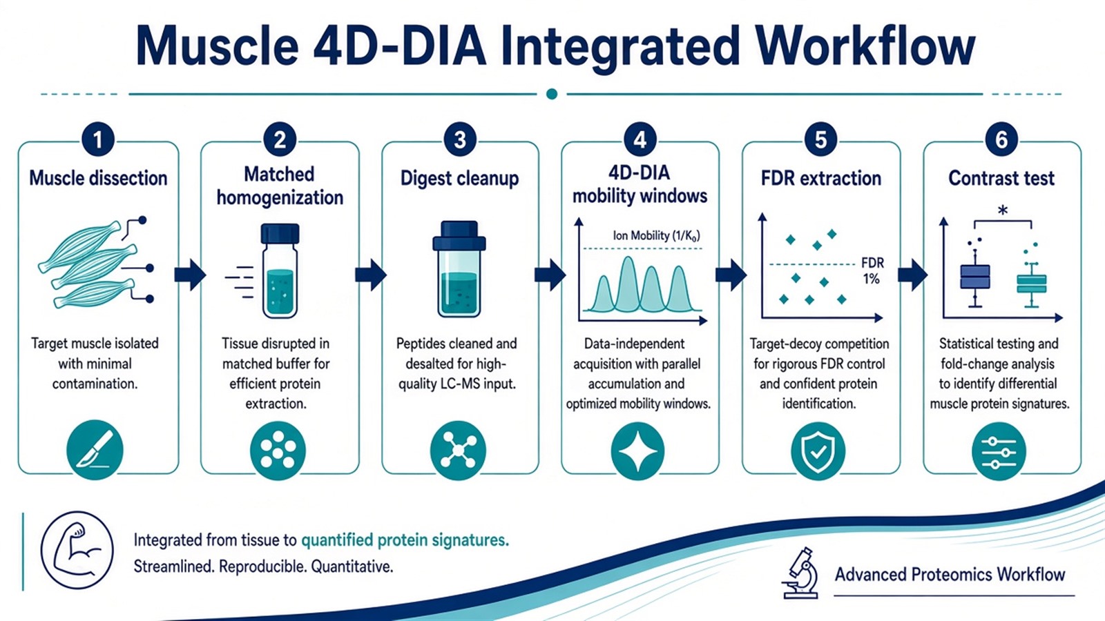
Figure 1. Muscle 4D-DIA couples fibrous-tissue prep discipline to locked mobility-aware acquisition before contrast claims.
Operational checkpoints appear in Workflow of 4D-DIA Quantitative Proteomics. Extraction conventions appear in DIA Data Analysis Methods in Proteomics. Tradeoffs appear in Advantages and Disadvantages of 4D-DIA Quantitative Proteomics.
Parameters and Limits in Muscle 4D-DIA Projects
Topic |
Muscle 4D-DIA sensitivity |
Failure mode if ignored |
|---|---|---|
Dissection map |
Site and fiber context declared |
Mixed regions as regulation |
Perfusion or blood control |
Limits plasma carryover |
Serum proteins track batch |
Homogenization depth |
Matched across arms |
Contractile ratio artifacts |
Input matching |
Weight or total protein aligned |
Load bias mimics biology |
Mobility and windows |
Locked mid cohort |
Incompatible acquisition blocks |
Extraction |
One declared version |
Mixed pipelines |
Missing values |
Documented policy |
Dropout tracks prep, not treatment |
Claim |
Relative contrast default |
Open intensities as amount |
Limits follow the same table. Muscle 4D-DIA does not by itself certify diagnostic assays, training prescriptions, or drug labels. Abundant myosin and actin isoforms can dominate ion statistics; low-abundance signaling proteins may remain missing when abundance and prep limit sampling (Lazar et al. 2016). Forced 4D scheduling without pilot interference evidence may add cost when mobility-free DIA or SWATH-style pilots already extract cleanly (SWATH Proteomics).
Enrollment and pilot checks appear in Exploring Quantitative Analysis in Proteomics.
How to Choose Muscle 4D-DIA for the Next Study
Use matrix pilots, contrast type, and follow-up goal together.
Project need |
Muscle 4D-DIA plan that often fits |
Recheck before committing |
|---|---|---|
Fibrous muscle, crowded MS/MS in pilot |
Label-free 4D-DIA after lock |
4D without interference pilot |
Clean mobility-free muscle pilot |
Mobility-free label-free DIA |
Forced mobility cost |
Sub-milligram biopsy discovery |
PCT pilot then 4D-DIA cohort |
Full cohort without yield QC |
Open enrollment, many files |
Interleaved 4D-DIA separate injections |
Sacrifice day tracks treatment |
Closed peptide list after discovery |
4D-PRM or PRM panel |
Full 4D when names fixed |
Amount on named muscle peptides |
SIS targeted assay |
Discovery intensities as concentration |
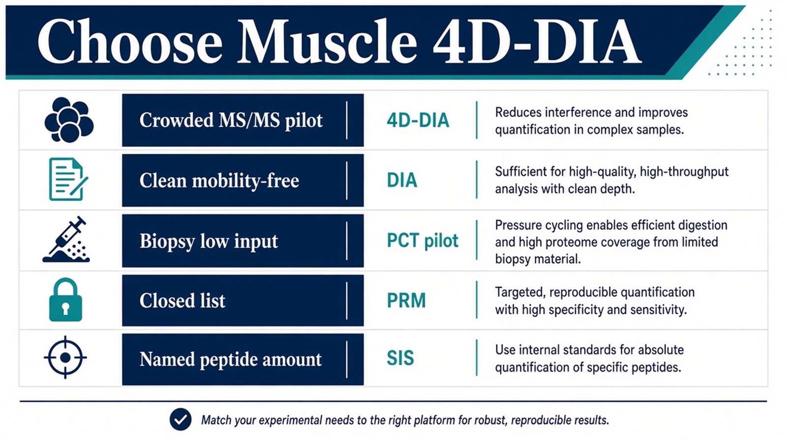
Figure 2. Choose muscle 4D-DIA when matched prep and mobility-aware pilots support the frozen contrast; mobility-free DIA, PCT prep, or PRM fit other evidence patterns.
Three-axis route selection appears in Protein Quantification Method.
Teams with muscle site, contrast design, harvest records, and pilot scope can review the MtoZ Biolabs Label-Free Quantitative Proteomics Service for muscle 4D-DIA planning.
Frequently Asked Questions
1. Is human and rodent muscle prep identical for 4D-DIA?
No. Species, perfusion access, and biopsy size change lysis and yield rules. Contrast arms still need matched routes within the study.
2. Does 4D-DIA remove myoglobin or contractile protein interference?
No. Mobility may reduce some MS/MS overlap. Abundant muscle proteins still dominate extraction and missing-value patterns.
3. Can exercise studies relabel time bins after acquisition?
No. Time arms should be frozen before contrast testing. New bins need a new analysis record.
4. Is muscle 4D-DIA the same as cardiac-only 4D-DIA?
Cardiac muscle shares fibrous prep constraints but adds perfusion and ischemia handling rules. Site-specific prep must be declared.
5. When should muscle projects skip 4D-DIA?
When mobility-free DIA pilots extract cleanly or when a closed PRM panel already defines the claim, full 4D-DIA may add little value.
MtoZ Biolabs can discuss muscle 4D-DIA planning when tissue type, contrast design, pilot data, and platform scope are documented.
If further evaluation of dissection maps, mobility pilot lock, or discovery-to-targeted staging is needed, project details can be submitted for a feasibility review.
Reference
- S.A. Gerber, J. Rush, O. Stemman, M.W. Kirschner, S.P. Gygi (2003). Absolute quantification of proteins and phosphoproteins from cell lysates by tandem MS. Proc. Natl. Acad. Sci. U.S.A., 100, 6940-6945. https://doi.org/10.1073/pnas.0832254100
- L.C. Gillet, P. Navarro, S. Tate, H. Rost, H. Selevsek, L. Reiter, R. Bonner, R. Aebersold (2012). Targeted data extraction of the MS/MS spectra generated by data-independent acquisition: a new concept for consistent and accurate proteome analysis. Mol. Cell. Proteomics, 11, O111.016717. https://doi.org/10.1074/mcp.O111.016717
- F. Meier, A.D. Brunner, O. Frank, M.T. Krauss, M. Koch, H. Teichert, C. Trede, J. Kosinski, T. Park, M. The, M. Mann (2020). diaPASEF: parallel accumulation-serial fragmentation combined with data-independent acquisition. Nat. Methods, 17, 1229-1236. https://doi.org/10.1038/s41592-020-00998-0
- V. Demichev, C. Messner, S. Vernardis, K.S. Lilley, M. Ralser (2020). DIA-NN: neural networks and interference correction enable deep proteome coverage in high throughput. Nat. Methods, 17, 41-44. https://doi.org/10.1038/s41592-019-0638-x
- C. Lazar, L. Gatto, M. Ferro, C. Bruley, T. Burger (2016). Accounting for the multiple natures of missing values in label-free quantitative proteomics data sets to compare imputation strategies. J. Proteome Res., 15, 1116-1125. https://doi.org/10.1021/acs.jproteome.5b00981
- G. Rosenberger, S. Liu, J. Röst, H.L. Ludwig, C. Buil, B. Borchers, R. Aebersold, L. Collins (2017). Statistical control of peptide and protein error rates in large-scale targeted data-independent acquisition analyses. Nat. Commun., 8, 14224. https://doi.org/10.1038/ncomms14224
- J.R. Wisniewski, A. Zougman, N. Nagaraj, M. Mann (2009). Universal sample preparation method for proteome analysis. Nat. Methods, 6, 359-362. https://doi.org/10.1038/nmeth.1322
