



Single-cell proteomics projects need relative protein measurements from picogram-scale protein input, where adsorption loss and incomplete recovery can erase low-abundance peptides before liquid chromatography-tandem mass spectrometry (LC-MS/MS). Tandem mass tag (TMT) labeling supports high-throughput quantification by assigning each single-cell digest a channel, mixing channels into one plex, and reading reporter ions after fragmentation (Thompson et al. 2003). A high-abundance carrier channel is often added so MS1 detection and peptide-spectrum matches improve for the mixed set, while single-cell channels still contribute channel-specific reporters (Budnik et al. 2018; Specht et al. 2021). The useful design question is whether isolation, micro-scale prep, carrier ratio, and reporter readout can be locked before mixing, not whether cells are heterogeneous in principle.
TMT single-cell tables are relative by default. Amount claims need matched standards and a separate targeted design. Carrier boosting can raise identification depth and can also distort single-cell reporter ratios if the carrier overwhelms cell channels or if co-isolation compresses fold changes (Ting et al. 2011). Unlabeled single-cell injections remain an alternative when multiplexing or carrier design does not fit the cohort.
In MtoZ Biolabs offerings, TMT labeling for ultra-low-input and single-cell projects is scoped when cell isolation status, channel map, carrier plan, and relative claim are declared. Service intake appears in TMT Quantitative Proteomics Analysis Service and Single Cell Proteomics Service.
What TMT Labeling Covers in Single-Cell Proteomics
Four technology layers belong in the brief and stay separated.
Isolation layer places one cell, or a declared mini-pool, into a low-loss vessel with a documented sorting or micromanipulation method.
Prep layer lyses, digests, and cleans peptides in micro-volumes under matched timing so surface loss does not become a false channel effect.
Labeling layer assigns TMT channels to single-cell digests, adds any carrier channel, quenches, and pools for one LC-MS/MS path.
Claim layer states relative protein or peptide change across single-cell channels inside that plex. Cell-state clustering language still depends on missingness rules and batch control. Diagnostic or precision-medicine wording needs separate validation beyond the TMT table.
TMT labeling in single-cell proteomics multiplexes freezable ultra-low-input digests. It does not convert sparse reporter tables into clinical cell classification.
Carrier-Channel Theory That Matters in Practice
In bulk TMT, each channel usually carries a similar protein load. In single-cell TMT, many channels carry near-empty digests, so MS1 precursors may never be selected. A carrier channel of higher peptide abundance from the same cell type or a matched background can raise precursor intensity and improve identification rates for the mixed plex (Budnik et al. 2018). Reporter ions for single-cell channels are still extracted from MS2 or MS3 spectra after fragmentation.
The same boost creates the main practice risk. Excess carrier can dominate isolation windows, compress single-cell fold changes, and leave cell channels near noise. Carrier-to-sample ratio, isolation width, and whether reporters are read at MS2 or MS3 must be locked with the channel map. Bottom-up single-cell constraints around picogram input and carrier tuning appear in Bottom-Up Proteomics in Single-Cell Proteomics: Challenges, Workflow, and Method Selection. Bulk TMT analysis principles for identification-to-reporter coupling appear in Principle of TMT-Based Quantitative Proteomics Analysis.
End-to-End Workflow for Single-Cell TMT Quantification
A practical single-cell TMT spine usually runs as follows.
- Freeze cell source, isolation method, channel map, carrier plan, and relative claim before lysis.
- Isolate single cells into low-binding vessels; record viability and sorting gates when used.
- Lyse in micro-volume buffers compatible with downstream digestion and labeling.
- Reduce, alkylate, and digest under matched micro-scale conditions; minimize transfers.
- Label each single-cell digest with one TMT channel; prepare the carrier digest on the declared channel.
- Quench, clean up excess reagent, and pool cell channels with the carrier at the locked ratio.
- Acquire high-sensitivity LC-MS/MS with declared MS2 or MS3 reporter readout and quality control (QC) injections.
- Search spectra, extract reporters, review carrier interference and missingness, and normalize under stated rules.
- Test only predefined single-cell contrasts or clustering plans; archive channel map, carrier ratio, method files, and software versions.
Steps 1 through 4 protect the ultra-low-input record. Steps 5 through 9 are the labeling, acquisition, and informatics record. Sample-prep loss control appears in Sample Preparation Protocol for Single Cell Protein Analysis. When MS2 ratios look compressed, MS3 options appear in TMT MS3 and SPS-MS3: How to Mitigate Ratio Compression and Enhance Quantitative Accuracy.
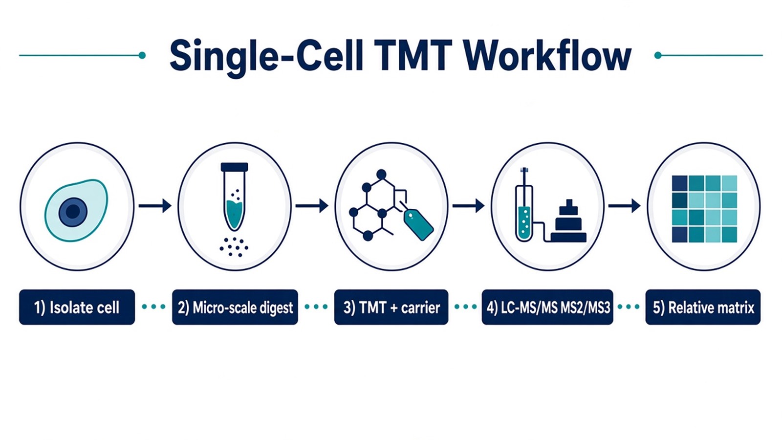
Figure 1. Single-cell TMT quantification depends on low-loss prep and a locked carrier-to-sample ratio before reporters are interpreted.
Parameters That Must Be Locked for Single-Cell TMT Claims
Layer |
Parameter that must be declared |
Failure mode if ignored |
|---|---|---|
Isolation |
One-cell definition and sorting record |
Mixed cells read as single-cell biology |
Vessel and timing |
Low-binding plastics and lysis schedule |
Adsorption loss as missing proteome |
Channel map |
Cell tags, blanks, and carrier channel |
Misassigned reporters after pooling |
Carrier ratio |
Carrier-to-sample load locked before mix |
Carrier dominance and ratio distortion |
Digestion |
Micro-volume enzyme and quench rules |
Incomplete digests as cell-state change |
Cleanup |
Miniaturized desalting route |
Residual reagent suppressing spray |
Reporter scan |
MS2 or MS3 rule |
Compressed single-cell fold changes |
Missingness |
Filter and imputation policy stated |
Imputed zeros as heterogeneity |
Batch |
Plex bridge or batch covariates |
Instrument day as cell cluster |
Deliverable |
Matrix, carrier ratio, method notes |
Report rebuilt after acquisition |
Limits follow the same table. Multiplex kit size raises cells per injection only when prep loss and carrier ratio stay controlled. High-throughput language does not replace isolation QC. Unlabeled DIA single-cell routes can reduce stochastic missingness in some designs; they do not remove the need for low-loss prep.
What a Single-Cell TMT Result Can Support
A reviewed single-cell TMT matrix supports relative comparison of proteins detected across channels that were mixed into one plex under a locked carrier plan. Cell-to-cell heterogeneity language requires enough quantitative completeness after missingness rules. Carrier-boosted identifications do not prove that every reported protein was abundant in every single cell.
Ratio compression and carrier interference can shrink real cell differences (Ting et al. 2011; Specht et al. 2021). A small observed fold change still needs isolation and carrier QC before it is called weak biology. Pathway enrichment is optional after the matrix passes quality checks. It does not repair adsorptive loss or invent diagnostic cell labels.
Bulk tissue TMT answers a different question. A later unlabeled single-cell digest cannot join an existing reporter matrix without a new plex design.
A single-cell TMT table supports relative channel contrasts inside a locked ultra-low-input plex. It does not certify precision-medicine cell typing.
How to Choose Single-Cell TMT Versus Adjacent Routes
Use input scale, throughput need, carrier feasibility, and claim unit together.
Project need |
Route that often fits |
Recheck before committing |
|---|---|---|
Many single cells, one shared LC-MS/MS path |
TMT plex with locked carrier plan |
Carrier ratio untested in the cell type |
Ultra-low input without multiplexing |
Unlabeled single-cell LC-MS/MS |
Force TMT without isolation QC |
Sparse missingness across many cells |
Evaluate unlabeled DIA or hybrid designs |
Assume TMT removes all missing values |
Compressed MS2 single-cell ratios |
TMT with MS3 review |
Keep MS2 without carrier QC |
Bulk tumor or culture proteome |
Standard bulk TMT |
Call bulk averages single-cell biology |
Named peptide confirmation |
Targeted PRM or MRM on suitable input |
Re-run the same sparse TMT table as confirmation |
Amount or concentration unit |
SIS-supported targeted assay |
Ordinary TMT reporters as absolute amount |
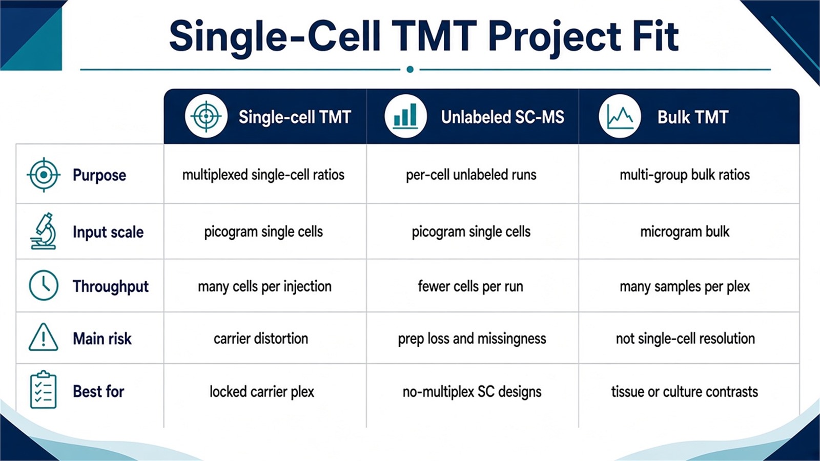
Figure 2. Choose single-cell TMT when ultra-low-input digests can freeze into a carrier-assisted plex; keep unlabeled or bulk routes when isolation, enrollment, or claim unit changes.
Teams with isolation records, channel maps, and carrier plans can review MtoZ Biolabs Single Cell Proteomics Service and TMT Quantitative Proteomics Analysis Service when those fields are declared in the project record.
Frequently Asked Questions
1. Why do single-cell TMT methods often use a carrier channel?
Carrier peptides raise precursor intensity so more peptides can be identified in the mixed plex. Single-cell channels still need detectable reporters after fragmentation. Excess carrier can distort cell-specific ratios.
2. Does TMT labeling always outperform unlabeled single-cell proteomics?
No. TMT raises cells per injection when prep and carrier design hold. Unlabeled routes can fit when multiplexing is unnecessary or when carrier interference would dominate the claim.
3. Must every single-cell TMT project use MS3?
No. MS2 can suffice for triage when directional consistency is enough. MS3 becomes useful when co-isolation or carrier mixing is expected to hide real cell differences.
4. Can single-cell TMT results be reported as absolute protein amounts?
No. Reporter ratios are relative. Amount reporting needs matched standards and a declared calibration outside ordinary TMT discovery tables.
5. What project fields are needed before scoping single-cell TMT?
Isolation method, estimated cell number, channel and carrier plan, relative claim, and any batch or bridge design help evaluate feasibility before lysis.
MtoZ Biolabs can provide TMT labeling and single-cell proteomics analysis for ultra-low-input multiplexed projects, including channel layout, carrier-assisted labeling, LC-MS/MS, and relative matrix reporting when isolation status and study design are available.
If further evaluation of carrier ratio, MS2 versus MS3 readout, or whether unlabeled single-cell acquisition fits better is needed, project details can be submitted for a feasibility review.
Reference
- A. Thompson, J. Schafer, K. Kuhn, S. Kienle, J. Schwarz, G. Schmidt, T. Neumann, R. Johnstone, A.K. Mohammed, C. Hamon (2003). Tandem mass tags: a novel quantification strategy for comparative analysis of complex protein mixtures by MS/MS. Anal. Chem., 75, 1895-1904. https://doi.org/10.1021/ac0262560
- B. Budnik, E. Levy, G. Harmange, N. Slavov (2018). SCoPE-MS: mass spectrometry of single mammalian cells quantifies proteome heterogeneity during cell differentiation. Genome Biol., 19, 161. https://doi.org/10.1186/s13059-018-1547-5
- H. Specht, E. Emmott, A.A. Petelski, R.G. Huffman, D.H. Perlman, M. Serra, P. Kharchenko, A. Koller, N. Slavov (2021). Single-cell proteomic and transcriptomic analysis of macrophage heterogeneity using SCoPE2. Genome Biol., 22, 50. https://doi.org/10.1186/s13059-021-02267-z
- L. Ting, R. Rad, S.P. Gygi, W. Haas (2011). MS3 eliminates ratio distortion in isobaric multiplexed quantitative proteomics. Nat. Methods, 8, 937-940. https://doi.org/10.1038/nmeth.1714
