



APEX protein labeling covalently tags nearby proteins, not interactions. An engineered ascorbate peroxidase, usually APEX2, is fused to a bait or a localization sequence. In living cells it oxidizes biotin-phenol in the presence of hydrogen peroxide (H₂O₂) and writes a biotin handle onto nearby electron-rich side chains, chiefly tyrosine. Streptavidin then recovers those biotinylated proteins for liquid chromatography–tandem mass spectrometry (LC-MS/MS). The product of the experiment is a set of labeled proteins. It is not a map of spatial organization, a transient-interaction inventory, or a protein network. A working radius near 20 nm is often quoted. It is an estimate, not a measured sphere that excludes distant proteins.
The pulse can be run in a living cell without fixation or permeabilization. Enrichment does not stay in that cell. After H₂O₂ the sample is still lysed and bound to a streptavidin bead. APEX-only, matched for localization, estimates enzyme background. A no-H₂O₂ or no-biotin-phenol arm estimates probe background. For the peroxidase pulse that produces the radical, APEX Proximity Labeling provides a complementary chemistry page.
Researchers who already have an APEX2 fusion and need a streptavidin table of labeled proteins can also review the MtoZ Biolabs Proximity Labeling Mass Spectrometry Service for project-specific feasibility and analysis planning.
What Is Written onto the Protein
The original plant ascorbate peroxidase (APEX) was built as a genetically encoded reporter. APEX2 is the variant usually used to label proteins in the cytosol and other compartments, because it is more active and more stable. Calling the method APEX protein labeling does not make first-generation APEX the default enzyme.
Biotin-phenol plus H₂O₂ produces a short-lived phenoxyl radical. That radical adds to nearby tyrosines and, less often, other electron-rich residues. The covalent product is a biotinylated protein. Streptavidin affinity is the recovery step. High biotin–streptavidin affinity allows a harsh wash. It does not raise mass-spectrometry sensitivity by itself. For how that streptavidin file is designed as a proteome, APEX Proteomics provides a complementary reading order.
Neurobiology, organelle, virology, cancer, and immune-signaling sentences are later overlays. Labeling synapse-adjacent proteins does not identify key regulators of neurotransmitter transmission. Labeling a mitochondrial, ER, or Golgi-targeted fusion does not enrich organelle-specific proteins under physiological conditions. The table reports who was near the peroxidase when H₂O₂ was added.
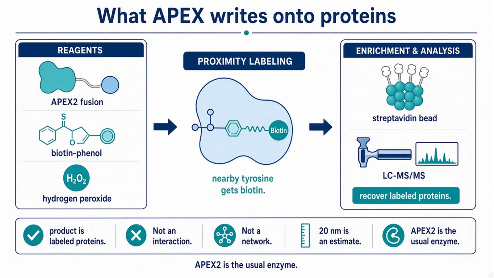
Figure 1. APEX2 plus biotin-phenol plus H₂O₂ tags nearby tyrosines. Streptavidin recovers those proteins. Proximity is not binding. APEX2 is the usual labeling enzyme.
Name the residue class and the recovery bead before the first pulse. A one-minute label does not convert a biotin handle into an interaction.
What the One-Minute Window Can Support
These advantages hold only with the condition in the same sentence.
A short H₂O₂ pulse, typically under one minute, can biotinylate proteins that would leave a complex after lysis. That is temporal control of when proteins receive the label. It is not high spatial specificity, and it does not minimize artifacts of cellular-state change. H₂O₂ is itself a change in cellular state. The original “seconds” wording and the one-minute wording describe the same short window. They are not two different resolutions.
The fusion can be directed to a compartment or a bait. That is genetic encoding of where the radical is generated. It is not a tool for dynamic protein networks. A protein complex is not demonstrated because APEX2 was fused to one subunit.
Relative to BioID, the protein-labeling window is much shorter. BioID typically needs many hours of biotin and marks lysines. APEX2 marks tyrosines in about a minute and requires peroxide. Shorter is not superior, and it is not particularly suitable for every rapidly occurring interaction. For the ligase comparison that does not use H₂O₂, BioID&TurboID provides a complementary ligase page. For the family that includes both enzyme classes, Proximity Labeling Mass Spectrometry provides a complementary overview.
APEX2 uses added H₂O₂, not ambient oxygen, as the oxidant for the phenolic probe. That fact is sometimes restated as function in low-oxygen compartments. It does not make APEX an ideal method for hypoxic interaction networks, and it does not group mitochondria and nuclear pore complexes as hypoxia models. Those two sites are compartment-targeting jobs. They still need a matched APEX-only arm.
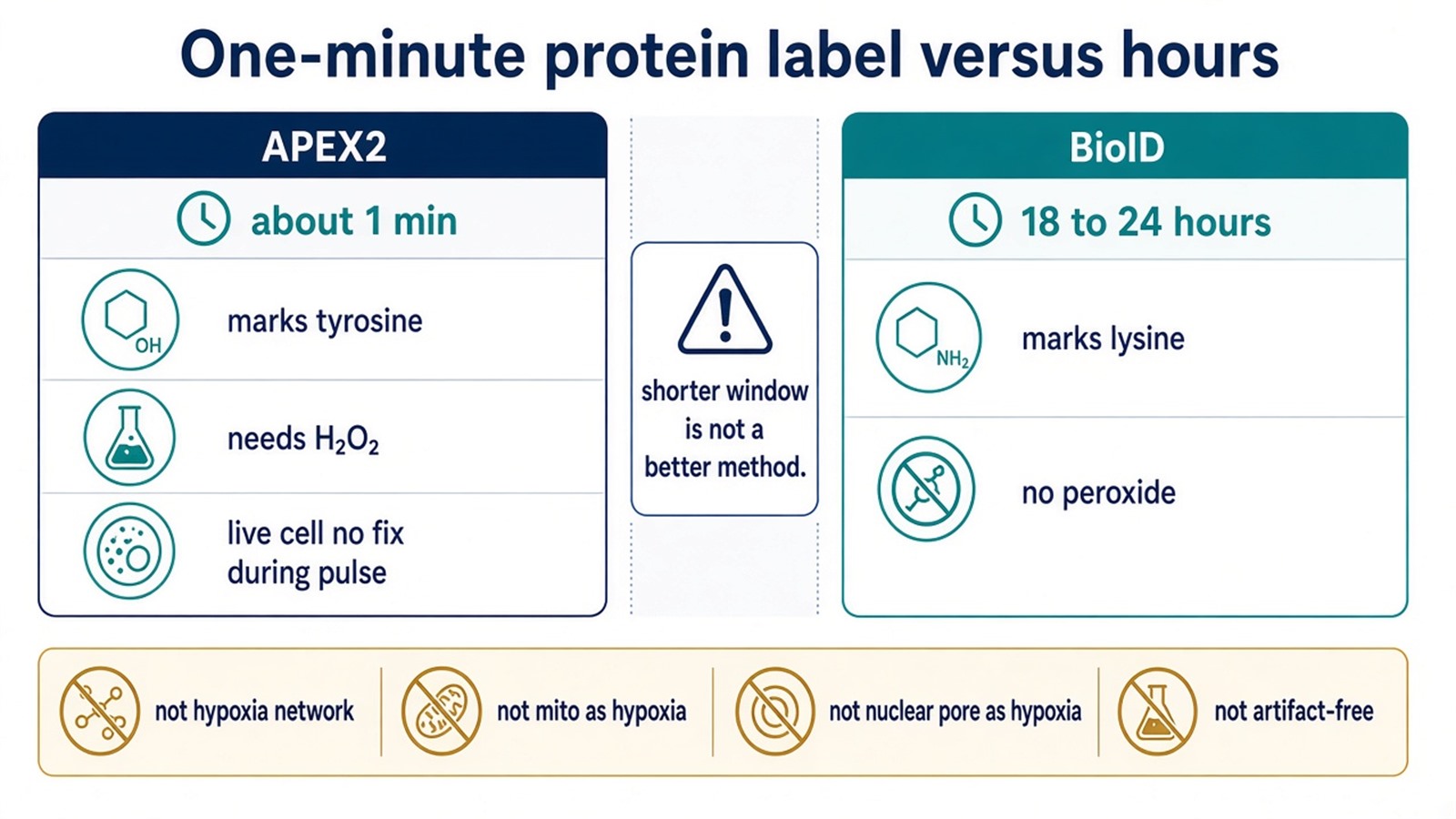
Figure 2. APEX2: about one minute, tyrosine, H₂O₂ required. BioID: hours, lysine, no peroxide. Mitochondria and nuclear pore are targeting choices, not hypoxic-network proofs.
APEX2, CRISPR Knock-In, and the Cost of the Label
APEX2 can label at a lower H₂O₂ dose than first-generation APEX in many setups. That is a catalytic improvement. It is not a guarantee of lower cellular toxicity, and it does not ensure specificity. Background biotinylation is expected. Specificity is the APEX-only comparison after the pulse, not the H₂O₂ titration alone.
CRISPR-Cas9 can place APEX2 at an endogenous locus so the fusion follows the native gene. That is a dose and localization choice. It does not produce endogenous-level labeling of target proteins in the sense of an untagged proteome, and it does not enhance the physiological relevance of the proteomic data by itself. The knock-in still writes biotin onto nearby tyrosines during an H₂O₂ pulse. APEX-only remains required.
H₂O₂ concentration and exposure time still have to be written down. Optimization reduces some oxidative damage. It does not eliminate non-specific labeling, and it does not convert the table into a physiological network.
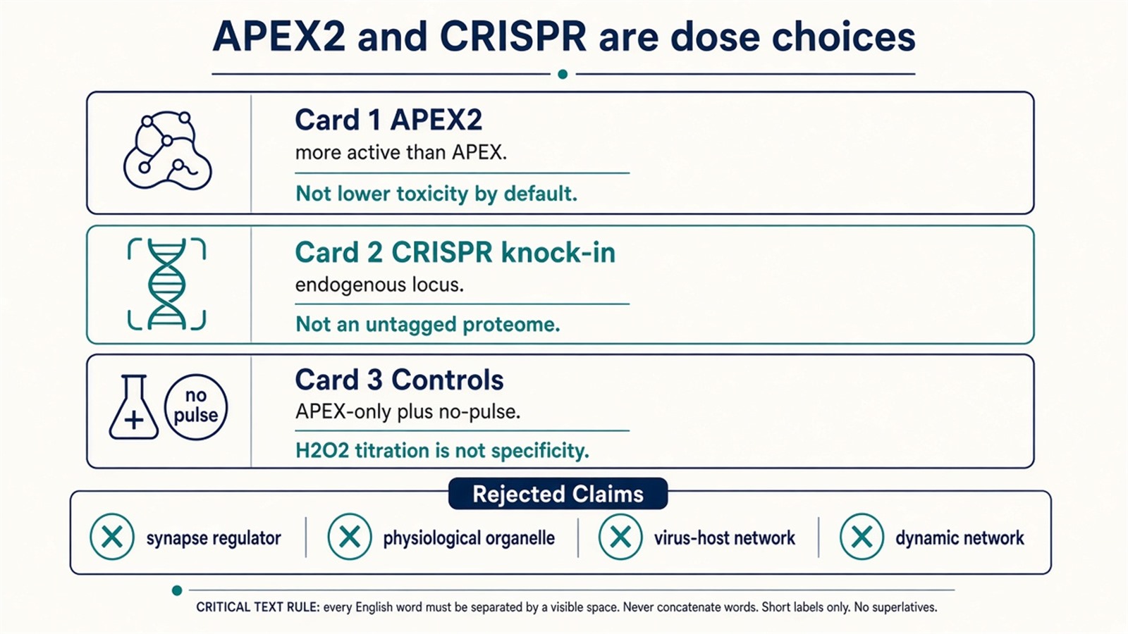
Figure 3. APEX2 is the usual enzyme. CRISPR knock-in is a dose control. APEX-only and no-pulse arms still travel through streptavidin and the search.
Experimental Limits
APEX protein labeling reduces a living cell to what a peroxidase fusion, a biotin-phenol/H₂O₂ pulse, streptavidin, and the search engine can report. Diagnostic, virus-host, or drug-network language sits outside ordinary research reporting unless a separately validated assay is in place.
Project Planning and Service Scope
A usable protein-labeling plan names APEX2 rather than first-generation APEX unless electron-microscopy history is the point, the APEX-only arm, the pulse length, and the claim that will be allowed, before the first H₂O₂ addition. MtoZ Biolabs scopes proximity-labeling MS with Orbitrap-class LC-MS/MS. Interpretability still follows those arms, not a one-minute-specificity slogan.
Once the project is a streptavidin proteome rather than a single labeled-protein claim, enzyme choice and the two proteomic jobs can be scoped together. Researchers who need that neighborhood or compartment table can review the MtoZ Biolabs Proximity Proteomics Service for project-specific sample evaluation, feasibility assessment, and workflow planning.
Frequently Asked Questions
1. What does APEX protein labeling write onto proteins?
A biotin handle, mainly on nearby tyrosines, during an H₂O₂ pulse. The recovered objects are biotinylated proteins, not demonstrated binders.
2. Is APEX or APEX2 the usual enzyme for protein labeling?
APEX2. First-generation APEX is the parent reporter. APEX2 is more active in many intracellular sites.
3. Does the one-minute pulse avoid cellular artifacts?
No. It shortens the labeling window. H₂O₂ is still an oxidant pulse.
4. Is APEX better than BioID because it is faster?
Faster is the window. BioID marks lysines over hours without peroxide. The methods answer different timing and residue questions.
5. Does CRISPR knock-in make the data more physiological?
It can lower overexpression. It does not remove H₂O₂, streptavidin, or the need for APEX-only.
Reference
- Rhee H.W., Zou P., Udeshi N.D., et al. (2013). Proteomic mapping of mitochondria in living cells via spatially restricted enzymatic tagging. Science, 339, 1328-1331. https://doi.org/10.1126/science.1230593
- Lam S.S., Martell J.D., Kamer K.J., et al. (2015). Directed evolution of APEX2 for electron microscopy and proximity labeling. Nat. Methods, 12, 51-54. https://doi.org/10.1038/nmeth.3179
- Hung V., Udeshi N.D., Lam S.S., et al. (2016). Spatially resolved proteomic mapping in living cells with the engineered peroxidase APEX2. Nat. Protoc., 11, 456-475. https://doi.org/10.1038/nprot.2016.018
- Martell J.D., Deerinck T.J., Sancak Y., et al. (2012). Engineered ascorbate peroxidase as a genetically encoded reporter for electron microscopy. Nat. Biotechnol., 30, 1143-1148. https://doi.org/10.1038/nbt.2375
- Roux K.J., Kim D.I., Raida M., Burke B. (2012). A promiscuous biotin ligase fusion protein identifies proximal and interacting proteins in mammalian cells. J. Cell Biol., 196, 801-810. https://doi.org/10.1083/jcb.201112098
