



Unlabeled mass spectrometry detection in proteomics means peptides from label-free digests are ionized and recorded as MS1 and MS/MS signals without isobaric or metabolic tags on the discovery path (Bantscheff et al. 2012). In project language, detection names what the mass spectrometer measures after liquid chromatography (LC) separation, not the full label-free quantification (LFQ) record that adds search, alignment, normalization, and contrast statistics (Cox et al. 2014). The guide below integrates unlabeled MS detection within LFQ workflows, locked parameters, limits, and project fit. It is not a disease application tour, not a technology trend essay, and not a promise of universal sensitivity across every matrix.
Unlabeled detection outputs are ion currents and fragment spectra tied to LC retention time. Relative protein claims still require a declared LFQ pipeline, biological replicates, and a frozen contrast. Detection depth does not by itself prove interaction, modification mapping, or drug mechanism without separate experimental design (Gerber et al. 2003).
In MtoZ Biolabs offerings, unlabeled MS detection sits on the same label-free LC-MS/MS spine used for DDA LFQ or unlabeled data-independent acquisition (DIA) when matrix and contrast scope fit the cohort.
LFQ record context appears in LFQ. LC-MS/MS experimental scope appears in Label-Free LC-MS/MS.
What Unlabeled MS Detection Covers in LFQ Projects
Four detection layers belong in the brief.
Ionization and transfer convert eluting peptides to gas-phase ions at the source. Spray stability and sheath conditions affect signal continuity across runs.
MS1 survey detection records precursor m/z and intensity over time. MS1 maps feed intensity-based LFQ after peptide identification in data-dependent acquisition (DDA).
MS/MS detection records fragment ions for sequence assignment in DDA or for windowed fragmentation in DIA and SWATH-style plans (Gillet et al. 2012).
Time alignment links detection events to LC retention. Drift without documented alignment breaks feature matching across injections.
Unlabeled MS detection is the signal acquisition layer. It does not replace tagging decisions, replicate design, or informatics version control.
Chemistry contrast appears in What Is Label-Free Analysis?.
Unlabeled Detection Versus Labeling and Targeted MS Routes
Detection chemistry and assay scope can diverge.
Label-free detection reads native peptide ions from separate injections. Quantification compares aligned intensities across runs after normalization.
Labeled detection still uses mass spectrometry but encodes state with SILAC, TMT, or iTRAQ reporters read in MS/MS. Multiplex fits closed arms labeled the same day, not every open cohort.
Targeted detection on parallel reaction monitoring (PRM) or multiple reaction monitoring (MRM) schedules named transitions on a closed list. It supports follow-up remeasurement, not open discovery breadth by itself.
Spectral counting uses MS/MS assignment counts as a coarse abundance proxy. MS1 area LFQ on the same DDA files usually supports group fold-change statistics more directly.
Route selection appears in Protein Quantification Method. DIA detection context appears in Label-Free DIA Quantitative Proteomics.
End-to-End Workflow Linking Detection to LFQ
A practical unlabeled detection-to-LFQ spine usually runs as follows.
- Confirm matrix, contrast, and relative claim unit before scaling injections.
- Prepare matched digests without discovery tags to injection-ready load.
- Lock LC gradient, column, and load from pilot when matrix is new.
- Acquire separate LC-MS/MS runs with interleaved order when cohort size allows.
- Record MS1 and MS/MS under a frozen acquisition template for the block.
- Search with declared database, enzyme, modifications, and false discovery rate (FDR).
- Extract label-free features from MS1 areas or DIA fragment traces per declared rules.
- Normalize within study groups; apply missing-value policy before statistics.
- Test predefined contrast only; archive instrument methods and software versions.
Steps 1 through 5 are unlabeled MS detection layers. Steps 6 through 9 build the LFQ record.
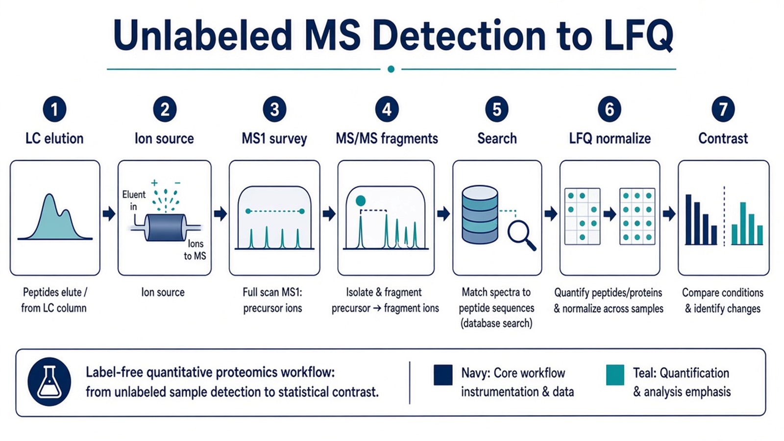
Figure 1. Unlabeled detection delivers raw ion traces; LFQ comparability depends on locked acquisition and one processing chain.
Quality integration appears in How to Improve Data Quality and Reproducibility in Label-Free Proteomics?. Normalization choices appear in How to Normalize Data in Label-Free Quantitative Proteomics?.
MS Detection Parameters That Must Be Locked for LFQ
Layer |
Parameter that must be declared |
Failure mode if ignored |
|---|---|---|
Source |
Ionization mode and spray settings |
Signal dropouts between days |
MS1 |
Resolution, scan rate, dynamic range policy |
Incomparable peak shapes |
DDA MS/MS |
Top-N, isolation width, collision energy |
ID bias and quant dropout |
DIA MS/MS |
Window layout and cycle time |
Mixed templates in one matrix |
LC |
Gradient, column ID, temperature |
Retention misalignment |
Load |
Mass on column |
Suppression and saturation |
Calibration |
Mass accuracy checks |
Drift in m/z matching |
Raw handling |
Converter and checksum policy |
Broken downstream import |
Search |
FDR and modification rules |
Inflated detection lists |
Quant bridge |
MS1 versus fragment metric |
Detection without LFQ rule |
Limits follow the same table. Unlabeled detection does not remove ion suppression in complex matrices. Low-abundance peptides may appear in MS/MS intermittently in DDA while missing from quant columns (Lazar et al. 2016). High-resolution MS1 alone does not fix confounded run order.
Depth expectations appear in How Many Proteins Can Label-Free Detect?. Processing tools appear in Tools for Label-free Peptide Quantification.
How to Choose Unlabeled Detection and LFQ Routes
Use matrix pilot, detection goal, acquisition evidence, and follow-up unit together.
Project need |
Detection and LFQ plan that often fits |
Recheck before committing |
|---|---|---|
Open cohort relative discovery |
Unlabeled DDA with MS1 LFQ |
MS/MS counts as sole metric |
High DDA missingness after pilot |
Unlabeled DIA fragment detection plus LFQ |
DDA when DIA adds little |
Closed multiplex arms |
Labeled MS/MS reporter detection |
Forced unlabeled when tags required |
Phospho or PTM-rich discovery |
Enrichment plus unlabeled detection |
PTM search drift mid block |
Interaction claims |
Dedicated interaction experiment design |
Single-shot LFQ as binding proof |
Named peptide follow-up |
Discovery detection then PRM |
DDA detection list as final panel |
Amount on peptides |
Targeted SIS detection |
Discovery intensities as concentration |
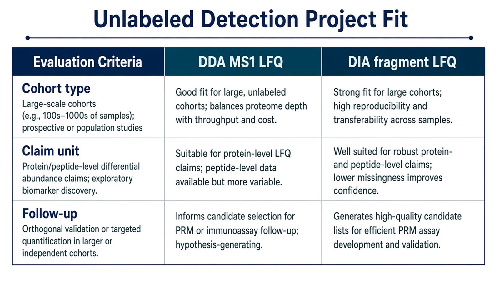
Figure 2. Unlabeled detection fits open separate-injection LFQ; labeling, DIA pivots, or targeted MS fit other enrollment and claim patterns.
Prep before detection appears in Quantitative Protein Analysis. Enrollment checks appear in Exploring Quantitative Analysis in Proteomics.
Teams with matrix documentation, locked acquisition templates, and contrast maps can review the MtoZ Biolabs Label-Free Quantitative Proteomics Service for unlabeled detection and LFQ alignment when declared in the project record.
Frequently Asked Questions
1. Is unlabeled mass spectrometry detection the same as label-free proteomics?
Label-free names unlabeled discovery chemistry on separate injections. Unlabeled MS detection names ion measurement at the instrument. Full label-free proteomics projects add LFQ informatics and contrast testing on top of detection.
2. Does unlabeled detection always mean higher sensitivity than labeling?
Not automatically. Label-free avoids tag chemistry loss but shares ion suppression and complexity limits. Labeled routes trade tagging steps for multiplex comparability within a set.
3. Can unlabeled detection map protein interactions from one LFQ run?
Routine open LFQ detection lists are not interaction assays. Binding or co-complex claims need dedicated enrichment, controls, and designs outside standard discovery detection.
4. Does high-resolution MS1 detection replace DIA for LFQ?
High-resolution MS1 can sharpen precursor integration in DDA LFQ. Unlabeled DIA adds fragment-level detection that may help quantifiable coverage in some matrices after pilot comparison.
5. When should detection settings change after a pilot?
Change gradient, load, or acquisition templates only with a new documented block. Mid-cohort detection drift splits LFQ comparability.
MtoZ Biolabs can discuss unlabeled MS detection and LFQ planning when raw format, locked methods, and contrast design are documented.
If further evaluation of DDA versus unlabeled DIA detection pilots, normalization choice, or discovery-to-targeted staging is needed, project details can be submitted for a feasibility review.
Reference
- M. Bantscheff, S. Lemeer, H.U. Haab, B. Kuster (2012). Quantitative mass spectrometry in proteomics: a critical review. Anal. Bioanal. Chem., 404, 939-965. https://doi.org/10.1007/s00216-012-5892-4
- J. Cox, M.Y. Hein, C.A. Luber, I. Paron, M. Mann (2014). Accurate proteome-wide label-free quantification by delayed normalization and maximal peptide ratio extraction, termed MaxLFQ. Mol. Cell. Proteomics, 13, 2513-2526. https://doi.org/10.1074/mcp.M113.031031
- L.C. Gillet, P. Navarro, S. Tate, H. Rost, N. Selevsek, L. Reiter, R. Bonner, R. Aebersold (2012). Targeted data extraction of the MS/MS spectra generated by data-independent acquisition: a new concept for consistent and accurate proteome analysis. Mol. Cell. Proteomics, 11, O111.016717. https://doi.org/10.1074/mcp.O111.016717
- C. Lazar, L. Gatto, M. Ferro, C. Bruley, T. Burger (2016). Accounting for the multiple natures of missing values in label-free quantitative proteomics data sets to compare imputation strategies. J. Proteome Res., 15, 1116-1125. https://doi.org/10.1021/acs.jproteome.5b00981
- S.A. Gerber, J. Rush, O. Stemman, M.W. Kirschner, S.P. Gygi (2003). Absolute quantification of proteins and phosphoproteins from cell lysates by tandem MS. Proc. Natl. Acad. Sci. U.S.A., 100, 6940-6945. https://doi.org/10.1073/pnas.0832254100
