



TMT quantification reads relative protein or peptide abundance from reporter ions after tandem mass tag (TMT) labeled digests are mixed and fragmented on liquid chromatography-tandem mass spectrometry (LC-MS/MS) (Thompson et al. 2003). Identification and quantification remain separate operations: peptide fragment ions name the sequence, and reporter intensities assign channel abundances. The useful project question is whether a freezable multi-sample set needs relative ratios inside one plex, not whether throughput slogans sound attractive. Oncology, immunology, and drug-screening themes do not change that quantitative definition.
TMT quantification is relative by default. Amount or concentration on named peptides needs a matched stable isotope-labeled internal standard (SIS) and a separate targeted design. Co-isolated precursors at MS2 can compress true fold changes; an additional MS3 reporter scan can reduce that mixing on compatible instruments (Ting et al. 2011). Incomplete labeling or unmatched protein load still appears as channel offset after any search software runs.
In MtoZ Biolabs quantitative offerings, TMT quantification is scoped when groups, replicates, channel map, and relative claim are declared before tagging. Service intake appears in TMT Quantitative Proteomics Analysis Service.
What TMT Quantification Covers
Four quantification layers belong in the brief and stay separated.
Design layer freezes experimental arms onto TMT channels, including biological replicates and any bridge channel across plexes.
Sample layer matches extraction, digestion, and protein load so channel differences are less likely to be prep artifacts.
Readout layer identifies peptides and extracts reporters at MS2 or MS3 from the mixed precursor after labeling and pooling.
Claim layer states relative change for the predefined contrast. Biomarker lists, pathway maps, or multi-omics overlays are optional after the ratio matrix passes quality checks. Clinical diagnosis or precision-medicine wording needs separate validation beyond the TMT table.
TMT quantification reports relative channel ratios inside a locked plex. It does not invent absolute concentration or diagnostic cutoffs.
How Reporter Readout Produces a Quantitative Matrix
After labeling chemistry attaches isobaric tags, channels share one precursor mass at MS1 until fragmentation releases channel-specific reporters (Thompson et al. 2003). Labeling operation detail appears in Tandem Mass Tag Labeling. Isobaric definition appears in What Is TMT Isobaric Labeling?.
Quantification starts only after a peptide-spectrum match is accepted and reporters are extracted from the designated scan. Impurity correction, unique-peptide rules, and protein rollup turn peptide reporters into a protein matrix. Identification-to-reporter coupling appears in Principle of TMT-Based Quantitative Proteomics Analysis. In MS2 TMT, co-isolated precursors can mix reporters and pull ratios toward one (Savitski et al. 2013). MS3 options against that compression appear in TMT MS3 and SPS-MS3: How to Mitigate Ratio Compression and Enhance Quantitative Accuracy.
MS1 peak area of the intact precursor does not separate channels. The quantitative object is the reporter ratio, not a copy-number ranking across different proteins.
End-to-End Workflow for TMT Quantification
A practical TMT quantification spine usually runs as follows.
- Freeze contrast, replicates, channel map, and relative versus amount claim before labeling.
- Match extraction and protein load across all arms that will enter the plex.
- Digest under matched conditions; confirm amine-compatible buffers for tagging.
- Label each digest with one TMT channel; quench and clean up residual reagent.
- Pool channels; fractionate when identification depth is required for the claim.
- Acquire LC-MS/MS with declared MS2 or MS3 reporter readout and quality control (QC) injections.
- Search spectra with locked FDR and modification settings; extract and correct reporters.
- Roll peptides to proteins; normalize within the plex under a stated assumption; test only the predefined contrast.
- Archive channel map, instrument method, search settings, and software versions with deliverables.
Steps 1 through 3 protect the sample record. Steps 4 through 9 are the labeling, acquisition, and quantification record.
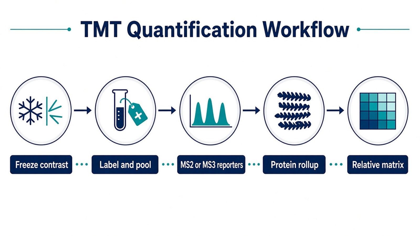
Figure 1. TMT quantification depends on a locked channel map and reporter extraction after identification before group contrasts are tested.
Parameters That Must Be Locked for a Quantification Claim
Layer |
Parameter that must be declared |
Failure mode if ignored |
|---|---|---|
Contrast |
Groups and replicates before labeling |
Post hoc regrouping read as biology |
Channel map |
Tag-to-sample layout and bridge plan |
Unlinked plexes treated as one matrix |
Load |
Matched protein input across digests |
Channel offset as treatment effect |
Labeling |
Completeness checks and quench |
Incomplete tags as abundance change |
Reporter scan |
MS2 or MS3 rule |
Compressed fold changes read as weak biology |
Search |
Database, FDR, unique-peptide rules |
Homolog mixing as differential signal |
Impurity correction |
Kit-specific factors applied |
Neighbor-channel leakage as biology |
Normalization |
Within-plex assumption stated |
Scale drift as differential biology |
Missingness |
Filter and imputation policy stated |
Imputed zeros as real change |
Deliverable |
Matrix, channel map, method notes |
Report rebuilt after acquisition |
Limits follow the same table. High-throughput multiplex size is a logistics parameter, not a biological result. Instrument access language does not replace matched prep. Low-abundance detection still depends on load, fractionation, and missingness rules.
What a TMT Quantification Result Can Support
A reviewed TMT matrix supports relative comparison of the same protein across channels that were mixed into one plex for a predefined contrast. Pathway enrichment and multi-omics overlays are optional after quality checks. They do not repair unmatched loads or invent clinical cutoffs.
Ratio compression can shrink a real treatment or phenotype effect when co-isolation is severe (Ting et al. 2011). A small observed fold change still needs isolation QC before it is called weak biology. Missing reporters do not prove protein absence. Shared peptides can pull homologs into one protein group.
A later unlabeled digest cannot join the same reporter matrix. A new plex needs a bridge or reference design. Named peptide confirmation usually needs targeted parallel reaction monitoring (PRM) or multiple reaction monitoring (MRM). Absolute concentration claims need SIS-supported targeted assays.
A TMT quantification table supports same-protein relative contrasts inside a locked plex. It does not certify biomarkers or therapy choice.
How to Choose TMT Quantification Versus Adjacent Routes
Use freezable group structure, reporter-scan need, claim unit, and follow-up intent together.
Project need |
Route that often fits |
Recheck before committing |
|---|---|---|
Multi-group relative ratios on a fixed set |
TMT quantification in one plex |
Samples still arriving after mixing |
Shared chromatography across arms |
Matched load before labeling |
Channel offsets already visible in bulk assays |
Open enrollment cohort |
Unlabeled LFQ or DIA |
Force late samples into an existing TMT mix |
Compressed MS2 ratios in complex digests |
TMT with MS3 review |
Keep MS2 without isolation QC |
Named peptide confirmation |
Targeted PRM or MRM follow-up |
Re-run the same TMT table as confirmation |
Amount or concentration unit |
SIS-supported targeted assay |
Ordinary reporter ratios read as ng per mL |
Multi-omics overlay |
TMT matrix plus matched other-omics design |
Treat transcript units as protein ratios |
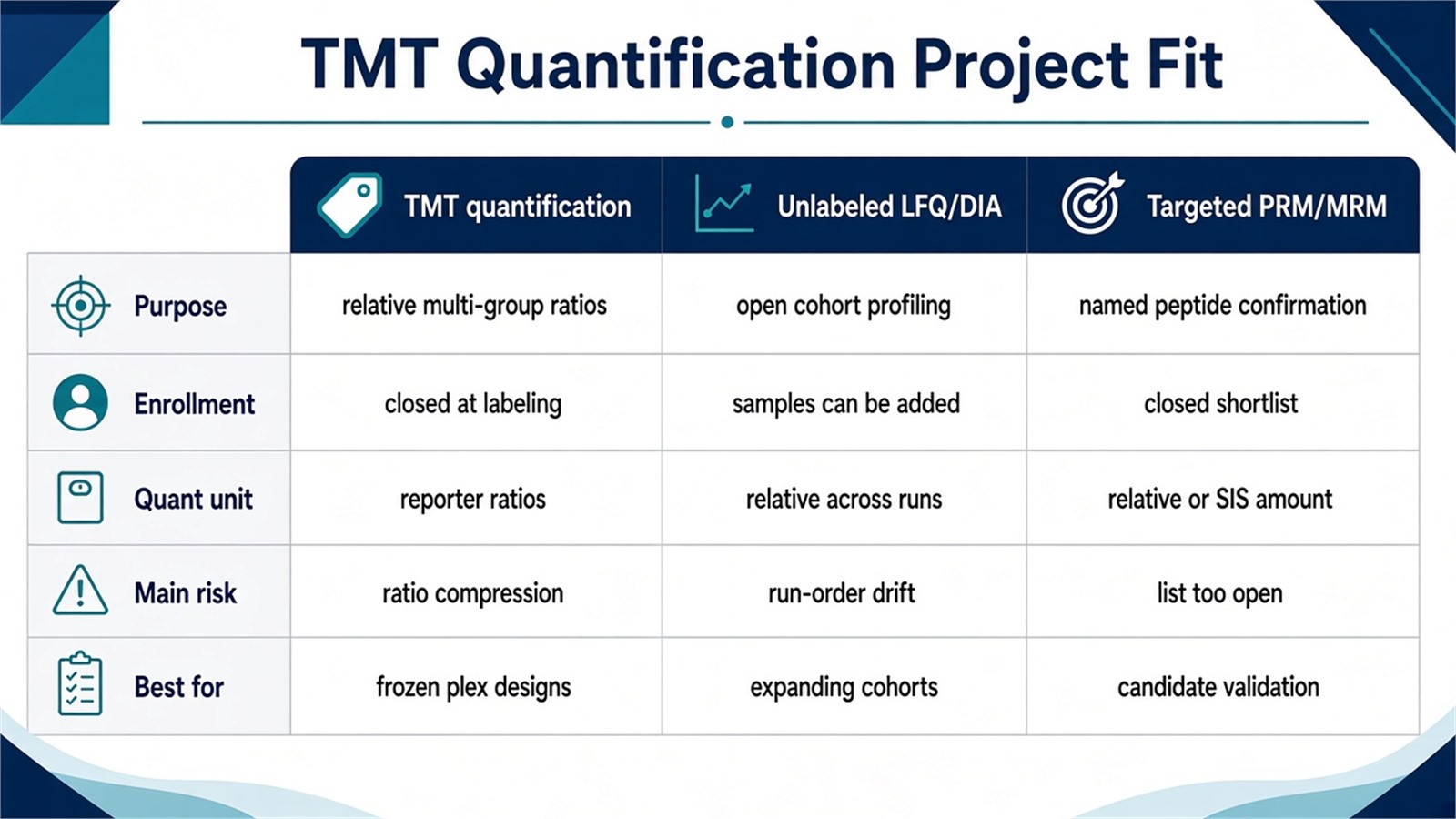
Figure 2. Choose TMT quantification when groups can freeze into one plex; keep unlabeled or targeted routes when enrollment or claim unit changes.
TMT quantification more often fits a multi-group or closed time-course design that can be labeled together. An exception is a small, well-buffered set that still needs unlabeled flexibility because a follow-up harvest is already booked. Unlabeled label-free quantification more often fits open enrollment. An exception is a short closed cohort that must share one injection to limit run-order drift. Application framing for those designs appears in Application of TMT Labeling in Quantitative Proteomics Detection.
Teams with defined groups, matched protein material, and a requested relative contrast can review MtoZ Biolabs TMT Quantitative Proteomics Analysis Service when those fields are declared in the project record.
Frequently Asked Questions
1. Is the MS1 peak area the TMT quantitative signal?
No. The mixed precursor is isobaric, so MS1 area does not separate channels. Relative abundance comes from reporter ions after fragmentation.
2. Does TMT quantification provide absolute protein concentration?
No. Standard TMT quantification is relative inside a plex. Amount units need a matched SIS and a declared calibration, usually in a targeted follow-up assay.
3. Must every TMT quantification project use MS3?
No. MS2 can suffice when directional consistency across replicates is enough for triage. MS3 becomes useful when co-isolation is expected to hide a real fold change.
4. Can one more sample join a TMT quantification matrix after mixing?
Not as an extra reporter channel in the same mix. A new plex with a bridge or reference sample can connect experiments.
5. What project fields are needed before scoping TMT quantification?
A versioned group map, sample matrix, matched protein material notes, relative versus amount claim, and any bridge plan help evaluate feasibility before labeling.
MtoZ Biolabs can provide TMT quantification for predefined multi-group proteomics projects, including plex layout, labeling, LC-MS/MS, and relative matrix reporting when sample type and study design are available.
If further evaluation of channel mapping, MS2 versus MS3 readout, or whether unlabeled quantification fits better is needed, project details can be submitted for a feasibility review.
Reference
- A. Thompson, J. Schafer, K. Kuhn, S. Kienle, J. Schwarz, G. Schmidt, T. Neumann, R. Johnstone, A.K. Mohammed, C. Hamon (2003). Tandem mass tags: a novel quantification strategy for comparative analysis of complex protein mixtures by MS/MS. Anal. Chem., 75, 1895-1904. https://doi.org/10.1021/ac0262560
- L. Ting, R. Rad, S.P. Gygi, W. Haas (2011). MS3 eliminates ratio distortion in isobaric multiplexed quantitative proteomics. Nat. Methods, 8, 937-940. https://doi.org/10.1038/nmeth.1714
- M.M. Savitski, T. Mathieson, N. Zinn, G. Sweetman, C. Doce, I. Becher, F. Pachl, B. Kuster, M. Bantscheff (2013). Measuring and managing ratio compression for accurate iTRAQ/TMT quantification. J. Proteome Res., 12, 3586-3598. https://doi.org/10.1021/pr400098r
