



Protein gel band identification names the proteins that were present in a stained SDS-PAGE slice. Chemical cross-linking mass spectrometry names residue pairs that a bifunctional reagent covalently joined. The two experiments share a later LC-MS/MS step and nothing else at intake. A polyacrylamide band is a sample format. It is not a cross-linker, and it is not silica or another solid reagent. In-gel digestion reports linear peptides from the slice. XL-MS needs its own reaction, quench, sequences, and a cross-linked peptide search. Stain type and the written question belong in the same plan as the search type. Mixing the two jobs turns a protein list into a false contact map.
Researchers who already have a visible, MS-compatible band can review the MtoZ Biolabs Gel and IP Sample Protein Identification Service for feasibility. Teams with a named pair, an amine-free buffer, and a restraint claim should start from the Crosslinking Mass Spectrometry Service. Method-level choice among enrichment and residue-level routes is covered in Measuring Protein-Protein Interactions.
A Gel Slice Is Not a Cross-Linking Reagent
A stained-slice request starts after electrophoresis has already separated a mixture. The slice is cut from a Coomassie, SYPRO Ruby, or MS-compatible silver-stained lane. The polymer holds denatured protein. It does not create covalent bonds between amino acids. NHS esters such as DSS, BS3, or DSSO create those bonds in solution or in cells before the gel exists.
Do not describe the gel matrix as a physical cross-linker. Silica strips, empty cassettes, and loading dye are not reagents for protein-protein capture. Reducing sample buffer and heat unfold chains for SDS-PAGE. They do not report who touched whom in the native complex.
A higher-mass band after a chemical reaction can still be only a protein-identification sample. The shift shows that a covalent product may have formed. Who sits in that band is a composition question. Which residues were joined is an XL-MS question and needs a different search.
If the band came from an IP or pull-down eluate, the identification table is a co-recovery list under that wash, not a residue map. Antibody-based product intake is scoped through the IP-Mass Spectrometry Service. Fusion-bait bands follow the Fusion Protein Interaction Analysis Service | Pull-Down and MS.
In-Gel Digestion Identifies Proteins in the Band
In-gel digestion is the usual path for gel band identification. The slice is destained, reduced, alkylated, and cut with trypsin inside the gel. Peptides are extracted, desalted, and searched as linear sequences (Shevchenko et al. 2006). Orbitrap-class LC-MS/MS and a UniProt or client FASTA search, typically in MaxQuant, return protein accessions that explain those peptides.
The report answers who was abundant enough to leave peptides in that molecular-weight window. It does not assign a binding site. Two names in one band can mean a heterodimer, a shared complex, a degradation product, antibody chains, or a comigrating contaminant. Keratin and the stain front should stay annotated.
A visible band is the planning guide. Invisible, over-fixed, or destain-incompatible silver bands often fail extraction. Gel slices can travel at room temperature or on ice packs. Do not ship dried, unlabeled, or heavily destained pieces without a lane map.
Cellular IP products that are still liquid or on beads are a different handoff. Those formats are covered in Cellular IP Product Mass Spectrometry Detection. Cut the band only when SDS-PAGE was already used to isolate the species of interest.
Chemical XL-MS Has Separate Reaction and Search Requirements
Chemical XL-MS is not in-gel identification of a shifted ladder. The experiment requires a chosen reagent, an amine-free buffer such as HEPES or PBS, a quench, digestion of the reacted sample, and a search that scores two peptides plus a linker mass (Leitner et al. 2016). Tris or glycine consume NHS esters and must be exchanged before the reaction. Planning amounts for purified proteins are 100 to 150 ug at purity of at least 90 percent, with 20 ug only when material is limited. Cell-level crosslinking typically plans 1x10^7 to 2x10^7 cells per condition.
The search must be a cross-linked peptide search, typically pLink 2 or MaxQuant, with the stronger XL result delivered. A linear protein search of a cut band does not recover that table. Intra-protein and inter-protein classes must stay split. Missing links do not exclude contact when lysines are buried or absent.
Acceptance of each row is a later QC step. Efficiency, a declared XL peptide FDR, site localization, and fragment support from both peptides belong on the Chemical Cross-Linking Mass Spectrometry Quality Control path. A linear band search cannot substitute for that reaction-and-search design.
If the interface is lysine-poor or too short-lived for a solution NHS reaction, chemical XL-MS should not be forced onto a stained slice. Photo-activated capture can be reviewed through the Photocrosslinking Mass Spectrometry Service.
The comparison below keeps the stained slice and the chemical reaction on separate tracks.
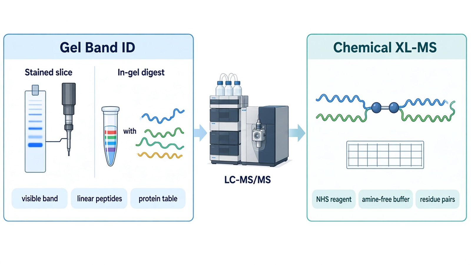
Figure 1. Gel Band Identification vs Chemical XL-MS.
Research Questions That Fit Each Route
The stained-slice route fits when the question is who occupies a visible molecular-weight window. Typical questions are an unknown expression product, a Co-IP or pull-down band, or a higher-mass species after a reaction whose composition is still unknown. It fits poorly when the publication sentence is residue proximity, a unique pose, or direct binding in cells.
Chemical XL-MS fits when a pair or complex is already named and the next sentence must be a spacer-limited residue pair. It fits poorly when the only material is an unlabeled gel cassette, when no sequences are available, or when the request is simply to identify a stained band.
|
Research question |
Route that fits |
Required material |
Typical output |
|---|---|---|---|
|
Who is in this stained slice? |
Gel band identification |
Visible MS-compatible band, species, lane map |
Protein accession table |
|
Who co-recovered in an IP or pull-down band? |
In-gel ID of that eluate |
Band plus capture history and controls |
IDs in that window versus controls |
|
Which residues were close enough to link? |
Chemical XL-MS |
Named pair or complex, amine-free buffer, reagent plan |
Intra- and inter-protein residue pairs |
|
Did a covalent product form after NHS chemistry? |
Efficiency gel, then XL-MS search |
Reacted protein or cells, not the empty cassette |
Shift plus a later XL table |
|
What is the atomic structure? |
Neither route |
Coordinate method outside this intake |
Not delivered by either search |
The table is a boundary rule, not a ranking of stains or reagents.
Two names in one untreated band do not become an XL-MS result by renaming the file. Two names after a controlled chemical reaction still need the XL search before they become residue pairs.
Samples, Overlap Cases, and Deliverables
Ship stained-slice material as a visible band with stain type, species, and the claim sentence. Coomassie, SYPRO Ruby, or an MS-compatible silver stain are the usual options. High-salt destain leftovers and unlabeled empty lanes should not be shipped. Liquid IP products, if the gel step is unnecessary, should follow the liquid planning amounts used for IP-MS, at least 20 ug at more than 0.5 ug/uL with SDS below 0.1 percent.
Ship XL-MS material as already reacted solutions or cells, or as two uncrosslinked recombinant proteins with sequences. Do not send a blank gel strip as the reagent. Solutions and cells travel snap-frozen on dry ice. A reacted sample that was later run on SDS-PAGE can still be used for composition ID of the shifted species. That slice answers composition. A parallel aliquot, or the same reaction before the gel, is what enters the XL search.
Intake is faster when the research question is mapped to one route before shipment. The diagram below links band visibility, stain, sequences, buffer amines, and the claim sentence to the correct request.
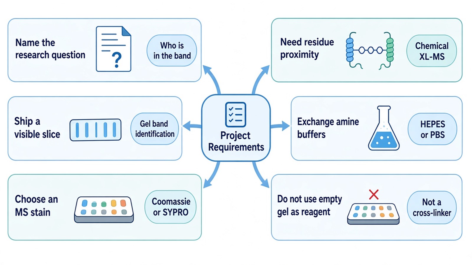
Figure 2. Matching Project Requirements With Gel Band Identification or XL-MS.
Deliverables for the slice route typically include a protein table, peptide support, and a short methods note on destain, digest, and search. Deliverables for XL-MS typically include intra- and inter-protein residue pairs after a dedicated search. Neither report includes a unique atomic model or a claim that two comigrating names are a functional complex. To start intake, send the question, the physical format, sequences if XL-MS is intended, and the stain or buffer notes to info@MtoZ-Biolabs.com or use the site inquiry form so the route can be confirmed before shipment.
Frequently Asked Questions
1. Does cutting a higher-mass band after crosslinking replace an XL-MS search?
No. The slice can identify who is in that window. Residue pairs require the chemical reaction plus a cross-linked peptide search on a suitable aliquot.
2. Can a silica strip or an empty gel cassette be used as the cross-linker?
No. The gel is a separation matrix. Chemical XL-MS uses a bifunctional reagent such as DSS, BS3, or DSSO in an amine-free buffer.
3. What stain is typically planned for an in-gel slice?
Plan Coomassie, SYPRO Ruby, or an MS-compatible silver stain, and keep the band visible. Harsh destain or incompatible silver recipes reduce peptide recovery.
4. How do typical amounts differ between a gel slice and chemical XL-MS?
A gel slice is planned around a visible band. Chemical XL-MS on purified protein is planned at 100 to 150 ug and at least 90 percent purity, with 20 ug only as a limited-material fallback.
5. If two proteins are identified in one untreated band, is that a contact site?
No. Comigration reports presence in the same molecular-weight window. A contact hypothesis still needs an XL-MS residue pair or an orthogonal pair test.
Researchers can keep gel band identification and chemical XL-MS on separate intake paths, then confirm stain, sequences, and the written question through MtoZ Biolabs Gel and IP Sample Protein Identification Service or the Crosslinking Mass Spectrometry Service.
Reference
- A. Shevchenko, H. Tomas, J. Havlis, J.V. Olsen, M. Mann (2006). In-gel digestion for mass spectrometric characterization of proteins and proteomes. Nat. Protoc., 1, 2856-2860. https://doi.org/10.1038/nprot.2006.468
- A. Leitner, M. Faini, F. Stengel, R. Aebersold (2016). Crosslinking and mass spectrometry: an integrated technology to understand the structure and function of molecular machines. Trends Biochem. Sci., 41, 20-32. https://doi.org/10.1016/j.tibs.2015.10.008
