



Absolute QUAntification (AQUA) detects named proteins by isotope dilution of proteotypic peptides, not by bulk dye binding or open proteome surveying. After protease digestion, usually with trypsin, a synthetic heavy peptide matching the target sequence is spiked at a known amount. The heavy and endogenous light peptides co-elute and ionize under closely related conditions, but separate by mass so their peak areas can be compared by targeted liquid chromatography-tandem mass spectrometry (LC-MS/MS) (Gerber et al. 2003). A calibration model converts that light-to-heavy ratio into peptide amount or concentration. The mechanism reports peptide-level detection under stated spike timing. It does not automatically equal intact-protein concentration, open discovery, or a clinical cutoff without separate design and validation.
Researchers who already have a closed target list and required reporting units can also review the MtoZ Biolabs Absolute Quantitative Analysis (AQUA) Service and Targeted Proteomics Service for project-specific feasibility and assay planning.
Mechanism at a Glance
Mechanism step |
What happens |
What it enables |
|---|---|---|
Proteotypic peptide selection |
Choose unique, detectable digest peptides for the named protein |
Sequence-specific detection instead of bulk protein dye assays |
Heavy SIS synthesis |
Incorporate stable isotopes (for example 13C or 15N) into the same sequence |
Mass-shifted internal standard with matched chemistry |
Digestion and spike |
Digest the sample; add the heavy peptide at a recorded step |
Light and heavy forms share later LC-MS path from that point |
Targeted MS detection |
Acquire light and heavy by MRM or PRM (Lange et al. 2008; Peterson et al. 2012) |
Peak areas for the scheduled pair only |
Calibration |
Convert light-to-heavy response through matrix-matched calibrators |
Amount or concentration inside the validated range |
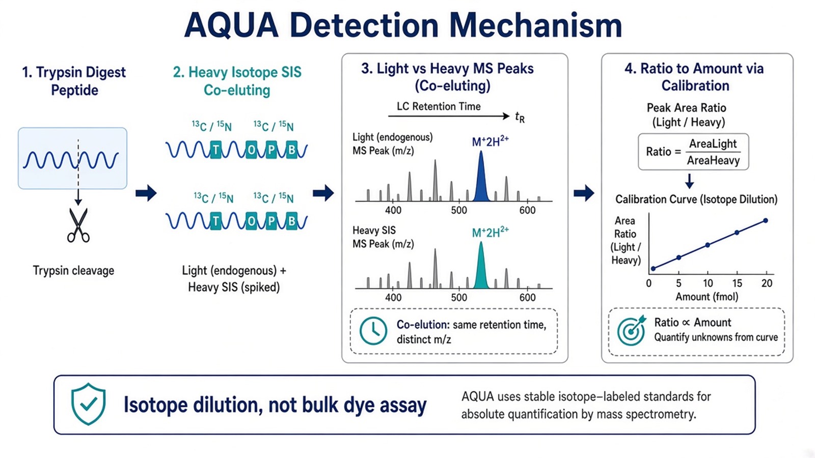
Figure 1. AQUA detection mechanism is isotope dilution of a proteotypic peptide: co-eluting light and heavy forms yield a calibrated amount, not a bulk protein colorimetric readout.
The heavy peptide is the quantitative reference. Acquisition mode alone does not create absolute units.
How Spike Timing Shapes the Mechanism
Spike timing is part of the detection mechanism because it defines which sample-prep losses are corrected.
Post-digestion spike (common)
The heavy peptide enters after proteins have already been digested. The assay then reports peptide recovery in that digest under calibration. Lysis loss and incomplete protein digestion that happened earlier stay outside the correction unless a different standard design is used.
Pre-digestion or enrichment-stage spike
Earlier spikes can bring more of the upstream path under isotope dilution when the scientific claim requires it. Protein-level or concatenated labeled standards can move the mechanism even earlier, at higher design cost.
Modification-matched standards
If the claim is a phosphorylated or otherwise modified peptide, the heavy standard must carry the same chemistry. An unmodified heavy peptide does not quantify a modified endogenous form.
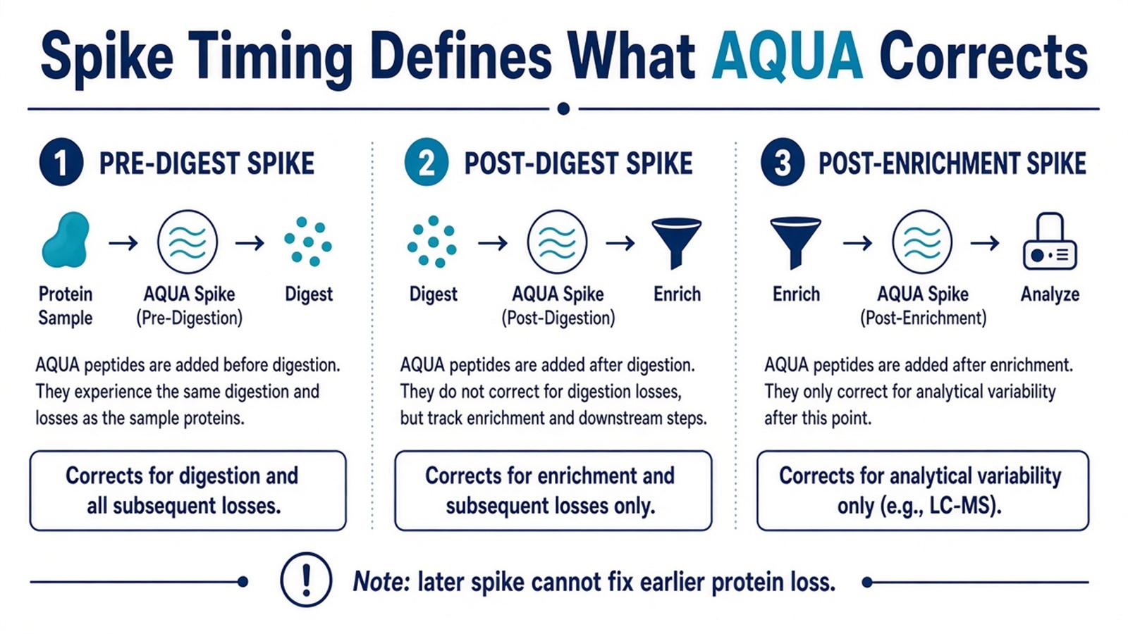
Figure 2. Spike timing is mechanistic: a later heavy peptide cannot reconstruct protein that was lost before the standard entered the sample.
For workflow and method-layer context beyond this mechanism page, see AQUA Proteomics and Absolute Quantification in Targeted Proteomics. For absolute method families, see Absolute Protein Quantification: Methods and Uses. For acquisition layers under AQUA, see SRM Mass Spectrometry and PRM and MRM in Targeted Proteomics: Advantages and Limitations.
Judgment Criteria: When This Detection Mechanism Fits
Study need |
AQUA mechanism often fits |
Reassess before ordering standards |
|---|---|---|
Claim |
Amount or concentration for named peptides |
Relative fold change alone |
Targets |
Closed proteotypic peptides with usable fragments |
Open discovery inventory still changing |
Spike design |
Spike timing matches what must be corrected |
Expect post-digest spike to fix lysis loss |
Matrix |
Matrix-matched calibrators can bracket expected range |
No recoverable peptide in that matrix |
Platform |
MRM or PRM can schedule light/heavy pairs |
Only unlabeled discovery acquisition planned |
Validation |
Research absolute assay |
Diagnostic cutoff without validated framework |
Prefer AQUA when the detection question needs calibrated peptide amount. Prefer relative MRM or PRM without full calibration when confirmation trends are enough. Prefer LFQ, DIA, or TMT while the inventory is still open. Prefer protein-level labeled standards when digestion recovery is part of the claim.
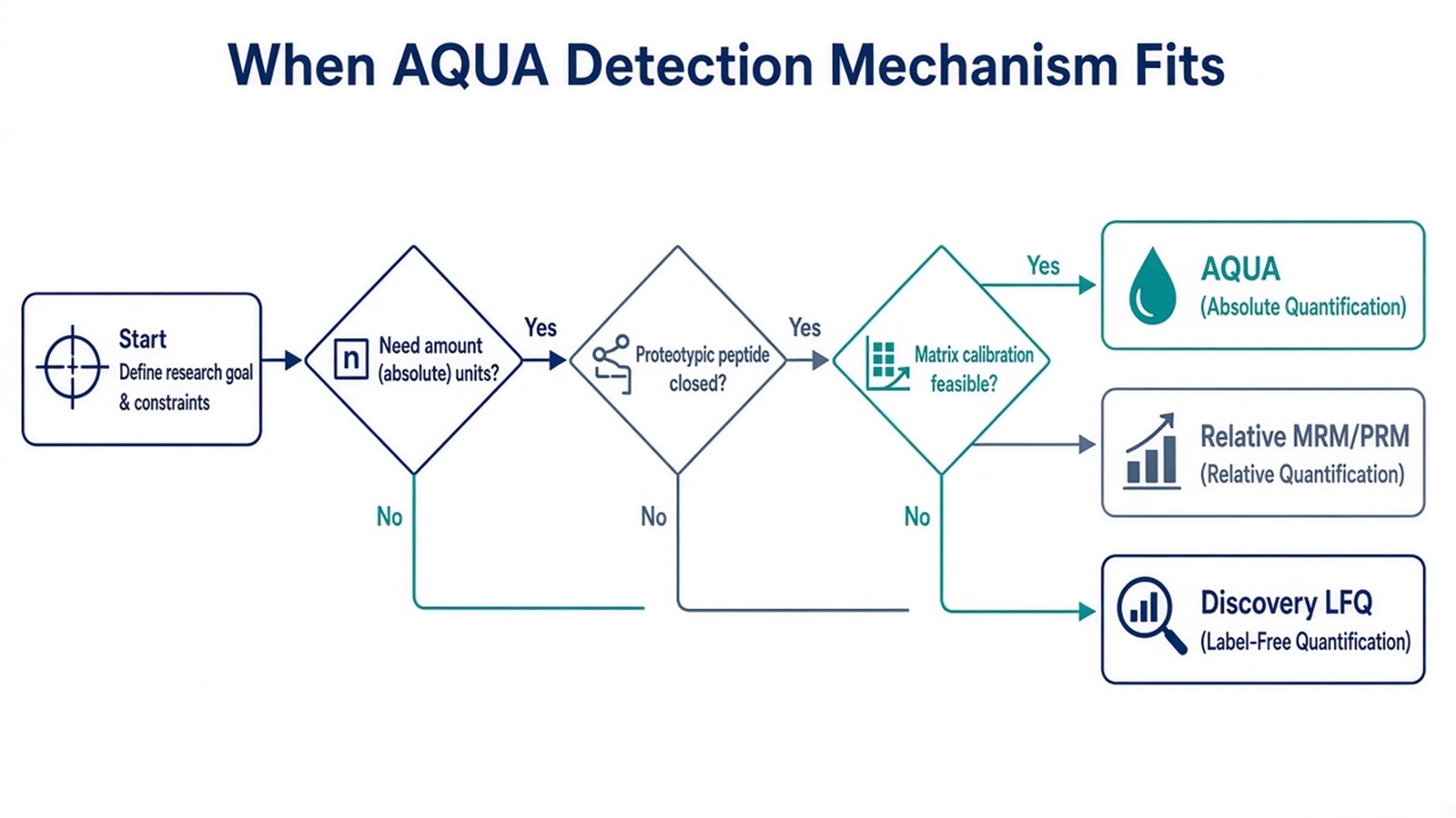
Figure 3. Use the AQUA mechanism when amount units and closed peptides are required; keep relative routes when the claim stops at fold change.
For design trade-offs, see Advantages and Disadvantages of AQUA Technique. For sample and pretreatment constraints across absolute and relative claims, see Absolute and Relative Quantitative Proteomics Analysis.
Limits and Application Boundaries
AQUA does not detect proteins outside the scheduled peptide list. Matrix-matched calibration reduces, but does not erase, ion suppression when calibrators poorly represent the sample. Femtomolar-style sensitivity claims without matrix, peptide, and instrument detail overstate method class alone. Shared peptides report gene-family ambiguity rather than a single protein.
Typical applications of this mechanism:
- Absolute amount for biomarker research peptides after discovery shortlisting.
- Stoichiometry or specification readouts for named process proteins.
- Modification-site amount when the heavy standard matches the modified sequence.
- Bridging MS peptide amount to later immunoassay development.
Diagnostic cutoffs and open proteome concentration maps sit outside ordinary research AQUA detection unless a separately validated assay framework is in place.
Once peptide selection, spike timing, calibration, and acquisition class are defined, the project can be evaluated as an AQUA detection assay. Researchers can review the MtoZ Biolabs Absolute Quantitative Analysis (AQUA) Service for standard design, feasibility assessment, and workflow planning.
Frequently Asked Questions
1. What is the core mechanism of AQUA in protein detection?
Isotope dilution of a proteotypic peptide: a known-amount heavy standard and the endogenous light peptide are compared by targeted MS and converted to amount through calibration.
2. Why do heavy and light AQUA peptides behave similarly in LC-MS?
They share sequence and chemistry, so retention and ionization are closely matched; stable isotopes create the mass difference used for detection.
3. Does the mechanism quantify intact protein copies automatically?
No. It quantifies the monitored peptide under the stated spike and digestion design. Protein inference needs explicit assumptions.
4. Can AQUA remove all matrix effects?
No. Matched standards and matrix-matched calibrators reduce bias; poor peptide recovery or unmatched calibrators still fail.
5. Is AQUA a discovery detection method?
No. Only closed, scheduled peptides are detected. Open inventories need discovery routes such as LFQ, DIA, or TMT.
Reference
- S.A. Gerber, J. Rush, O. Stemman, M.W. Kirschner, S.P. Gygi (2003). Absolute quantification of proteins and phosphoproteins from cell lysates by tandem MS. Proc. Natl. Acad. Sci. U.S.A., 100, 6940-6945. https://doi.org/10.1073/pnas.0832254100
- V. Lange, P. Picotti, B. Domon, R. Aebersold (2008). Selected reaction monitoring for quantitative proteomics: a tutorial. Mol. Syst. Biol., 4, 222. https://doi.org/10.1038/msb.2008.61
- A.C. Peterson, J.D. Russell, D.J. Bailey, M.S. Westphall, J.J. Coon (2012). Parallel reaction monitoring for high resolution and high mass accuracy quantitative, targeted proteomics. Mol. Cell. Proteomics, 11, 1475-1488. https://doi.org/10.1074/mcp.O112.020131
