



A laboratory that already named a protein often still needs a mass or molar amount in the digest. Label-free quantification (LFQ) compares the same protein across samples from unlabeled mass spectrometry signals. Absolute protein quantification reports concentration or amount only when a matched stable isotope-labeled internal standard (SIS) and a declared calibration are in place.
Absolute QUAntification (AQUA) is the peptide-level form of that logic. A synthetic heavy peptide of known amount is spiked, and the light-to-heavy chromatographic ratio is converted to peptide amount (Gerber et al. 2003). SIS chemistry and acquisition are separate decisions. Selected reaction monitoring (SRM) or multiple reaction monitoring (MRM) records predefined precursor-fragment transitions (Lange et al. 2008). Parallel reaction monitoring (PRM) records a high-resolution fragment spectrum for each scheduled precursor (Peterson et al. 2012). In MtoZ Biolabs offerings, AQUA panels are typically acquired as high-resolution PRM. An SRM/MRM panel remains valid when a scheduled triple-quadrupole assay already exists.
What an Absolute Amount Claim Requires
An amount claim needs a closed peptide list, a proteotypic sequence that maps to the intended protein, and an SIS whose assigned concentration is documented. The report unit must be stated, for example peptide fmol in the injected digest or protein mass inferred from that peptide. A ratio of unlabeled intensities between two groups is a relative result even when the software prints a number with many digits.
LFQ, isobaric tagging, and metabolic labeling answer relative questions unless a SIS of known quantity is added to the same assay. Discovery intensities cannot be converted into ng per mL by applying a universal response factor after the run. Intensity-based copy-number estimates from unlabeled matrices are planning tools for ranking detectability. They are not AQUA amounts.
If the protein list is still open, keep work on a discovery matrix until names freeze. Researchers can refer to A Guide to Label-Free Quantitative Proteomics: From Experiment to Data Analysis when the remaining task is relative LFQ matrix construction and group contrast.
How AQUA Turns a Proteotypic Peptide into a Reported Amount
The protein sequence is inspected for candidate peptides. A proteotypic peptide is unique to the intended protein or protein group, is typically generated by the planned protease, and ionizes well enough to be scheduled. Missed-cleavage motifs, ragged termini, and easily modified residues are poor first choices unless the biology is the modification itself.
The matching SIS peptide is synthesized with heavy amino acids so chromatographic behavior stays close to the endogenous (light) peptide while the mass shift allows clean extraction. The SIS concentration must be assigned before it is treated as a calibrant. Purity, isotopic enrichment, and stock quantification all flow into the reported amount. Spiking after digestion compares two peptides that already exist. Spiking before digestion can capture some processing losses, yet a peptide SIS still does not unfold and cleave as the intact protein does (Brun et al. 2007).
AQUA reports peptide amount from a light-to-heavy ratio only after the SIS concentration, spike point, and extraction rule are declared.
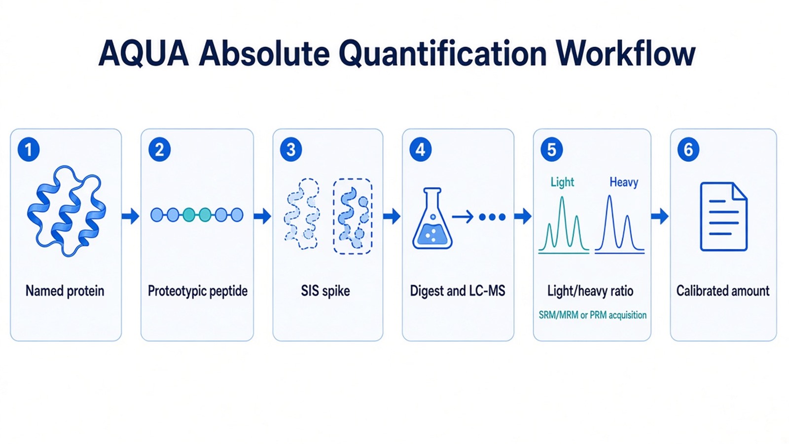
Figure 1. AQUA converts a named protein into a calibrated peptide amount after a matched SIS is spiked and light and heavy peaks are extracted together.
Acquisition then isolates the light and heavy precursors on a chromatographic schedule. SRM/MRM uses a short transition list. PRM stores full fragment spectra and allows fragment choice after acquisition. Mixing unlabeled discovery files with an AQUA panel in one statistical model does not create an amount for proteins that never received an SIS.
Peak areas are extracted, a light-to-heavy ratio is formed, and calibration converts that ratio into amount. A single-point spike can be sufficient when the SIS level sits in the linear range of the endogenous peptide. A multi-point curve is the stronger design when the expected range is wide or when a lower limit of quantification must be shown (Carr et al. 2014). The protein-level number is an inference from the peptide, not an independent measurement of every residue.
Researchers who need the scheduled isolation and fragment-extraction details can refer to PRM LC-MS Targeted Proteomics for how a closed precursor list is acquired after names exist.
Parameters That Control Calibration, Spike Timing, and the Number
Peptide uniqueness controls which gene products and isoforms are included. A shared peptide reports a protein group. A site-specific modified SIS reports that modified peptide, not unmodified occupancy, unless the unmodified pair is measured in the same design.
SIS quality controls the scale. If the heavy stock concentration is wrong, every sample is wrong in the same direction. If isotopic impurities leak into the light channel, low endogenous levels are inflated. The spike amount should sit near the expected endogenous level so that both traces are measured, not one saturated and one missing.
Digestion and extraction control recovery. Incomplete cleavage, adsorption, and matrix suppression can drop the light peptide without an identical loss on a late-spiked SIS. Buffer salts, detergents, and residual hemoglobin or albumin can hide a low-abundance target even when the SIS is clean in solvent.
Calibration and quality rules should be locked before the study set is quantified. Record the SIS lot and assigned concentration, the spike point relative to digestion, the fragments or transitions used, the regression model if a curve is fitted, and the rule for points below the lower limit of quantification. Carryover checks belong on blank injections after high standards. A discovery screen and a longitudinal biomarker panel do not share the same documentation burden (Carr et al. 2014).
When chromatograms or load still explain most of the variance before any SIS is added, researchers can refer to Quantitative Proteomics: Strategies, Advantages, and Challenges for how those failure patterns are traced in unlabeled or labeled discovery runs.
Limits of Peptide-Level SIS for Protein Copy Number
AQUA is specific because the peptide is specific. That same property limits protein copy-number claims. Proteolytic processing, splice isoforms, and post-translational modifications can split one gene product across several peptides. Measuring one unmodified peptide does not report the modified fraction. Measuring a phosphorylated AQUA peptide does not report total protein unless the unmodified partner is quantified on the same scale.
A peptide SIS is added as a peptide. It does not test whether the parent protein was fully extracted from tissue or fully digested in that matrix. Protein-level isotope-labeled standards, sometimes called PSAQ-style standards, are designed to enter extraction and digestion with the analyte protein (Brun et al. 2007). That materials strategy is separate from peptide AQUA and is not required for every amount claim.
AQUA does not inventory an unknown proteome. Proteins without a validated proteotypic peptide stay unreported. Cross-protein ranking from LFQ intensity remains a relative statement. Amount in the injected digest is not amount in the original tissue unless extraction recovery is measured.
How to Choose AQUA Versus Relative Label-Free Quantification
The useful test is whether names are closed, whether the claim is amount, and whether an SIS can be designed for a unique peptide.
Study need |
Check first |
Route that often fits |
|---|---|---|
Open protein inventory |
Identification depth and group contrast |
Relative LFQ or unlabeled DIA matrix |
Same-protein fold change, no amount unit |
SIS availability versus budget |
Relative LFQ, or relative PRM/MRM without SIS |
Named protein, mass or molar unit required |
Unique peptide and SIS stock |
AQUA with targeted LC-MS |
Site occupancy or modified peptide |
Modified and unmodified pairs |
Site-specific SIS peptides, evaluated per sequence |
Later samples will be added |
Whether the list can stay closed |
Relative LFQ while the list is open; AQUA after names freeze |
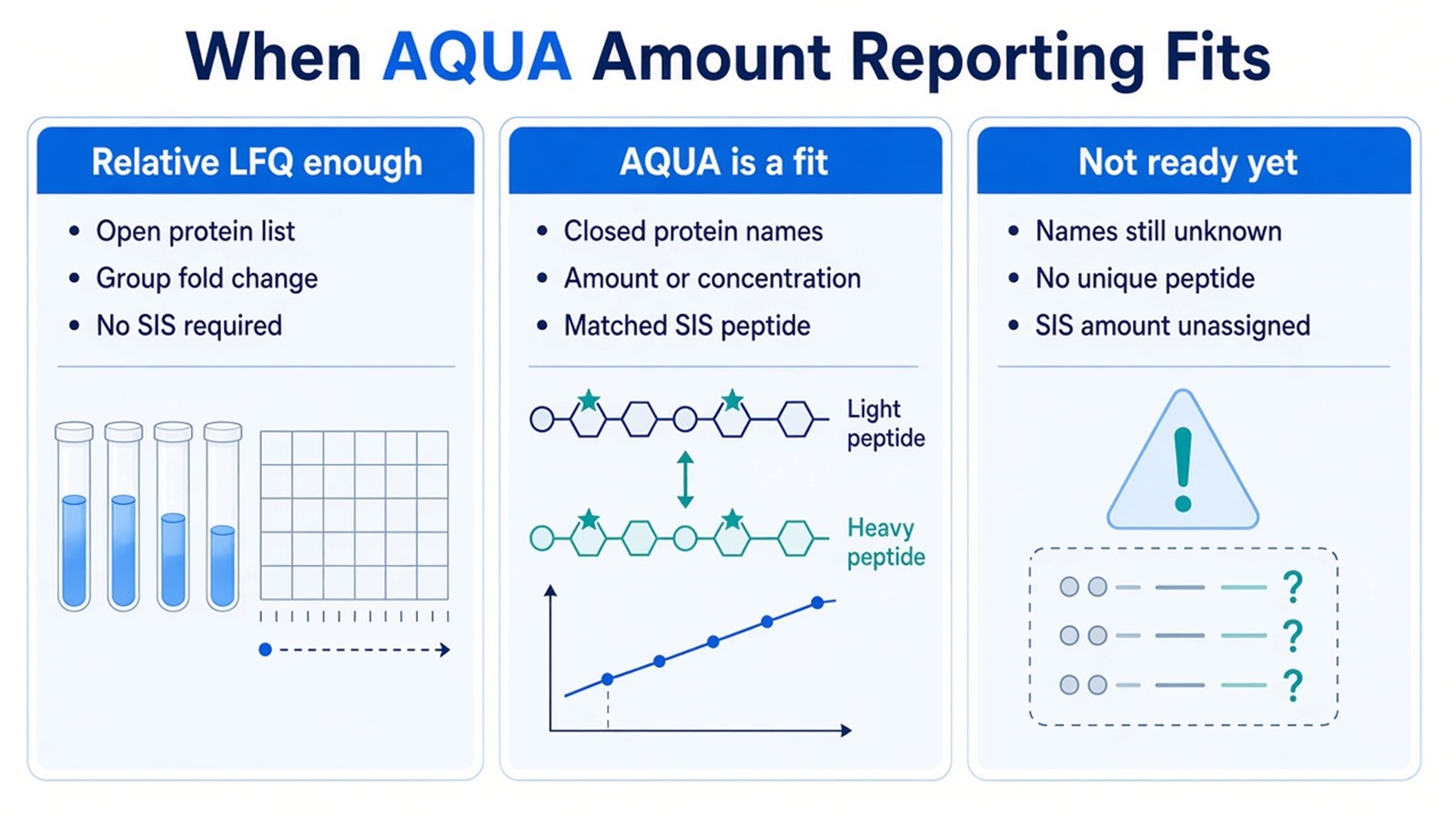
Figure 2. AQUA fits when protein names are closed, an amount unit is required, and a matched SIS peptide can be assigned; relative LFQ remains the fit for an open list.
Researchers who already have named proteins, a requested amount unit, and sample type information can also review the MtoZ Biolabs Absolute Quantitative Analysis (AQUA) Service for project-specific peptide evaluation, SIS planning, and calibration design.
Frequently Asked Questions
1. Can LFQ intensities be converted to ng per mL after the run?
No reliable conversion exists without a matched SIS and a declared calibration for that peptide. A later universal factor does not repair ion suppression, digestion yield, or protein inference that were never measured.
2. Should the heavy peptide be spiked before digestion?
Pre-digest spiking can capture some handling losses. A peptide SIS still does not unfold and cleave as the intact protein does. If digestion completeness is the main risk, a protein-level standard is the closer materials match, and the report should still name the spike point.
3. If PRM already runs without an SIS, is the result absolute quantification?
No. PRM without an SIS is a targeted relative assay. Absolute reporting starts when a matched SIS of assigned amount and a calibration rule are present. The acquisition mode does not by itself create concentration units.
4. Does one AQUA peptide report every isoform of a protein?
Only the products that contain that peptide. Shared or isoform-specific sequences must be declared in the protein inference. A second peptide is often required when splice forms or processing products are part of the biological question.
5. Can AQUA replace an immunoassay for the same protein name?
Mass spectrometry reports a defined peptide sequence. An immunoassay reports an epitope. The two numbers can diverge when proteoforms, matrix, or antibody cross-reactivity differ. Treat them as orthogonal measurements unless the epitope and the peptide are shown to match.
MtoZ Biolabs can provide AQUA-based absolute protein quantification for closed protein or peptide lists, including proteotypic peptide evaluation, SIS-supported calibration, and targeted LC-MS reporting when project files and study design are available.
If further evaluation of peptide uniqueness, SIS spike point, calibration range, or whether relative LFQ is sufficient is needed, project details can be submitted for a feasibility review.
Reference
- V. Brun, A. Dupuis, A. Adrait, M. Marcellin, D. Thomas, M. Court, F. Vandenesch, J. Garin (2007). Isotope-labeled protein standards: toward absolute quantitative proteomics. Mol. Cell. Proteomics, 6, 2139-2149. https://doi.org/10.1074/mcp.M700163-MCP200
- S.A. Carr, S.E. Abbatiello, B.L. Ackermann, C. Borchers, B. Domon, E.W. Deutsch (2014). Targeted peptide measurements in biology and medicine: best practices for mass spectrometry-based assay development using a fit-for-purpose approach. Mol. Cell. Proteomics, 13, 907-917. https://doi.org/10.1074/mcp.M113.036095
- S.A. Gerber, J. Rush, O. Stemman, M.W. Kirschner, S.P. Gygi (2003). Absolute quantification of proteins and phosphoproteins from cell lysates by tandem MS. Proc. Natl. Acad. Sci. U.S.A., 100, 6940-6945. https://doi.org/10.1073/pnas.0832254100
- V. Lange, P. Picotti, B. Domon, R. Aebersold (2008). Selected reaction monitoring for quantitative proteomics: a tutorial. Mol. Syst. Biol., 4, 222. https://doi.org/10.1038/msb.2008.61
- A.C. Peterson, J.D. Russell, D.J. Bailey, M.S. Westphall, J.J. Coon (2012). Parallel reaction monitoring for high resolution and high mass accuracy quantitative, targeted proteomics. Mol. Cell. Proteomics, 11, 1475-1488. https://doi.org/10.1074/mcp.O111.013698
