



Protein-ligand interaction analysis helps researchers investigate whether a known protein and ligand bind, quantify binding affinity, characterize association and dissociation kinetics, and examine ligand-associated structural changes. The appropriate method depends on the research question and the type of evidence required. This article compares major binding assays, explains factors that affect measurement reliability, and outlines how to interpret results when different methods produce different findings.
Researchers planning a defined pair can use the MtoZ Biolabs Protein Ligand Binding Assay Service to discuss whether the available protein, ligand, controls, and concentration range support affinity or kinetic analysis. For a broader overview of protein interaction types, their molecular partners, and the methods used to study them, see Protein Interaction Analysis: What It Reveals, Key Methods, and Applications.
Defining the Binding Question for a Known Protein and Ligand
The choice of assay should follow the intended research outcome. It is which claim the experiment must test. A yes or no binding screen asks whether the pair produces a concentration-dependent signal above matched controls. An affinity study asks what equilibrium dissociation constant, or KD, is consistent with the observed response under defined conditions. A kinetic study asks how quickly the complex forms and dissociates. Competition experiments ask whether one molecule changes the binding of another, while structural response studies ask where ligand exposure alters protein dynamics or solvent accessibility.
The molecular context matters. A purified soluble domain may be suitable for an initial biophysical test, but it may not reproduce a membrane environment, multimeric state, cofactor requirement, or post-translational modification. An isolated peptide can help investigate a proposed binding motif, but the result should be interpreted in the context of the full-length protein and its molecular environment.
Binding should also be separated from function. A reproducible in vitro interaction can support physical association under the tested conditions, but it does not show that the ligand changes signaling, enzyme activity, localization, or phenotype. Functional assays answer those downstream questions. This distinction is especially important when comparing compounds that bind with similar affinity but differ in cellular exposure or mechanism.
What Different Assays Can Tell You About Binding
Affinity and Kinetics
Surface plasmon resonance, or SPR, immobilizes one partner on a sensor surface and follows the response as the other partner associates and dissociates. Properly designed concentration series can provide KD together with association and dissociation rate constants. Biolayer interferometry, or BLI, also provides real-time binding traces from a sensor surface and can support affinity, kinetic, and competition measurements. Both methods require attention to immobilization, transport effects, nonspecific surface binding, and whether the selected model adequately describes the data.
Microscale thermophoresis, or MST, measures changes in molecular movement within a microscopic temperature gradient. It is commonly used to estimate affinity in solution with one fluorescently observed partner. MST avoids surface immobilization, which can help when attachment compromises activity, but fluorescence labeling, intrinsic fluorescence, adsorption, and aggregation can still alter the response. MST is an affinity method, not a routine source of association and dissociation rate constants.
Stability and Structural Response
Differential scanning fluorimetry, or DSF, follows protein unfolding as temperature increases and reports a melting temperature, Tm. A ligand-associated change in Tm can be useful for compound screening or buffer optimization. However, a positive or negative thermal shift depends on unfolding thermodynamics, assay chemistry, and the conformational state sampled. A change in Tm is not an absolute KD, and no detectable shift does not by itself exclude binding.
Hydrogen-deuterium exchange mass spectrometry, or HDX-MS, compares deuterium uptake across protein peptides. Ligand-associated protection or increased exchange can identify regions whose solvent accessibility or dynamics change after ligand exposure. These regions may include a contact surface, a propagated allosteric response, or both. HDX-MS therefore provides peptide-level structural and dynamic evidence, but it does not automatically identify exact contacting residues or quantify affinity.
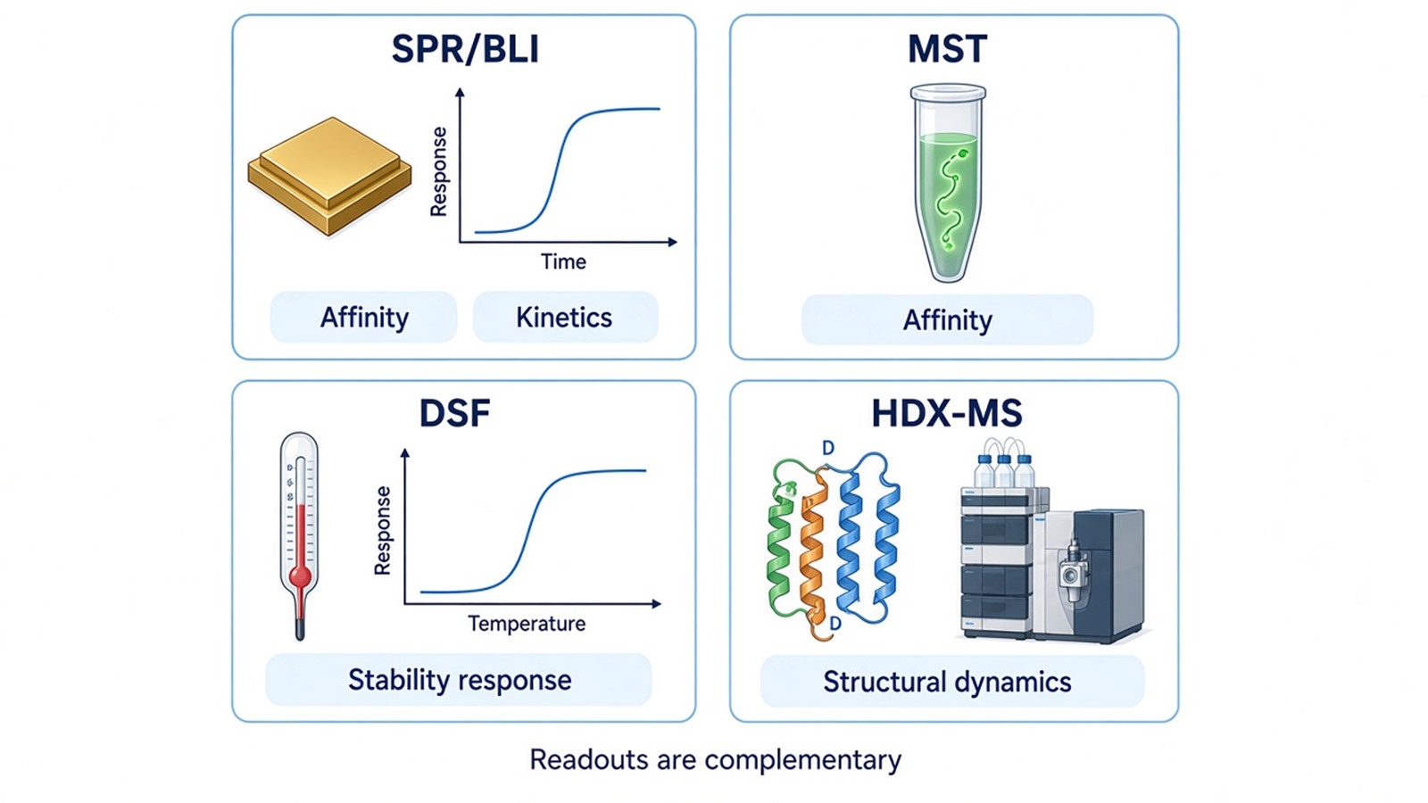
Figure 1. Selecting the appropriate assay based on the intended readout: binding affinity, kinetics, stability, or structural response.
Experimental Factors That Affect Assay Interpretability
Protein quality is a shared dependency across these methods. Heterogeneous oligomers, partial degradation, inactive fractions, or unstable constructs can create curved baselines, weak concentration dependence, or misleading fitted values. Before a binding experiment, researchers should confirm that the construct is soluble and compatible with the planned buffer over the concentration range required by the assay.
Ligand behavior is equally important. Poor aqueous solubility can produce an apparent plateau that reflects precipitation rather than saturation. Colloidal aggregation can create nonspecific responses. Solvent concentration should be matched across samples and controls because solvent can alter protein stability, refractive index, fluorescence, or thermophoresis. For colored or fluorescent compounds, optical interference should be assessed with ligand-only and protein-only controls where the assay permits.
Surface methods add orientation and activity questions. Immobilization can obstruct a binding site or select a subpopulation of protein molecules. Reversing which partner is immobilized, changing capture chemistry, or using a solution method can help test this possibility. In MST, the labeled partner should retain binding competence, and capillary adsorption or fluorescence changes unrelated to thermophoresis should be checked. In DSF, compounds can interfere with the reporter dye or alter the unfolding curve independently of specific binding.
Concentration coverage determines whether affinity can be resolved. A narrow range that never approaches saturation may indicate a response without supporting a stable KD estimate. Controls should address matrix effects, nonspecific surfaces, inactive analogs when scientifically justified, and reference proteins or ligands.
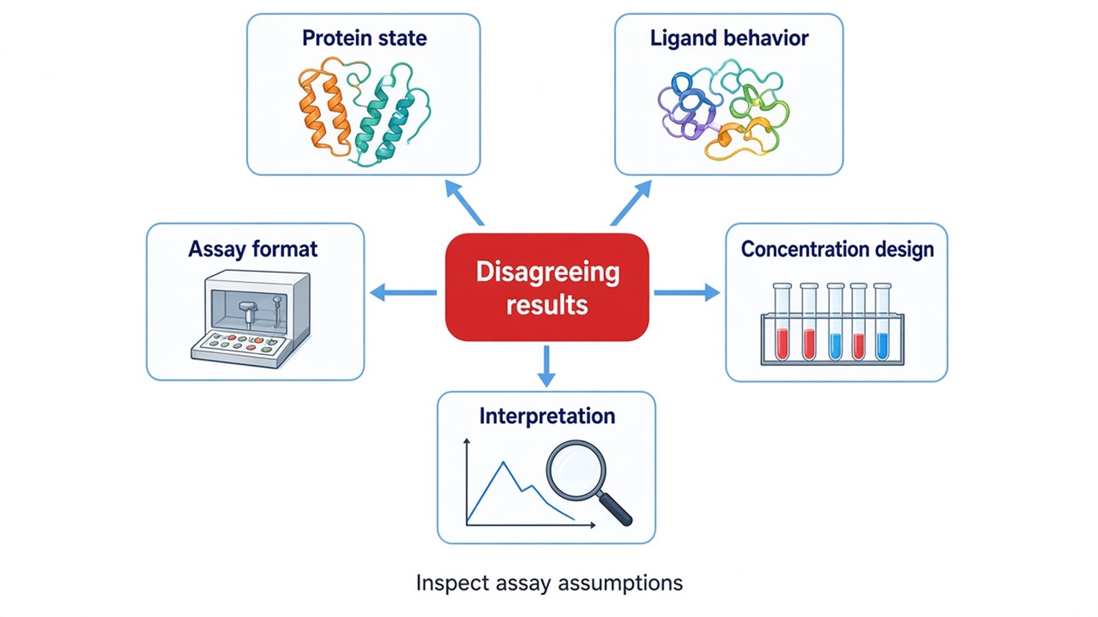
Figure 2. Protein quality, ligand behavior, assay format, concentration design, and data analysis can all influence protein-ligand binding measurements.
Interpreting Conflicting Evidence and Deciding What to Verify
Different assays may produce apparently inconsistent results because they measure different properties of the protein-ligand system. Interpreting these findings requires identifying what each measurement establishes and selecting a follow-up experiment that addresses the unresolved question.
When affinity measurements are positive but DSF shows no thermal shift, the ligand may bind without substantially changing the dominant unfolding transition, or the change may fall below the practical resolution of the assay. If SPR or MST provides a reproducible concentration-dependent response with appropriate controls, the DSF result can be reported as no detectable thermal response under the tested conditions.
When DSF shows a thermal shift but affinity has not been established, the compound can be prioritized for further investigation. The next step may be a concentration-dependent SPR, BLI, or MST experiment to examine binding using a different measurement principle. If surface-based and solution-based results disagree, review protein activity, immobilization orientation, mass transport, labeling, ligand solubility, and nonspecific interactions before changing the analysis model.
When HDX-MS identifies ligand-responsive regions but affinity remains unknown, a separate affinity measurement can quantify binding, while competition or mutagenesis experiments can investigate hypotheses about the responsive regions. Functional assays can then determine whether the interaction is associated with a biological effect.
The final interpretation should distinguish three outcomes: evidence of binding under defined conditions, quantitative affinity or kinetic parameters supported by the experiment, and structural or functional effects associated with ligand exposure. Keeping these outcomes separate makes it easier to identify which questions have been answered and which require further investigation.
When the protein target is unknown, the research objective changes from characterizing a defined protein-ligand pair to discovering candidate targets. A target-identification strategy such as Drug Affinity Responsive Target Stability addresses this different research question.
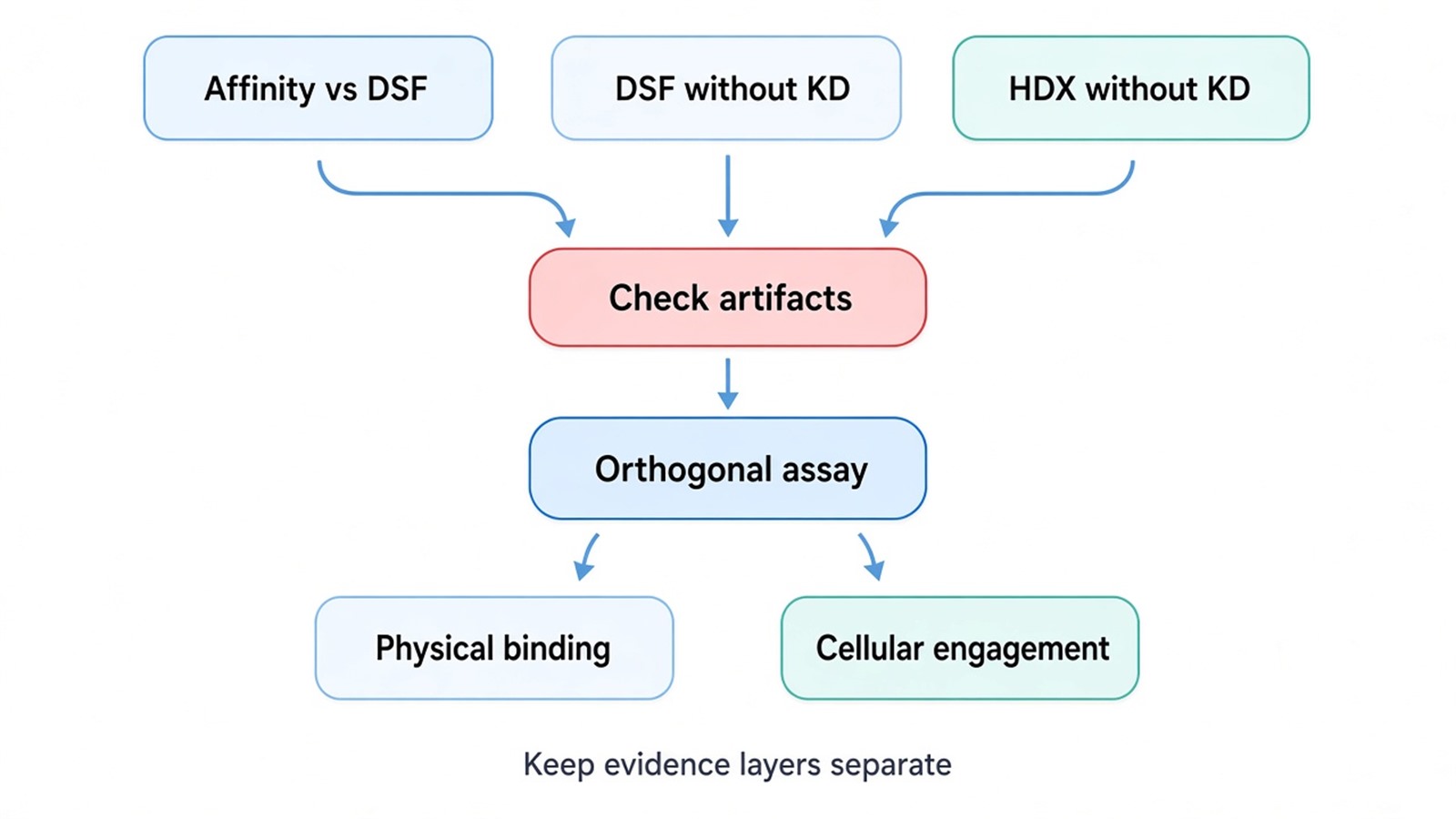
Figure 3. Conflicting readouts are resolved by checking assay-specific artifacts, repeating with matched controls, and adding an orthogonal physical principle before advancing the claim.
Frequently Asked Questions
1. Which method is suitable when a small molecule is expected to bind weakly?
Start from the expected affinity range, compound solubility, protein availability, and signal size rather than choosing by sensitivity claims alone. SPR may be suitable when low sample consumption and real-time kinetics are important, while MST can provide a solution-based affinity estimate if fluorescence behavior is acceptable. BLI can be useful for parallel comparisons, but low molecular mass and weak responses may require careful feasibility assessment.
2. Can a fluorescently labeled peptide be used directly for affinity measurement?
It may be suitable for MST if the fluorophore position does not disrupt the interaction and free dye is controlled. Compare labeled and unlabeled material when possible, inspect initial fluorescence across the concentration series, and include a nonbinding control. A label already present on the peptide can be advantageous, but it does not remove the need to confirm retained binding competence.
3. What information is needed before requesting an affinity study?
Provide the identities and construct boundaries of both partners, purity and concentration information, buffer composition, ligand solubility, any existing binding evidence, and the biological question. Positive controls, inactive analogs, or known competitors can materially improve interpretation. The final method should be selected after checking whether the protein and ligand are compatible with immobilization, labeling, and the required concentration range.
4. Why might two laboratories report different KD values for the same pair?
Different constructs, immobilization chemistries, temperatures, buffers, active protein fractions, concentration ranges, and fitting models can all affect apparent KD. The values should be compared together with experimental conditions and confidence in the fits. A difference does not automatically indicate error, and neither value should be treated as the cellular affinity without additional evidence.
Conclusion
Protein-ligand interaction analysis should begin with the claim that needs evidence. SPR and BLI can resolve affinity and kinetics, MST can estimate affinity in solution, DSF reports thermal stability responses, and HDX-MS maps ligand-associated changes in protein dynamics at peptide resolution. Their outputs are complementary rather than interchangeable. Careful controls, compatible molecular preparations, informative concentration ranges, and orthogonal confirmation are central to distinguishing measurable binding from stability, structural, and functional effects.
MtoZ Biolabs offers free project consultations. Contact us with your target protein and ligand information, research objectives, available sample details, and any preliminary binding data to discuss a suitable approach for protein-ligand binding affinity and kinetic analysis.
Reference
- Y.N. Abdiche, D.S. Malashock, J. Pinkerton, J. Pons (2008). Determining kinetics and affinities of protein interactions using a parallel real-time label-free biosensor, the Octet. Analytical Biochemistry, 377, 209-217. https://doi.org/10.1016/j.ab.2008.03.035
- C.J. Wienken, P. Baaske, U. Rothbauer, D. Braun, S. Duhr (2010). Protein-binding assays in biological liquids using microscale thermophoresis. Nature Communications, 1, 100. https://doi.org/10.1038/ncomms1093
- M.W. Pantoliano, E.C. Petrella, J.D. Kwasnoski, V.S. Lobanov, J. Myslik, E. Graf, T. Carver, E. Asel, B.A. Springer, P. Lane, F.R. Salemme (2001). High-density miniaturized thermal shift assays as a general strategy for drug discovery. Journal of Biomolecular Screening, 6, 429-440. https://doi.org/10.1177/108705710100600609
- Z. Zhang, D.L. Smith (1993). Determination of amide hydrogen exchange by mass spectrometry: a new tool for protein structure elucidation. Protein Science, 2, 522-531. https://doi.org/10.1002/pro.5560020404
