



The process of IP-MS is eight laboratory steps that turn a lysate into a peptide-identified eluate table: sample preparation, antibody–bead coupling, lysate binding, capture, wash, elution, proteolytic digestion, and LC-MS/MS. Immunoprecipitation–mass spectrometry (IP-MS), including the tandem-MS readout used on most current instruments, reports proteins that survived that written sequence. It does not, by itself, report a protein–protein interaction, a binding site, or an absolute copy number. The useful first decision is whether the antibody is pre-bound to the resin or added to the lysate first. Merging those two routes on paper does not merge them at the bench.
IP-MS is used in two related jobs. One job is to identify or quantify a bait after antibody enrichment. The other is to inspect proteins that remain with that bait after a non-denaturing wash, which is the Co-IP-MS reading of the same process. Both jobs share the wet-lab order below. They do not share the same claim after the file is opened.
Researchers who already have an IP-grade antibody, a tagged line, or a lysate ready for this sequence can also review the MtoZ Biolabs IP-Mass Spectrometry Service for project-specific feasibility and analysis planning.
Why the Eight-Step Order Is Kept
The original workflow splits capture into coupling, binding, and precipitation because those three actions can be scheduled differently. Magnetic beads are collected with a magnet. Agarose is collected by centrifugation. The physics changes. The requirement does not: bait and control must use the same resin, the same wash, and the same elution or on-bead digestion. For why Co-IP is coupled to MS at all, rather than how the steps are ordered, How to Combine Co-IP with Mass Spectrometry for Protein Interaction Analysis? provides a complementary coupling discussion.
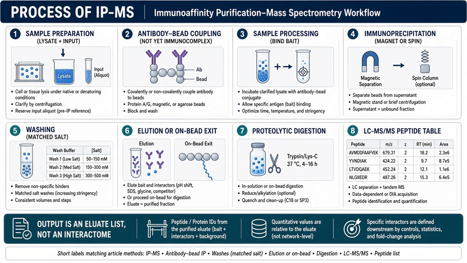
Figure 1. Sample preparation, antibody–bead coupling, binding, capture, wash, elution, digestion, and LC-MS/MS. Later software cannot rebuild a wash or a control that was not processed.
Write the capture route and the matched control before the first lysis. The spectrometer reads only what this process delivered.
Analysis Workflow
1. Sample preparation
Prepare a cell or tissue lysate that still contains the intended bait. Buffer class, salt, detergent, and mechanical shear decide whether complexes stay assembled or fall apart before the antibody sees them. Non-denaturing lysis, commonly NP-40 or a similar mild detergent with protease and phosphatase inhibitors, is the usual starting point when partners are the goal. SDS or harsh sonication can raise total protein yield and still dissolve the complex the IP was meant to keep. Collect a matched input from the same material. Input is part of the process record. It is not a substitute for an IgG or bead-only arm.
2. Immunocomplex preparation
Couple the antibody to a solid carrier such as Protein A/G magnetic beads or agarose. This step produces an antibody–bead conjugate. It is not yet an immunocomplex. The immunocomplex forms only after the antigen binds. Pre-binding the antibody to beads is one valid route. The other is to incubate free antibody with lysate and add beads afterward. Choose one route for bait and control. Switching routes between arms creates a background that looks like enrichment.
3. Sample processing
Add the antibody–bead conjugate to the lysate, or add the antibody-bound lysate to empty beads, and allow the bait to bind. Time, temperature, and lysate mass must be written and matched. A longer bait incubation than the control incubation is not a more complete IP. It is an unmatched process. Low-abundance baits need more input or a better antibody, not a silent change in this step.
4. Immunoprecipitation
Collect the resin. Magnetic beads are pulled to a magnet. Agarose is pelleted by centrifugation. The pellet, or the magnet-held slurry, is the enriched fraction. Proteins that remain here include the bait, proteins that stayed bound through lysis, proteins that bound the antibody scaffold, and proteins that bound the resin. Calling every protein in this fraction an interactor skips the later ranking. For the difference between recovering the antigen and recovering a neighborhood, Differences Between Immunoprecipitation and Co-Immunoprecipitation provides a complementary pellet-rule discussion.
5. Washing
Wash the captured material to reduce proteins that did not survive the stated salt and detergent. Wash stringency is a trade-off. A light wash leaves abundant bead binders. A harsh wash can strip true partners with the background. Write the wash, then keep it identical on the control. Raising salt only on the bait IP is not a specificity upgrade.
6. Elution
Release proteins from the resin with acid, peptide competition, or a denaturing buffer. The eluate is what digestion and MS will see if on-bead digestion is not used. Incomplete elution looks like a weak bait. It can also hide partners that remained on the bead. When elution is inefficient, on-bead digestion is an alternative. Bait and control must use the same exit.
7. Proteolytic digestion
Reduce, alkylate, and digest the eluted proteins, or the washed beads, into peptides. Trypsin is the common protease. Cleanup such as a trap or stage-tip improves peptide recovery when capacity matches input. It does not repair an unmatched IP. Peptide amount loaded on the column should be recorded. Overloading one arm and not the other recreates the process error as a file error.
8. Mass spectrometry analysis
Separate peptides by nano-LC and acquire tandem spectra, typically on an Orbitrap-class or equivalent high-resolution instrument, in data-dependent or data-independent mode. Search identifies peptide sequences and assembles protein groups. Relative intensity, label-free quantification, or a TMT reporter can be attached when the acquisition was designed for it. Mass-to-charge values are the measurement. Interaction language is not. For how that file is ranked after this process exists, How to Analyze IP Mass Spectrometry Results? provides a complementary analysis order.
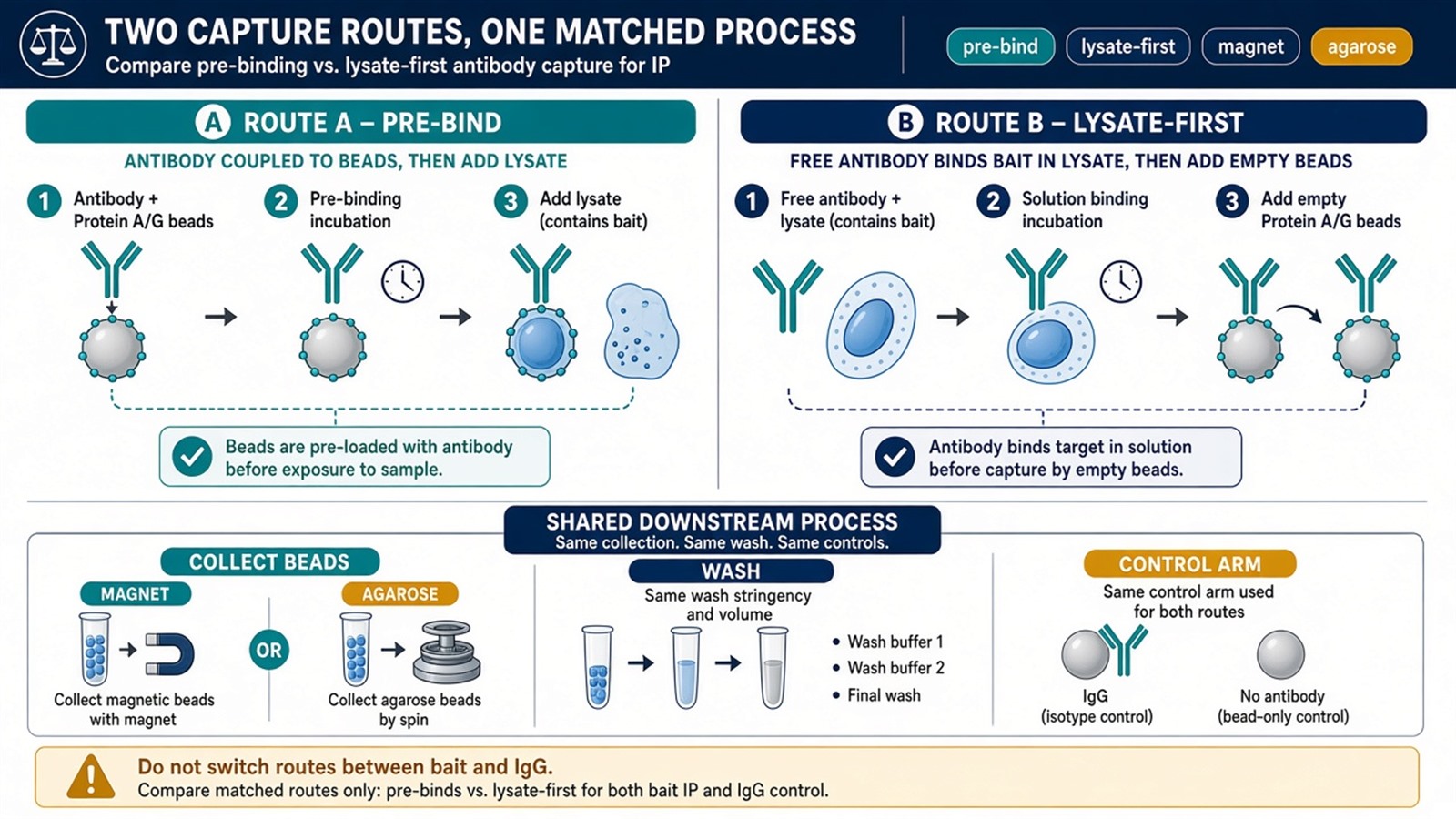
Figure 2. Pre-bind antibody to beads, or bind antibody in lysate then add beads. Magnetic collection and agarose pelleting change the physics, not the need for a matched control.
What Must Travel in Parallel
The eight steps describe one arm. A usable IP-MS process runs a matched IgG or isotype, a beads-only or empty-tag arm, and biological replicates through the same order. Adding those arms after a single bait IP cannot reconstruct the missing baseline. For which arms belong to Co-IP-MS versus in-cell crosslinking MS, What Controls Are Needed for Co-IP-MS and In-Cell Crosslinking MS? provides a complementary control-menu discussion.
A Western or equivalent bait check after elution, and before committing the rest of the peptide to a deep run, belongs in the same process. An empty bait blot is a process stop. It is not a reason to search harder.
What This Process Does Not Finish
The eight steps deliver a peptide-identified table from a written capture. They do not prove direct binding, map a contact residue, or assign pathway membership. Annotation is an overlay after enrichment is scored. Treating step 8 as quantification of every protein in the cell over-reads an enriched eluate.
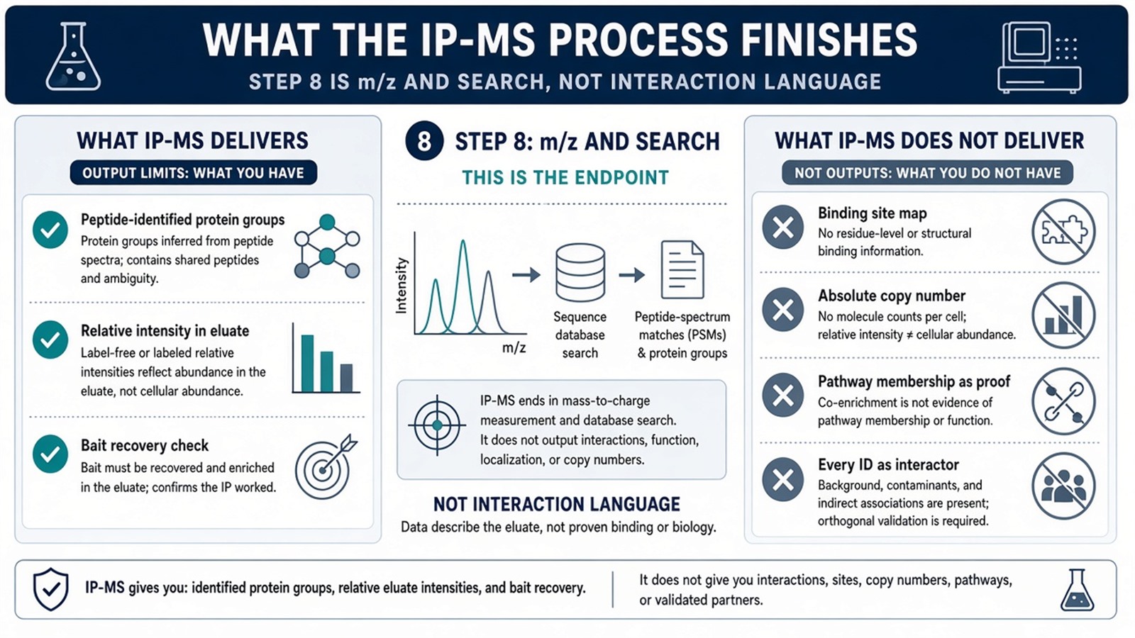
Figure 3. After digestion and LC-MS/MS the file lists protein groups and relative intensity in the eluate. Interaction, site, and pathway claims start only after bait recovery and control ranking.
Common Process Mistakes
Calling the antibody–bead conjugate an immunocomplex before antigen is bound hides the two-route decision.
Collecting the bait IP on a magnet and the IgG on agarose, or washing them differently, makes background look like a partner list.
Skipping elution QC and sending an empty bait to a long gradient wastes instrument time without creating interactors.
Reading every identification from step 8 as a co-precipitated partner skips ranking.
Experimental Limits
A correct process reduces, rather than removes, nonspecific capture. The same eight steps on a weak antibody still return bead binders. Diagnostic language sits outside ordinary research reporting unless a separately validated assay framework is in place.
Project Planning and Service Scope
A usable IP-MS plan names the capture route, the resin, the wash, the elution or on-bead exit, the matched control, and how the file will be ranked, before the first lysis. MtoZ Biolabs scopes this sequence on Orbitrap-class LC-MS/MS. Interpretability still follows the written steps, not the instrument name.
Once the process is specified, the project can be run as bait identification or as Co-IP-MS partner discovery. Researchers who need an open partner table after a defined capture can review the MtoZ Biolabs Co-IP-MS Protein Interaction Analysis Service for project-specific sample evaluation, feasibility assessment, and workflow planning.
Frequently Asked Questions
1. What is the process of IP mass spectrometry?
Eight steps: prepare lysate, couple antibody to resin, bind bait, collect the resin, wash, elute, digest, and acquire LC-MS/MS. The output is a peptide-identified eluate table.
2. Is antibody–bead coupling already an immunocomplex?
No. Coupling makes an antibody–bead conjugate. The immunocomplex forms when the antigen binds.
3. Can on-bead digestion replace elution?
Yes, when elution is inefficient. Bait and control must use the same exit. Switching exits between arms is a process error.
4. Does step 8 quantify interacting proteins?
It can report relative intensity in the eluate when the run was quantitative. It does not prove that a protein interacted, and it does not report an absolute copy number unless a standard was designed.
5. How does this process differ from Co-IP-MS analysis?
This page orders the wet-lab steps. Analysis ranks the file against a control after those steps exist. The two jobs are sequential, not interchangeable.
Reference
- Gingras A.C., Gstaiger M., Raught B., Aebersold R. (2007). Analysis of protein complexes using mass spectrometry. Nat. Rev. Mol. Cell Biol., 8, 645-654. https://doi.org/10.1038/nrm2208
- Dunham W.H., Mullin M., Gingras A.C. (2012). Affinity-purification coupled to mass spectrometry: basic principles and strategies. Proteomics, 12, 1576-1590. https://doi.org/10.1002/pmic.201100523
- ten Have S., Boulon S., Ahmad Y., Lamond A.I. (2011). Mass spectrometry-based immuno-precipitation proteomics: the choice of method matters. Proteomics, 11, 1153-1159. https://doi.org/10.1002/pmic.201000548
- Mellacheruvu D., Wright Z., Couzens A.L., et al. (2013). The CRAPome: a contaminant repository for affinity purification-mass spectrometry data. Nat. Methods, 10, 730-736. https://doi.org/10.1038/nmeth.2557
- Turriziani B., Garcia-Munoz A., Pilkington R., et al. (2014). On-beads digestion in conjunction with data-dependent mass spectrometry: a shortcut to quantitative and dynamic interaction proteomics. Biology, 3, 320-332. https://doi.org/10.3390/biology3020320
