



Co-IP detects a protein–protein interaction only as co-recovery. An antibody occupies an epitope on the bait. Protein A or Protein G on beads occupies the Fc. Proteins that remain attached to the bait through lysis and wash travel with that immune complex onto the resin, are eluted, and become a Western band or a peptide identification. Co-immunoprecipitation (Co-IP) does not measure the contact surface, a binding constant, or a live-cell distance. The mechanism of detection is this conversion of a wash-stable association into a reporter signal. Calling the method immunoprecipitation, or calling the signal a direct interaction, collapses that conversion.
The original sections below are kept because they are the detection chain: epitope binding, immune-complex assembly, bead separation, elution and readout, then controls that say which proteins were not explained by Fc or resin. For the three events named without this detection chain, Principle of Co-Immunoprecipitation in Protein Interaction Analysis provides a complementary principle discussion.
Researchers who already have an IP-grade antibody and a pair they want to detect by this mechanism can also review the MtoZ Biolabs Co-Immunoprecipitation Protein Interaction Analysis Service for project-specific feasibility and analysis planning.
Specific Antibody-Antigen Binding
Detection starts with an epitope that is accessible in the lysate. The antibody may be a monoclonal, a polyclonal, or a recombinant reagent. Origin in mouse or rabbit is not the mechanism. Epitope access is. A denatured blot epitope can be invisible in a native lysate. A buried interface epitope can be invisible when the partner is bound. Strong affinity raises occupancy of the bait. It does not raise specificity of the later pellet. Proteins that share a related epitope, or that stick to the antibody scaffold, enter the same chain. Specificity of the antibody–antigen pair is necessary for bait capture. It is not a filter for partners.
Formation of the Immune Complex
Once the antibody is bound, the immune complex is the bait, the antibody, and whatever still occupies the bait. Partners are not recognized by the antibody unless they share the epitope. They remain if their off-rate is slower than the lysis and incubation time in that buffer. Mild detergent and 4 °C slow dissociation. They do not preserve every weak pair, and they do not prevent post-lysis contacts from forming. The mechanism of “detecting an interaction” at this stage is occupancy of the same complex, not proof that the contact existed in the intact cell.
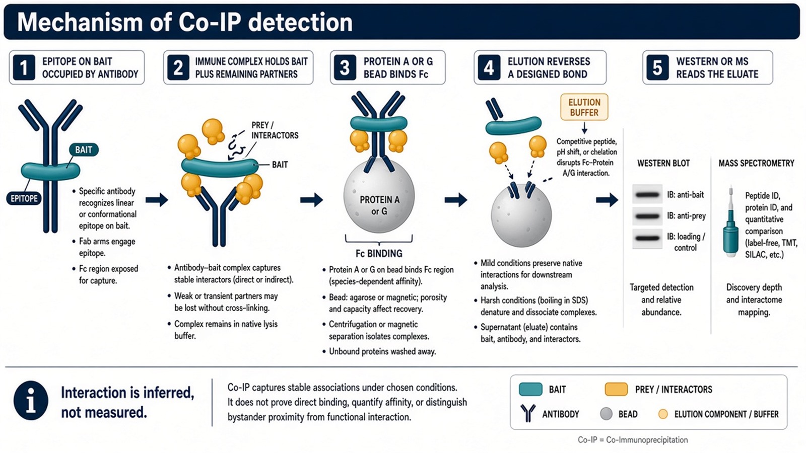
Figure 1. Epitope capture, immune-complex occupancy, Protein A/G collection, elution, then Western or MS. The interaction itself is not the measured quantity.
Detect co-recovery first. Infer an interaction only after a matched control and a written wash.
Separation of the Immune Complex
Protein A (from Staphylococcus aureus) and Protein G (from Streptococcus) bind immunoglobulin Fc. They are two bacterial proteins with different species and subclass preferences, not one reagent. Beads coated with A, G, or A/G collect the immune complex. Magnetic collection and centrifugation change the physics. They do not change what is bound. A secondary antibody is not required for this step. It is an optional bridge when the primary antibody binds A/G poorly. Adding a secondary reagent adds another scaffold that can collect background.
The separated slurry holds four classes of protein: the bait, wash-stable partners, antibody-scaffold binders, and resin binders. Separation is enrichment. It is not purification of an interaction.
For how IP and Co-IP judge this slurry differently, Differences Between Immunoprecipitation and Co-Immunoprecipitation provides a complementary pellet-rule discussion.
Elution and Analysis
Elution reverses one or more of the designed bonds. Low pH or peptide competition can release antigen from antibody. High salt or detergent can loosen partners first. SDS releases almost everything, including antibody chains. The elution chemistry therefore decides what the detector is allowed to see. Incomplete elution looks like a missing interaction.
Western blotting detects nominated chains with a second antibody after SDS-PAGE. It answers whether those chains were in the eluate. Mass spectrometry detects peptides from protein groups that were abundant enough to identify. Neither detector measures the interface. A band or an ID is the reporter. The interaction is the inference. For the bench order that delivers this eluate, Workflow of Co-Immunoprecipitation for Protein Interaction Analysis provides a complementary workflow. For an open list, How to Combine Co-IP with Mass Spectrometry for Protein Interaction Analysis? provides a complementary coupling discussion.
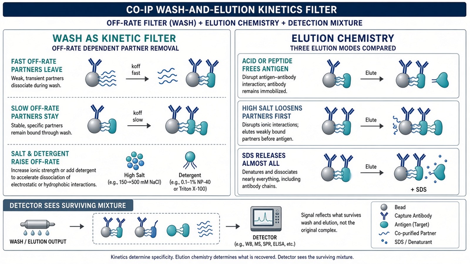
Figure 2. Partners with a faster off-rate leave during wash. Acid, peptide, or SDS elution reverse different bonds. The detector reads the surviving mixture, not the original contact.
Control Experiments and Data Analysis
A non-immune isotype matched by species, subclass, and micrograms estimates scaffold and Fc background. Beads-only estimates resin background. Empty-tag estimates handle binders when the bait is tagged. These arms are part of the detection mechanism. Without them, the reporter cannot say which of the four protein classes produced the band. Downstream MS can quantify relative intensity in the eluate. It does not convert an unmatched IP into a specific interaction. For ranking after the file exists, How to Analyze IP Mass Spectrometry Results? provides a complementary analysis order.
What This Mechanism Does Not Detect
The contact residue, the buried surface, and the on- and off-rates in the living cell are outside the reporter. Reciprocal Co-IP repeats the same mechanism from the other epitope. Crosslinking MS, SPR, or a structure is a different measurement. Treating a successful bait blot as detection of every protein in the pellet skips the conversion step.
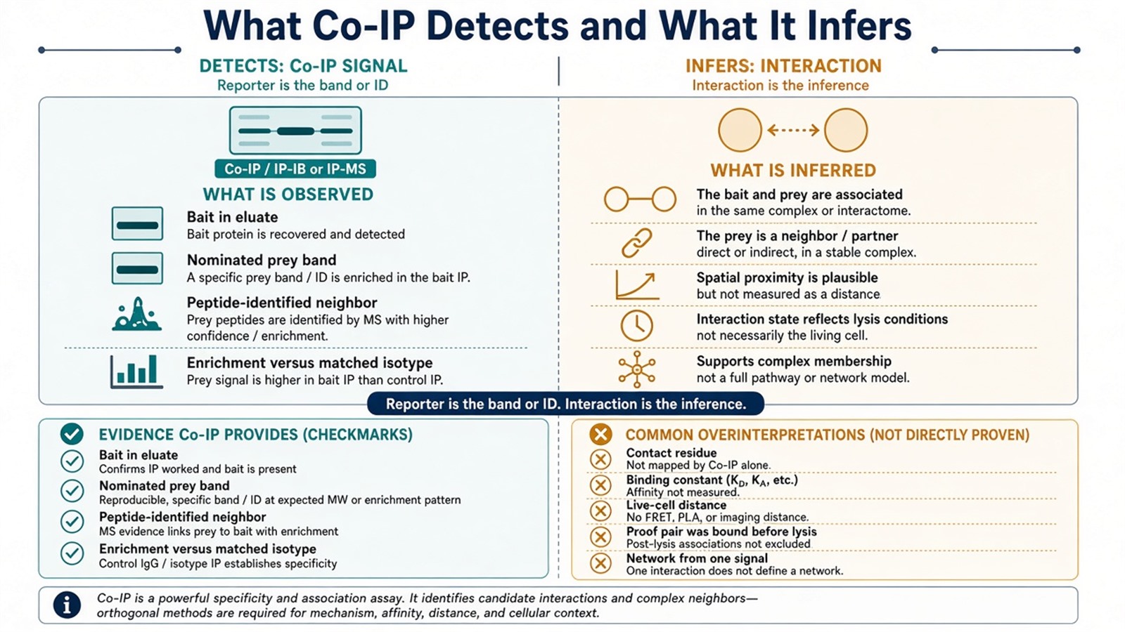
Figure 3. Detected: bait in the eluate, a nominated prey band, or a peptide-identified neighbor. Not detected: the interface, a binding constant, or proof that the pair was bound before lysis.
Experimental Limits
The mechanism reduces a lysate to what the epitope, the Fc binder, and the wash leave behind. It does not remove background, and it does not restore a complex that lysis already opened. Diagnostic language sits outside ordinary research reporting unless a separately validated assay framework is in place.
Project Planning and Service Scope
A usable detection plan names the epitope, the A versus G resin, the wash as a kinetic filter, the elution chemistry, the reporter (Western or MS), and the matched control, before the first lysis. MtoZ Biolabs scopes endogenous or tagged Co-IP, and Co-IP-MS, on that written chain. Interpretability still follows the conversion from occupancy to reporter, not the instrument name.
Once the mechanism is specified and the reporter is an open list, the project can be scoped as Co-IP-MS. Researchers who need LC-MS/MS after a defined detection chain can review the MtoZ Biolabs Co-IP-MS Protein Interaction Analysis Service for project-specific sample evaluation, feasibility assessment, and workflow planning.
Frequently Asked Questions
1. How does Co-IP detect a protein–protein interaction?
It converts a wash-stable association into a reporter signal. The antibody captures the bait, beads collect the Fc, partners that remain are eluted, and Western or MS reads the eluate.
2. Is the mechanism the same as IP?
The first two designed bonds are the same. Co-IP keeps lysis and wash in a range meant to retain neighbors and then asks about those neighbors. IP asks whether the bait was recovered.
3. Do Protein A and Protein G work by the same mechanism?
Both bind Fc. They prefer different species and subclasses. Choosing the wrong one lowers bait occupancy and looks like a missing interaction.
4. Does elution detect the interaction mechanism?
No. Elution reverses a designed bond so that a detector can see the surviving proteins. The interface is not measured.
5. Why is a control part of the detection mechanism?
Because the reporter cannot tell bait-dependent occupancy from Fc or resin binding unless a matched arm was processed.
Reference
- Harlow E., Lane D. (1999). Using Antibodies: A Laboratory Manual. Cold Spring Harbor Laboratory Press.
- Lin J.S., Lai E.M. (2017). Protein-protein interactions: co-immunoprecipitation. Methods Mol. Biol., 1615, 211-219. https://doi.org/10.1007/978-1-4939-7033-9_17
- Phizicky E.M., Fields S. (1995). Protein-protein interactions: methods for detection and analysis. Microbiol. Rev., 59, 94-123. https://doi.org/10.1128/mr.59.1.94-123.1995
- ten Have S., Boulon S., Ahmad Y., Lamond A.I. (2011). Mass spectrometry-based immuno-precipitation proteomics: the choice of method matters. Proteomics, 11, 1153-1159. https://doi.org/10.1002/pmic.201000548
- Gingras A.C., Gstaiger M., Raught B., Aebersold R. (2007). Analysis of protein complexes using mass spectrometry. Nat. Rev. Mol. Cell Biol., 8, 645-654. https://doi.org/10.1038/nrm2208
