



A mitochondrial proteomics design starts with the sample matrix, not with the instrument method. Whole-cell samples measure a total extract and place mitochondrion-annotated proteins inside a broader cellular map. Isolated or enriched mitochondria concentrate organelle material before digestion and shift depth toward mitochondrial-associated proteins. The claim the study needs to defend should select the matrix before the first harvest is frozen.
Mixing whole-cell lysate in one arm with enriched mitochondria in another creates preparation differences that can mimic biology. Lock one starting material across every comparison group, then plan amount, enrichment chemistry, and biological replicates from that single choice.
What Each Starting Material Measures
Whole-cell samples are processed as a total cellular proteome, capturing proteins from multiple cellular compartments, including the cytosol, nucleus, mitochondria, and other organelles. This starting material is suitable for cellular proteomics studies that aim to profile overall protein expression patterns without focusing on a specific subcellular compartment. Because proteins from different compartments are analyzed together, low-abundance proteins from specific organelles may compete with more abundant cellular proteins and can be less efficiently detected.
Isolated or enriched mitochondria are prepared through approaches such as differential centrifugation, density gradient separation, immunoaffinity enrichment, or other matched workflows. This starting material is suitable for cellular proteomics studies focusing on mitochondrial protein composition and mitochondrial-associated changes when isolation quality is appropriately controlled. The resulting dataset represents mitochondrial enrichment rather than absolute purity, and residual proteins from other compartments, such as the cytosol, endoplasmic reticulum, lysosome, or nucleus, should be evaluated using appropriate marker proteins.
For more information on how cellular samples are used for LC-MS/MS-based protein profiling and how sample selection supports different research goals, see Cellular Proteomics.
Sample-Matrix Comparison for Study Design
|
Design dimension |
Whole-cell samples |
Isolated or enriched mitochondria |
|---|---|---|
|
Claim scope |
Whole-cell or broad cellular profiling |
Organelle-focused comparison of the mitochondrial preparation |
|
Mitochondrial coverage |
Diluted by abundant non-mitochondrial protein |
Concentrated on mitochondrial material |
|
Preparation load |
Total lysate; simpler front end |
Isolation or enrichment required |
|
Typical stage |
Early exploration of a new system |
Focused organelle comparison |
|
Interpretation limit |
Weak for low-abundance mitochondrial proteins |
Enrichment profile, not absolute purity |
|
Cross-arm rule |
Same total-extract workflow in every group |
Same enrichment chemistry in every group |
A mitochondrion-annotated protein in a whole-cell table still sits in a cellular context. Reading that row as an organelle-specific finding overstates the design. If the manuscript sentence names the mitochondrion as the measured compartment, the starting material should match that scope from collection onward.
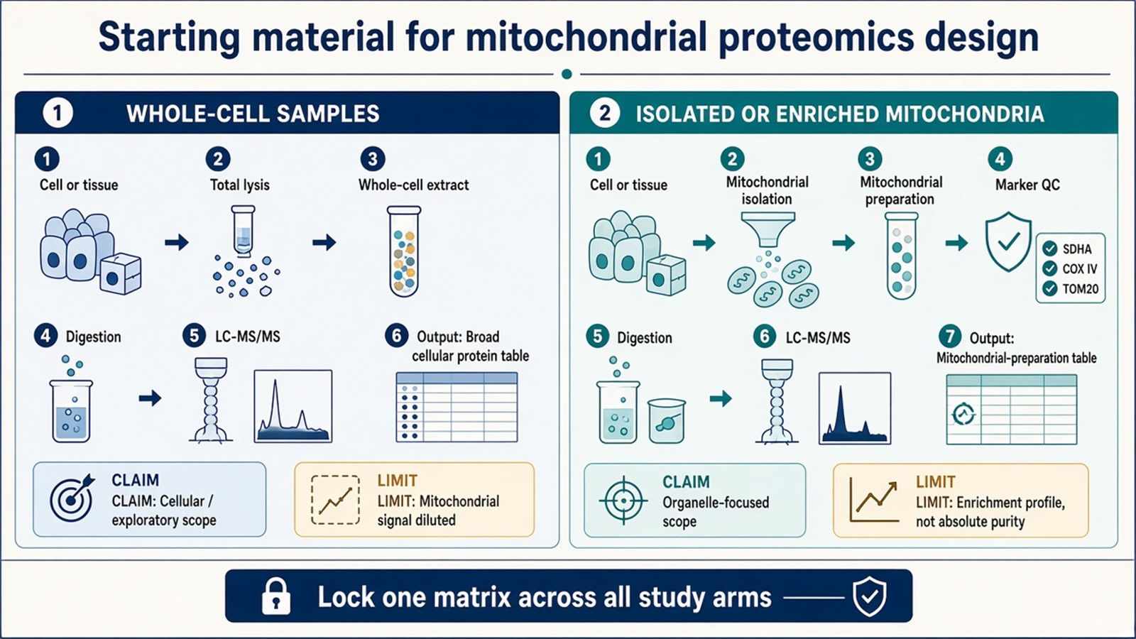
Figure 1. Whole-cell samples defend broad cellular profiling; isolated mitochondria defend preparation-level organelle focus after matched enrichment.
Starting-Material Decision Path
Write the claim in one sentence before collection. A cellular or exploratory sentence can fit whole-cell samples. A mitochondrion-specific sentence moves the design toward isolated or enriched mitochondria.
Check whether low-abundance mitochondrial proteins carry the biology. When the proteins of interest remain strong in total lysate, whole-cell work may still detect them. When the signal sits near the detection limit in bulk material, enrichment is usually required for a stable comparison.
Confirm the same matrix across all arms. Every group must share the same starting material and the same enrichment chemistry. One arm processed differently breaks cross-group interpretation.
Select the submission path and plan amount from the locked matrix.
|
Submission path |
Planning amount |
Design note |
|---|---|---|
|
Cells for in-house enrichment |
About 5 × 10^7 cells per sample |
Enrichment performed from cultured material |
|
Animal tissue for in-house enrichment |
About 200 mg per sample |
Enrichment performed from tissue |
|
Client-isolated mitochondrial protein |
At least 50 μg; 80–100 μg preferred; concentration at least 0.5 μg/μL, preferably at least 1 μg/μL |
Submitted ready for proteomics; record isolation method and buffer |
Those values are planning baselines from routine mitochondrial proteomics sample guidance. Low-yield isolations, unusual tissues, or scarce clinical material should be confirmed before collection. Apply the chosen path identically across arms.
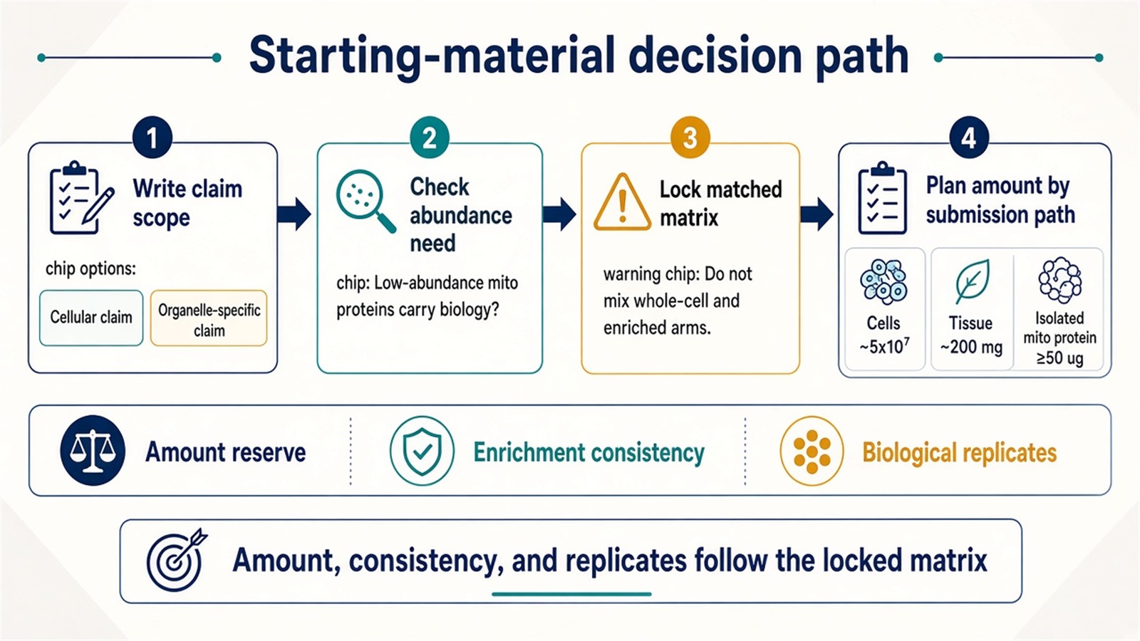
Figure 2. Claim scope and matched enrichment decide the matrix; amount planning follows after the matrix is locked.
Researchers who already have cells, tissues, or isolated mitochondria and a stated organelle claim can also review the MtoZ Biolabs Mitochondrial Proteomics Service for submission-path and amount planning.
Design Consequences: Amount, Consistency, and Replicates
Amount planning follows the path. Enrichment from cells or tissue consumes material before digestion, so each biological unit needs enough input to survive isolation and still reach LC-MS/MS. Client-isolated protein at the lower end of the recommended range may succeed, yet it leaves little reserve if a step needs repeating.
Enrichment consistency is a design rule. When groups are compared on organelle proteomes, isolation buffers, timing, and handling must be matched, or one arm can look organelle-enriched while another resembles bulk lysate. Uneven enrichment becomes a batch effect that later software cannot cleanly remove.
Replicate definition stays biological. Independent animals or independent culture flasks are units. Multiple aliquots from a single isolation are technical splits of one preparation. Technical repeats report instrument variation. They do not replace biological replication for a between-group claim.
Isolation purity, recovery, and contaminant markers should be documented with the cohort. Practical QC failure modes are reviewed in How Mitochondrial Isolation Quality Affects Proteomics Results. Control, replicate, and comparison structure for mitochondrial projects is outlined in How to Design a Mitochondrial Proteomics Study: Controls, Replicates, and Comparisons.
Sample Planning Checklist Before Collection
A usable design record names claim scope as cellular or organelle-specific, the locked starting material, the submission path, planned amount per biological unit, enrichment chemistry if used, biological replicate definition, group labels, and storage history. Collection and freezing conditions should stay consistent across arms. Unequal blood content, tissue region, or wash steps can shift the protein profile independently of the experimental factor.
Whole-cell and mitochondrial-enriched samples can appear in one project only as separate analytical blocks. They should not be merged into one statistical contrast as if they were the same matrix. Collection and documentation guidance for mitochondrial projects is summarized in Preparing Samples for Mitochondrial Proteomics Analysis.
Interpretation Limits Set by the Matrix
A whole-cell change in a mitochondrion-annotated protein reports remodeling in the total extract. It does not prove that the change occurred inside mitochondria alone. Absence of a mitochondrial protein from a whole-cell list is not proof that the organelle lacks that protein.
A mitochondrial-preparation change reports remodeling in the enriched material. It does not prove exclusive mitochondrial residence unless depletion of other fractions or an orthogonal assay supports that sentence. Enrichment improves focus; it does not create absolute purity. Pathway enrichment annotates detected names and does not demonstrate pathway activity inside mitochondria. Amount claims need matched standards outside ordinary discovery reporting.
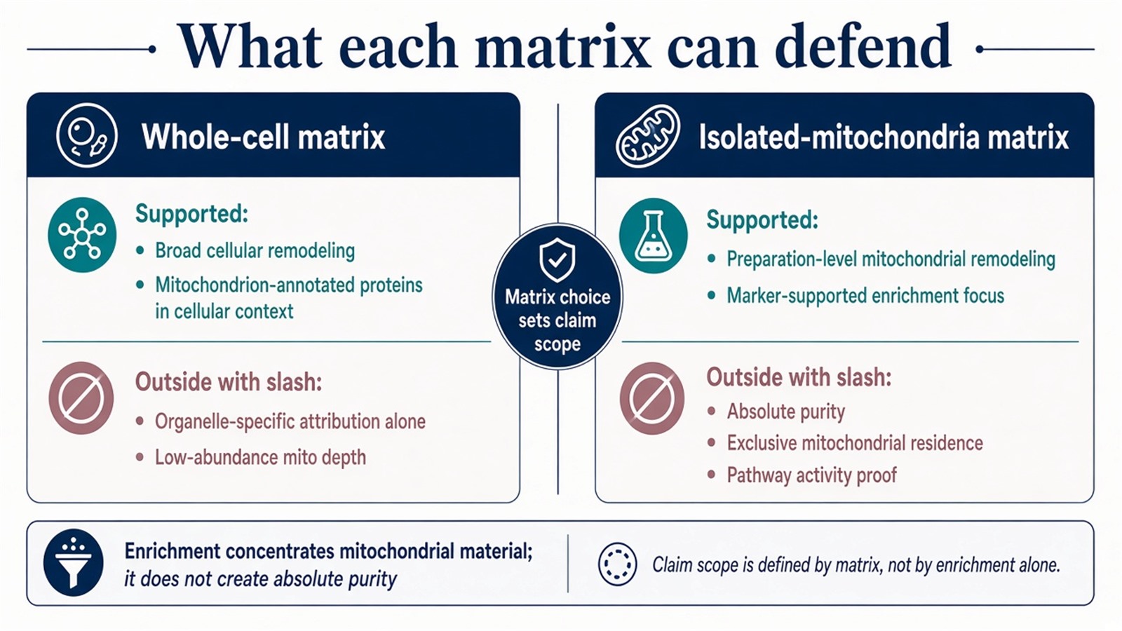
Figure 3. Each starting material defends a different claim scope; enrichment remains a profile, and exclusive localization needs additional evidence.
Frequently Asked Questions
1. Should a study use whole-cell samples or isolated mitochondria?
Use whole-cell samples for broad cellular profiling or early exploration. Use isolated or enriched mitochondria when the claim is organelle-focused and lower-abundance mitochondrial proteins must be recovered more reliably.
2. Why are mitochondrial proteins hard to see in a whole-cell lysate?
In a total lysate, mitochondrial proteins compete with the full cellular proteome. Abundant non-mitochondrial proteins dilute the organelle fraction during detection and quantification.
3. How much material is typically planned for isolated-mitochondria work?
Plan around about 5 × 10^7 cells or about 200 mg tissue for in-house enrichment, or client-isolated mitochondrial protein at least 50 μg with 80–100 μg preferred and concentration at least 0.5 μg/μL, preferably at least 1 μg/μL. Unusual matrices should be confirmed early.
4. Can whole-cell and mitochondrial-enriched samples sit in the same study?
Yes as separate analytical datasets. They should not be treated as directly comparable matrices in one contrast.
5. Does enrichment give a pure mitochondrial proteome?
No. Enrichment produces an enrichment profile. It concentrates mitochondrial and mitochondria-associated proteins when handling is matched across groups.
MtoZ Biolabs can provide proteomics study design support for whole-cell samples or isolated mitochondria, including starting-material selection, submission-path and amount review, and guidance on matched enrichment across study arms when project samples and study design are available.
If further evaluation of research claim, sample type, available amount, or preparation status is needed, project details can be submitted for a feasibility review.
Reference
- D.N. Itzhak, S. Tyanova, J. Cox, G.H.H. Borner (2016). Global, quantitative and dynamic mapping of protein subcellular localization. Elife, 5, e16950. https://doi.org/10.7554/eLife.16950
- T.P.J. Dunkley, R. Watson, J.L. Griffin, P. Dupree, K.S. Lilley (2004). Localization of organelle proteins by isotope tagging (LOPIT). Mol. Cell. Proteomics, 3, 1128-1134. https://doi.org/10.1074/mcp.T400009-MCP200
- A.I. Nesvizhskii, R. Aebersold (2005). Interpretation of shotgun proteomic data: the protein inference problem. Mol. Cell. Proteomics, 4, 1419-1440. https://doi.org/10.1074/mcp.R500012-MCP200
- Y. Zhang, B.R. Fonslow, B. Shan, M.C. Baek, J.R. Yates 3rd (2013). Protein analysis by shotgun/bottom-up proteomics. Chem. Rev., 113, 2343-2394. https://doi.org/10.1021/cr3003533
- J.R. Wisniewski, A. Zougman, N. Nagaraj, M. Mann (2009). Universal sample preparation method for proteome analysis. Nat. Methods, 6, 359-362. https://doi.org/10.1038/nmeth.1322
