



TMT-based single-cell proteomics is a multiplex relative workflow for one-cell or few-cell channels inside an isobaric set. Tandem mass tags (TMT) label single-cell digests and a higher-input carrier (booster) channel so peptides share one precursor mass on liquid chromatography-tandem mass spectrometry (LC-MS/MS), then release reporter ions for within-plex ratios (Thompson et al. 2003; Budnik et al. 2018). In project language, the carrier mainly supports identification; single-cell channels carry the quantitative claim. The method does not report mass or molar concentration per cell unless a matched stable isotope-labeled internal standard (SIS) design is added, and it does not replace bulk TMT discovery or closed-panel parallel reaction monitoring (PRM) verification. The guide below maps single-cell TMT scope, workflow, locked parameters, limits, and project fit. It is not a transcriptomics primer, not a precision-medicine catalog, and not a claim that a carrier channel cancels adsorption loss or ratio compression.
Carrier excess can improve precursor signal while distorting single-cell reporters through ion interference and compression; MS3 or synchronous precursor selection MS3 (SPS-MS3) can reduce co-isolation mixing on compatible instruments but does not fix unmatched lysis volumes (Ting et al. 2011; Specht et al. 2021). Empty or negative-control channels help detect contamination before heterogeneity is reported.
In MtoZ Biolabs quantitative offerings, TMT-based single-cell proteomics is scoped after cell type, isolation method, carrier design, channel map, relative claim, and quality control (QC) rules are written in the brief. Bulk TMT intake appears in TMT Quantitative Proteomics Analysis Service. Single-cell protein analysis framing appears in Single-Cell Protein Analysis.
What Single-Cell TMT Projects Must Cover
Four layers belong in the brief and stay separated.
Cell-intake layer names species or line, isolation platform, viability or sorting gates, and whether the unit is truly one cell or a small pool. Rare-cell slogans without isolation documentation are outside the locked record.
Carrier-and-channel layer names which tag is the carrier, approximate carrier cell equivalents, which tags are single-cell channels, and which tags are empty or control. Carrier load is a design parameter, not a biological phenotype.
Nano-prep layer names low-volume lysis, digest in sub-microliter regimes when used, low-retention surfaces, labeling completeness checks, quench, and mixing rules. Surface adsorption dominates failure modes before instrument class matters.
Acquisition-and-report layer names MS2 versus MS3 reporter strategy, search and reporter software, normalization rules for sparse single-cell matrices, and the predefined contrast or clustering claim.
Single-cell TMT is still within-plex relative quantification. Carrier-aided identification does not convert reporter height into absolute protein copies per cell.
Bulk plex design without single-cell constraints appears in Protein Quantification Technology-TMT Labeling Quantitation. This page stays on comprehensive single-cell TMT framing.
How Carrier Channels Help and Where They Hurt
Carrier strategy and quantitative claim must be planned together.
A carrier channel (often tens to a few hundred cell equivalents in published SCoPE-style designs) raises precursor abundance so more peptides can be identified while single-cell channels occupy other tags (Budnik et al. 2018; Specht et al. 2021). That does not mean single-cell reporters are immune to bleed, contamination, or compression.
If the carrier fraction is too high relative to single-cell channels, reporter interference and ratio compression can erase real cell-to-cell differences. Empty channels monitor contamination. MS3 / SPS-MS3 can improve reporter purity on compatible instruments; detail appears in TMT MS3 and SPS-MS3: How to Mitigate Ratio Compression and Enhance Quantitative Accuracy.
Label-free single-cell or ultra-low-input routes remain alternatives when chemical multiplexing or carrier design does not fit the isolation workflow. They are a different experiment class, not an automatic upgrade.
End-to-End Workflow for TMT-Based Single-Cell Proteomics
A practical single-cell TMT spine usually runs as follows.
- Freeze cell type, isolation method, biological n, and the relative or heterogeneity claim.
- Draft the channel map: carrier, single-cell tags, empty or control tags, and plex bridges if needed.
- Pilot carrier-to-single-cell load ratios; stop if empty-channel contamination or extreme compression appears.
- Lyse and digest in low-volume, low-retention conditions matched across single-cell channels.
- Label, quench, and mix; verify labeling completeness on pilot aliquots before full cohorts.
- Acquire MS2 or MS3 reporter methods with isolation settings locked from the pilot.
- Extract reporters; apply declared normalization; review missingness, empty channels, and carrier-driven outliers.
- Test only predefined contrasts or clustering plans; archive channel map and method files.
- Escalate named proteins to bulk TMT depth or PRM only when a follow-up claim is declared.
Steps 1 through 5 protect the nano-sample record. Steps 6 through 9 produce the single-cell TMT quantitative record.
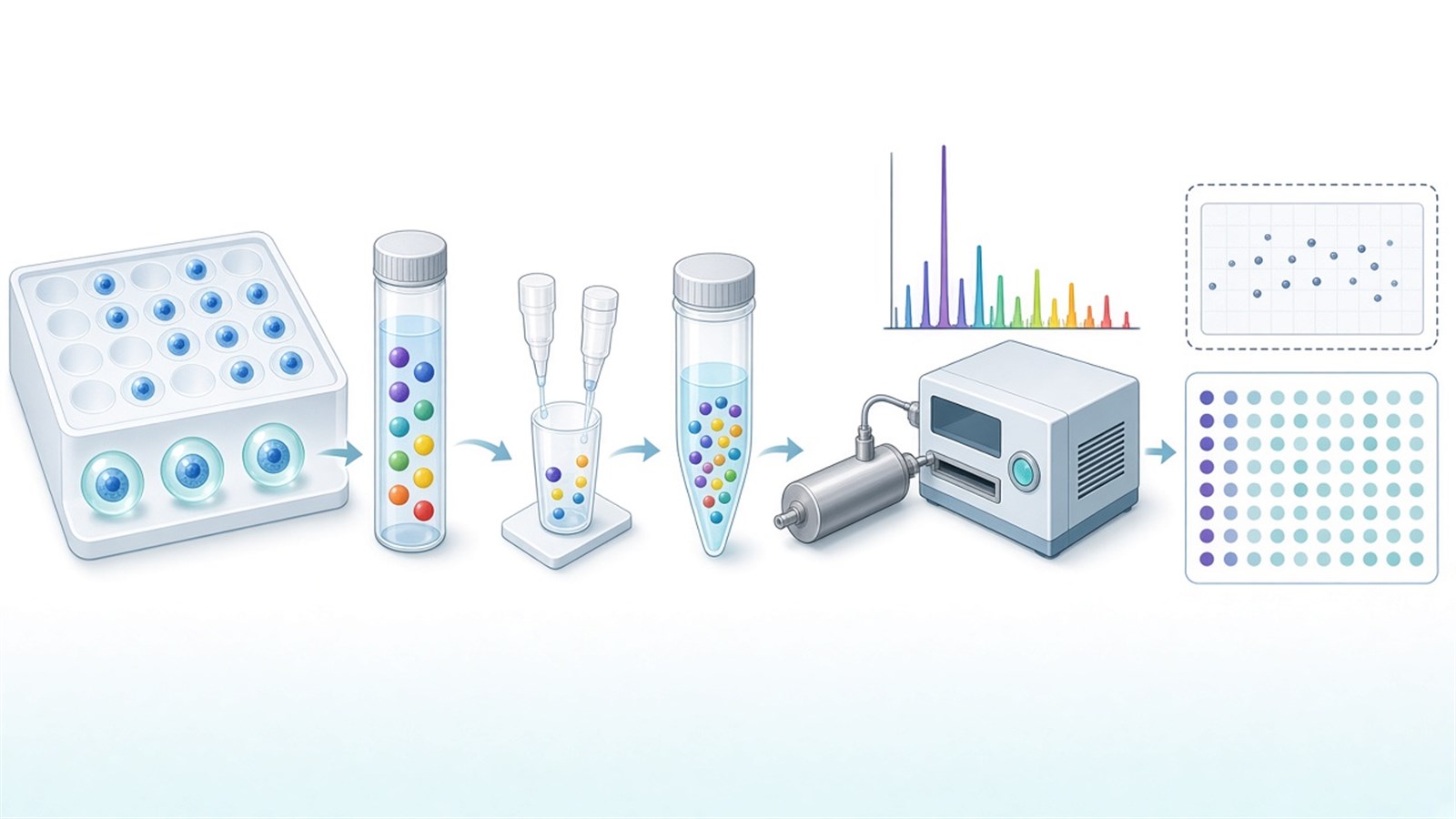
Figure 1. Single-cell TMT depends on a locked carrier map and low-loss prep before reporter differences are read as cell biology.
Parameters That Must Be Locked for Single-Cell TMT
Layer |
Parameter that must be declared |
Failure mode if ignored |
|---|---|---|
Isolation |
Platform, gate, one-cell vs pool |
Mixed cells sold as single-cell |
Carrier |
Tag and cell-equivalent load |
Identification without usable reporters |
Controls |
Empty or negative channels |
Contamination as heterogeneity |
Volume |
Lysis and digest volume regime |
Adsorption loss and dropouts |
Surfaces |
Low-retention consumables |
Trace peptide loss |
Labeling |
Completeness check and quench |
Split tagged forms at nano scale |
Isolation MS |
MS2 window or MS3 / SPS-MS3 |
Severe ratio compression |
Missingness |
Filters for sparse single-cell matrices |
Noise clusters as subtypes |
QC |
Carrier, empty-channel, and instrument QC |
Undetected prep or MS drift |
Deliverable |
Channel map and method file |
Report rebuilt after acquisition |
Limits follow the same table. Multiplex mixing reduces run variance inside a plex; it does not create microgram-like coverage from every cell. Tumor, immune, stem-cell, and drug-screening application language needs separate study design beyond the single-cell TMT table. Automated dispensers help isolation reproducibility; they do not remove the need for carrier and empty-channel design.
How to Choose TMT-Based Single-Cell Proteomics
Use input scale, carrier tolerance, claim unit, and follow-up intent together.
Project need |
Route that often fits |
Recheck before committing |
|---|---|---|
One-cell or few-cell channels, multiplex relative profiles |
TMT-based single-cell proteomics |
Force bulk TMT on sorted pools without saying so |
Sorted bulk populations, microgram input |
Standard bulk TMT |
Carrier single-cell design without true single cells |
Carrier pilots show extreme compression |
Lower carrier, add MS3, or redesign |
Raise carrier until biology disappears |
Absolute copies per cell |
SIS-capable targeted designs |
Reporter height as copy number |
Closed peptide verify after discovery |
PRM or related targeted follow-up |
Single-cell TMT as targeted assay |
Isolation not yet stable |
Fix sorting and nano-prep first |
Full plex on failing recovery |
No multiplex need, unlabeled ultra-low input |
Non-TMT low-input route |
Buy carrier chemistry without channel plan |
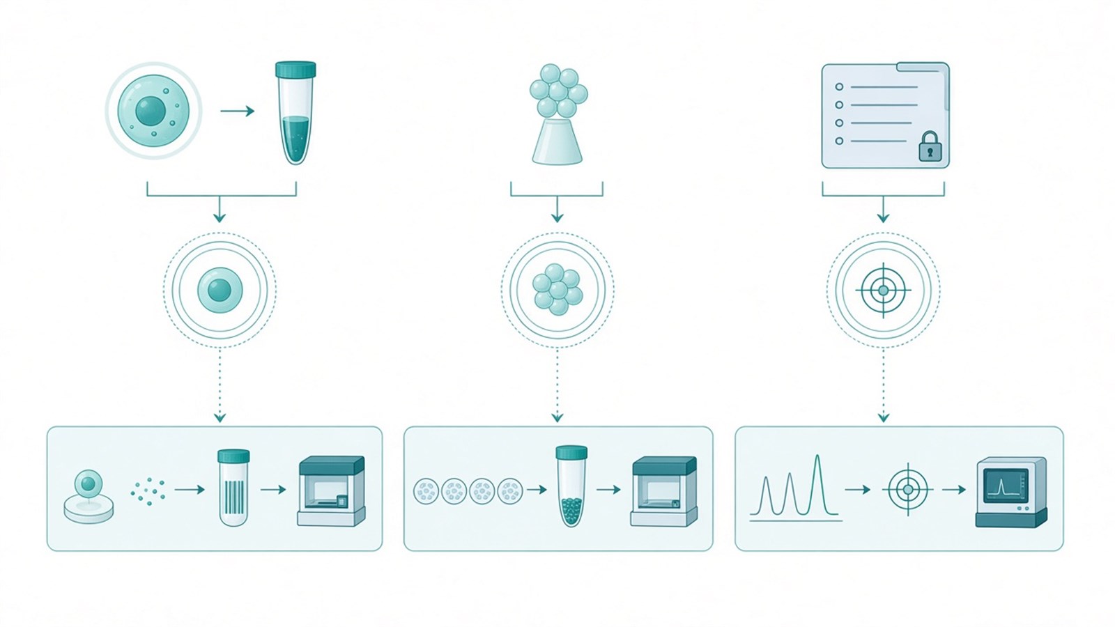
Figure 2. Choose single-cell TMT when carrier-aided multiplex relative profiles fit; keep bulk TMT or PRM when input scale or claim unit changes.
Teams with isolation documentation, carrier maps, and QC-inclusive plex plans can review MtoZ Biolabs TMT and single-cell protein analysis entries when those fields are declared in the project record.
Frequently Asked Questions
1. Does the carrier channel quantify the single cells?
No. The carrier mainly boosts precursors for identification. Quantitative claims for individual cells come from their own reporter channels after interference and missingness rules are applied.
2. Is label-free quantification always too weak for single cells?
Label-free ultra-low-input methods are a different class. TMT multiplexing with a carrier is one common strategy; it is not proof that every label-free single-cell attempt fails.
3. Can MS3 remove the need to tune carrier load?
No. MS3 can reduce co-isolation compression. Excessive carrier relative to single-cell channels can still distort reporters.
4. Are empty TMT channels optional?
They are strongly recommended. Empty or control channels help detect contamination before heterogeneity is interpreted.
5. When should projects stay on bulk TMT instead?
When the scientific unit is a sorted population with conventional protein input and single-cell isolation is not part of the claim.
MtoZ Biolabs can discuss TMT-based single-cell proteomics scoping when isolation method, carrier design, and contrast are documented.
If further evaluation of carrier ratios, MS3 options, or bulk versus single-cell fit is needed, project details can be submitted for a feasibility review.
Reference
- A. Thompson, J. Schäfer, K. Kuhn, S. Kienle, J. Schwarz, G. Schmidt, T. Neumann, R. Johnstone, A.K. Mohammed, C. Hamon (2003). Tandem mass tags: a novel quantification strategy for comparative analysis of complex protein mixtures by MS/MS. Anal. Chem., 75, 1895-1904. https://doi.org/10.1021/ac0262560
- B. Budnik, E. Levy, G. Harmange, N. Slavov (2018). SCoPE-MS: mass spectrometry of single mammalian cells quantifies proteome heterogeneity during cell differentiation. Genome Biol., 19, 161. https://doi.org/10.1186/s13059-018-1547-5
- H. Specht, E. Emmott, A.A. Petelski, R.G. Huffman, D.H. Perlman, M. Serra, P. Kharchenko, A. Koller, N. Slavov (2021). Single-cell proteomic and transcriptomic analysis of macrophage heterogeneity using SCoPE2. Genome Biol., 22, 50. https://doi.org/10.1186/s13059-021-02267-5
- L. Ting, R. Rad, S.P. Gygi, W. Haas (2011). MS3 eliminates ratio distortion in isobaric multiplexed quantitative proteomics. Nat. Methods, 8, 937-940. https://doi.org/10.1038/nmeth.1714
