



A Far-Western blot reports whether a labeled bait protein was retained on a membrane-bound prey after wash. That band is screening evidence. It is not a dissociation constant. Grayscale intensity mixes prey amount, transfer efficiency, renaturation, probe activity, and exposure time. Those variables do not rearrange into Kd. Positive and negative controls on the same membrane decide whether the overlay is interpretable. A later SPR, BLI, or ITC run on a prepared pair is what can return a biosensor or calorimetric constant. Prey load and probe activity belong in the same plan as the control lanes. The blot does not replace that step.
Researchers who already have a purified bait probe, a prey lysate or recombinant target, and a written overlay claim can review the MtoZ Biolabs Far-Western Blot Analysis Service for feasibility. Method-level choice among pair assays is covered in Measuring Protein-Protein Interactions.
A Far-Western Band Is Screening Evidence, Not a KD
Far-Western blot replaces the primary antibody of a Western blot with a labeled bait protein. Prey proteins are separated, transferred, often denatured and refolded on the membrane, then incubated with the probe (Wu et al. 2007). A chemiluminescent or fluorescent band means the probe stayed on that molecular-weight window after wash. The assay can compare several lysates or mutants on one membrane without an antibody to the prey.
The assay does not measure free and bound concentrations in solution. It does not record association and dissociation in real time. Calling the overlay a quantitative analysis of protein binding affinity collapses those two jobs. A stronger band can mean more prey was loaded, the epitope survived SDS-PAGE, or the film was exposed longer. A missing band can mean the contact needs a fold that was lost on the blot.
If the next sentence is co-recovery from a physiological lysate, use a later Co-IP rather than restating the overlay as a cellular complex. Antibody capture is scoped through the IP-Mass Spectrometry Service. If the next sentence is in-vitro retention of a soluble pair, a defined pull-down remains a separate path on the GST Pull Down Assay Service.
Positive and Negative Controls on the Same Membrane
Controls belong on the membrane that will be scored, not in a later methods note. A positive overlay uses a prey known to retain the same bait, or a recombinant fragment that still binds after transfer. A negative overlay uses an empty vector lysate, an unrelated protein at a similar load, or a bait mutant that should lose the contact. A no-prey lane and a no-probe membrane check antibody or streptavidin background when the bait is tagged or biotinylated.
A loading blot with an antibody to the prey, or a stain of a sister membrane, shows whether a weak overlay signal is low prey or failed binding. Do not normalize a saturated band to a loading control and treat the ratio as affinity. Saturation means the detector left the linear range.
Block and wash must be identical across lanes. Probe concentration should be high enough to see the positive control and low enough that the negative control stays dark. One universal probe dilution does not exist. The positive and negative pair sets the usable window for that bait.
Sensitivity and Conformation Limits After Transfer
Detection is limited by prey abundance, transfer, and whether the binding surface survives SDS-PAGE. Planning amounts for lysates are typically 1x10^7 cells per sample or at least 50 mg of tissue. Low-abundance prey can sit below the chemiluminescence floor even when the pair is real in solution. Weak or rapidly exchanging contacts are often lost during wash. Those negatives are sensitivity limits, not proof of non-binding.
Denaturation is the second limit. Many pairs need a folded domain, a modification, or a cofactor that the gel removes (Wu et al. 2007). Renaturation on the membrane can restore some contacts and can also create non-native overlays. Native PAGE avoids SDS but changes resolution and still immobilizes the prey. An overlay that depends on native fold should state the gel chemistry with the claim.
Do not describe the method as generally more sensitive than a biosensor. The blot is convenient for many lanes. It is a poorer detector for a scarce, conformation-dependent, or transient pair. Those pairs should move to a solution assay rather than be forced through longer exposure.
The comparison below keeps overlay screening and later affinity measurement on separate tracks.
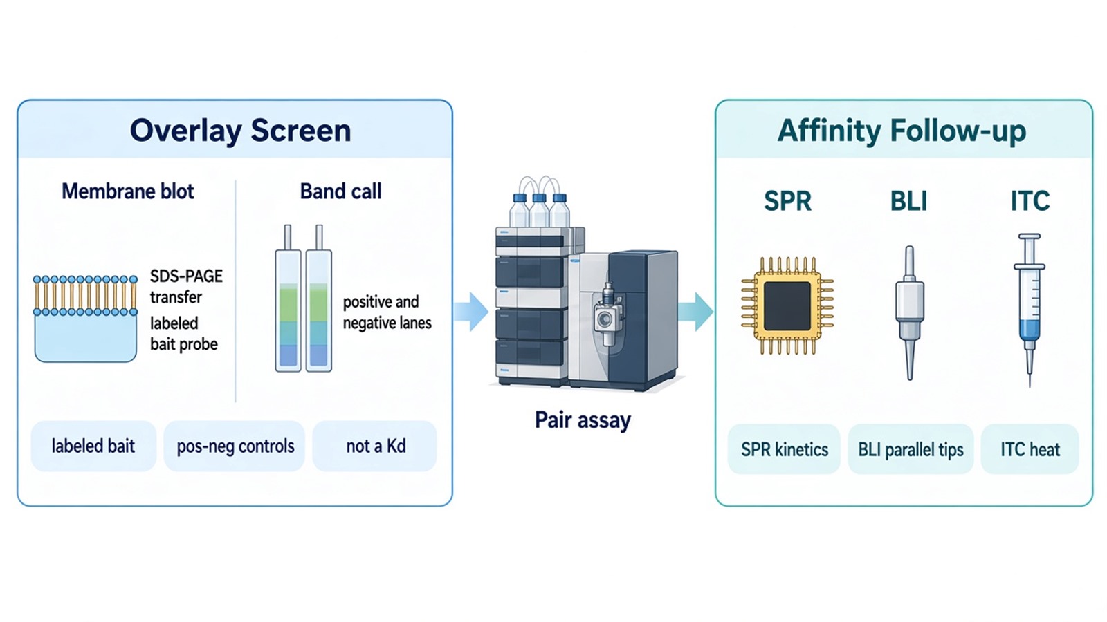
Figure 1. Far-Western Screening vs Quantitative Affinity Assays.
Why Band Grayscale Cannot Be Converted to Kd
Kd is a concentration ratio of free components at equilibrium. A Far-Western blot does not measure those concentrations. Pixel intensity is a detector response to residual probe on a solid support. Building a "standard curve" from serial probe dilutions still reports how much probe stuck to that blot under that exposure. It does not report the solution equilibrium of the pair.
Pooling several blots into a binding curve does not repair the missing free-ligand term. Exposure time, transfer bubbles, and local blocking create lane-to-lane offsets larger than most affinity ranks worth publishing. Semi-quantitative comparison is allowed only when load, transfer, probe batch, and imaging are matched, and when the claim is "stronger or weaker than the sister lane," not a nanomolar constant.
Kinetics cannot be read from a washed, dried membrane. There is no association window and no dissociation window once the blot has been developed.
SPR, BLI, and ITC as the Quantitative Follow-Up
When the publication sentence needs ka, kd, or KD, move the named pair off the membrane. SPR immobilizes one partner and records association and dissociation of the other. Planning amounts are typically 50 to 100 ug of protein at 90 percent purity or higher. Intake is described on the Surface Plasmon Resonance Technology page. If the method is still open, SPR is the usual first affinity route.
BLI uses a dipping sensor and can run several tips in parallel. Sensitivity is lower than SPR, and weak or low-mass analytes often fail. Scarce samples that need a tighter kinetic window should not stay on BLI after a noisy overlay. The Biolayer Interferometry (BLI) Service is the parallel-sensor option when both proteins are available and the signal is expected to be large.
ITC titrates one partner into the other in solution and reports heat, an equilibrium constant, and stoichiometry when both proteins are soluble and unlabeled. It does not use a membrane and it does not return ka and kd. Confirm amounts and buffer match at intake through the Isothermal Titration Calorimetry (ITC) Analysis Service. Do not treat an ITC constant as cellular affinity.
Claim the blot is asked to support |
Far-Western blot can supply |
Quantitative follow-up |
|---|---|---|
Probe retained on this prey band |
Overlay versus controls |
Not required for a screen |
Mutant weaker than wild type on one membrane |
Matched-lane rank |
SPR if a constant is required |
Solution KD, ka, or kd |
No |
SPR or BLI |
Heat and stoichiometry in solution |
No |
ITC |
Cellular complex membership |
No |
Co-IP or IP-MS |
The table is a handoff rule, not a ranking of imaging dyes.
Ship lysates or tissue snap-frozen on dry ice, plus the purified bait sequence, label chemistry, and the control set. State whether the gel will be SDS-PAGE or native. Mechanism-level pair work after a named contact is discussed in Mechanism of Protein-Protein Interaction. Broader pair intake can start from Protein-Protein Interaction Analysis.
The diagram below links bait, controls, prey load, and the affinity transfer to an overlay request.
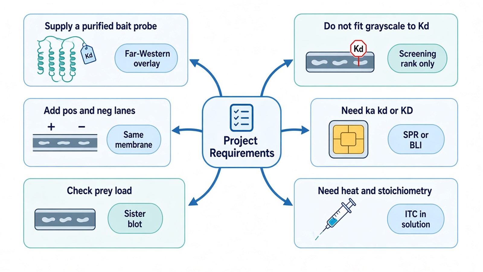
Figure 2. Matching Project Requirements With Far-Western Screening or Affinity Follow-Up.
Deliverables for the overlay typically include the developed membrane image, the loading or sister-membrane blot, and a lane-level call versus the stated controls. Deliverables do not include a Kd fitted from grayscale, a kinetic constant, or a claim of cellular binding. To start intake, send the bait, prey format, control plan, and whether SPR, BLI, or ITC should follow to info@MtoZ-Biolabs.com or use the site inquiry form so the screen and the affinity step can be split before shipment.
Frequently Asked Questions
1. Can Kd be calculated from Far-Western blot band intensity?
No. Pixel values report residual probe on a membrane. Kd requires free and bound concentrations in a controlled solution assay such as SPR, BLI, or ITC.
2. What controls should travel with the overlay run?
Include a known positive prey or fragment, a negative lysate or mutant, a no-probe or tag-only detection lane, and a loading blot for the prey. Score the overlay only when that set is on the same run.
3. Why can a real pair be invisible on the blot?
The prey may be below the imaging floor, the contact may need a fold or modification lost in SDS-PAGE, or the wash may have removed a weak overlay. Those limits are reasons to move the pair to SPR or ITC, not to raise exposure until the negative lane appears.
4. When is a lane-to-lane intensity comparison still useful?
When load, transfer, probe, and imaging are matched, a mutant-versus-wild-type rank on one membrane can support a screening claim. It still does not become a nanomolar constant.
5. How should SPR, BLI, and ITC be chosen after a positive overlay?
Choose SPR first when kinetics are required or the method is still open. Choose BLI when several tips will be run and the pair is expected to give a strong sensor signal. Choose ITC when both proteins are soluble and unlabeled and the next sentence is heat and stoichiometry.
Researchers can keep Far-Western blot as an overlay screen, then confirm bait, controls, and the affinity follow-up through MtoZ Biolabs Far-Western Blot Analysis Service or Surface Plasmon Resonance Technology.
Reference
- Y. Wu, Q. Li, X.Z. Chen (2007). Detecting protein-protein interactions by Far western blotting. Nat. Protoc., 2, 3278-3284. https://doi.org/10.1038/nprot.2007.459
