



A mechanism of protein-protein interaction is a testable account of how a named pair recognizes one another. The account should state which domain or motif is required, how tight the association is under defined conditions, whether binding changes conformation, and which residues can be mutated to weaken the readout. Partner lists from Co-IP, AP-MS, pull-down, or proximity labeling do not supply that account. Screening reports co-recovery, retention, or proximity under the submitted experimental workflow. Mechanism work starts after a pair is named and then asks for recognition logic, affinity, interface constraints, and a mutation or structural experiment that can fail.
Routine network screens remain useful for candidate generation. They should not be restated as interface maps or as proof that a domain drives function. Researchers who already have a purified pair, a domain hypothesis, and a written claim can review MtoZ Biolabs Protein-Protein Interaction Analysis for feasibility. Method-level screening choices are covered in Measuring Protein-Protein Interactions.
What Mechanism Evidence Is Distinct From Interaction Screening
Interaction screening answers who associates with a bait under the chosen lysis, wash, or labeling conditions. A mechanism claim answers how a specified pair engages. The two tasks share protein names, not evidence type. A co-enriched protein can be a bridging partner, a bead binder, or a true contact. A retained pull-down band can reflect an in vitro surface that does not exist in the cell.
Mechanism planning should write four items before samples ship. The recognition hypothesis names a domain, linear motif, or predicted surface. The affinity question states whether a kinetic or equilibrium value is required. The interface question states whether peptide-level protection, residue distance constraints, or an atomic model is the next need. The validation question states which mutant, truncated construct, or independent structure will be used to test the hypothesis.
Yeast two-hybrid, Co-IP, protein arrays, and complementation imaging can populate or confirm a pair list. They do not explain hydrophobic packing or allosteric rearrangement. Physical descriptions remain models until affinity, interface, and mutation data are collected on the same pair.
Molecular Recognition: Domains, Motifs, and Specificity
Molecular recognition is the opening mechanism layer. Many pairs engage through a folded domain, a short linear motif, or a composite surface assembled only after modification. Complementary shape and chemistry at the contact surface set specificity, while nearby residues can contribute little binding energy (Jones and Thornton 1996). A domain truncation that loses retention supports a recognition hypothesis. A truncation that also unfolds the bait does not.
Domain tests should keep the rest of the protein as intact as the claim allows. Isolated domains can bind in vitro and still miss a cofactor, membrane, or competing partner. Motif peptides can occupy a pocket without proving that the full-length protein uses the same register. Post-translational marks can create or hide a surface, so the modification state should be declared.
Defined-pair pull-down fits when a recombinant bait or GST fusion can be immobilized and a named prey is available. Retention of the wild-type prey versus a domain-deleted or motif-mutated prey is the typical comparison. Intake for that in vitro pair test is scoped through the GST Pull Down Assay Service. The report can support domain dependence under the submitted buffer. It does not assign a cellular occupancy or a residue-level interface.
Affinity and Kinetics as Mechanism Readouts
Affinity and kinetics convert a qualitative pair into a quantitative binding model. SPR records association and dissociation of a prepared analyte on an immobilized ligand and can report equilibrium or kinetic constants when the surface and activity are controlled (Homola 2008). BLI and MST can serve a similar role when the pair and buffer fit those formats. The fitted KD belongs to the chip, the immobilization chemistry, and the analyte activity used in that run.
Affinity is mechanism evidence only when the proteins match the biological claim closely enough. A domain fragment can bind more tightly than the full-length pair. Dense immobilization can create avidity that is absent in solution. Aggregated analyte can inflate apparent affinity. The report should keep the KD attached to those conditions and should not restate it as cellular occupancy.
SPR intake typically needs purified ligand and analyte, a stated immobilization route, and a regeneration plan. Ligand concentration guidance on common chips is often in the microgram-per-milliliter range. Real-time binding can be scoped through the Surface Plasmon Resonance Technology service. A kinetic shift after a designed mutation is stronger support than a single wild-type KD.
Conformational Change and Interface Mapping
Interface work asks which regions change solvent access or satisfy a distance constraint when the pair is mixed. HDX-MS compares deuterium uptake of free and bound states at peptide resolution. Slower exchange can reflect a contact, a distal allosteric tightening, or both (Engen 2009). Faster exchange can reflect opening remote from the contact. Protection maps are mechanism clues, not residue-level contact lists.
XL-MS captures residue pairs that lie within the reach of a chosen crosslinker. The output constrains architecture and nearby residues. It does not produce an atomic structure on its own (Leitner et al. 2016). Crosslink absence can mean the residues were not close, not reactive, or not recovered. Crosslink presence still needs an independent model if a docked interface is required.
HDX-MS and XL-MS fit after a pair is purified enough for controlled mixing and a recognition hypothesis exists. They fit poorly as screens of unknown partners. Peptide-level protection can be reviewed through the [Protein-Protein Interactions Characterization Service |
HDX-MS](https://www.mtoz-biolabs.com/protein-and-protein-binding-site-analysis.html). Distance constraints can be reviewed through the [Crosslinking Mass Spectrometry Service](https://www.mtoz-biolabs.com/crosslinking-mass-spectrometry-service.html). |
|---|
Domain tests, affinity values, and interface maps answer different mechanism questions. The comparison below groups those layers without treating any one readout as a complete mechanism.
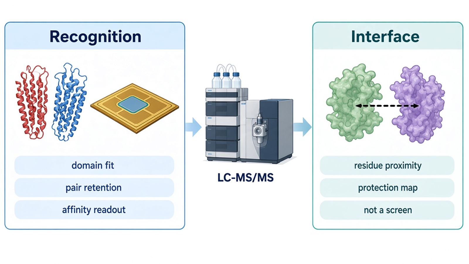
Figure 1. Domain Recognition vs Affinity Readout vs Interface Evidence.
Mutagenesis and Structural Experiments That Test the Claim
A mechanism claim is incomplete until a designed change can reduce the signal predicted by the model. Site-directed mutants at a predicted hot-spot or motif should be compared with a surface control mutant that is not expected to matter. Loss of pull-down retention, a weaker SPR signal, or a local HDX change that tracks the mutation all strengthen the account. Global unfolding or aggregation weaken the interpretation.
Structural experiments sit after the same logic, not at the start of screening. Crystallography or cryo-EM can supply an atomic interface when the complex is homogeneous enough. HDX-MS and XL-MS can precede or follow those methods as solution constraints. Mutagenesis should be planned from the peptide-level or structural map, then returned to a binding assay on the same pair.
Mechanism question |
Experiment often scoped |
Typical deliverable |
What the result does not prove |
|---|---|---|---|
Which domain or motif is required? |
Truncation or motif mutant pull-down |
Retention of named prey vs control constructs |
Cellular function or residue contacts |
How tight is the prepared pair? |
SPR, BLI, or MST |
Kinetic or equilibrium values under stated conditions |
In-cell occupancy or an interface map |
Which peptides change protection? |
HDX-MS of free vs bound states |
Peptide-level deuterium uptake differences |
Atomic contacts or a single binding pose |
Which residues satisfy a distance limit? |
XL-MS |
Crosslinked residue pairs |
A refined atomic structure |
Does a predicted residue carry the effect? |
Site-directed mutant plus a binding assay |
Loss or shift of the chosen readout |
That no other surface contributes |
The table is a planning aid, not a fixed order. Affinity can be measured before interface mapping when the pair is already clean. Interface mapping can come earlier when a mutation set is still undefined. Screening-scale Co-IP or array work should stay outside this table unless a named pair has already been isolated.
A mutant that fails every orthogonal assay is more informative than a long list of untested contacts. If only a lysate bait is available, mechanism work should pause at pair confirmation and return after purification or construct design.
Samples, Deliverables, and Feasibility Intake
Mechanism projects usually require purified or well-characterized proteins rather than crude IP eluates. Declare constructs, tags, estimated purity, buffer, detergent, and whether the pair stays soluble when mixed. For SPR, state preferred immobilization and any His, GST, biotin, or antibody handle. For HDX-MS, keep detergent load compatible with LC-MS and report quench constraints during intake. For XL-MS, state buffer amines that can quench the reagent and whether an existing structural model will be used.
Intake is faster when the mechanism question is mapped to an experiment before shipment. The diagram below links common claims to domain tests, affinity work, HDX-MS, XL-MS, mutagenesis, and structural routes used in feasibility review.
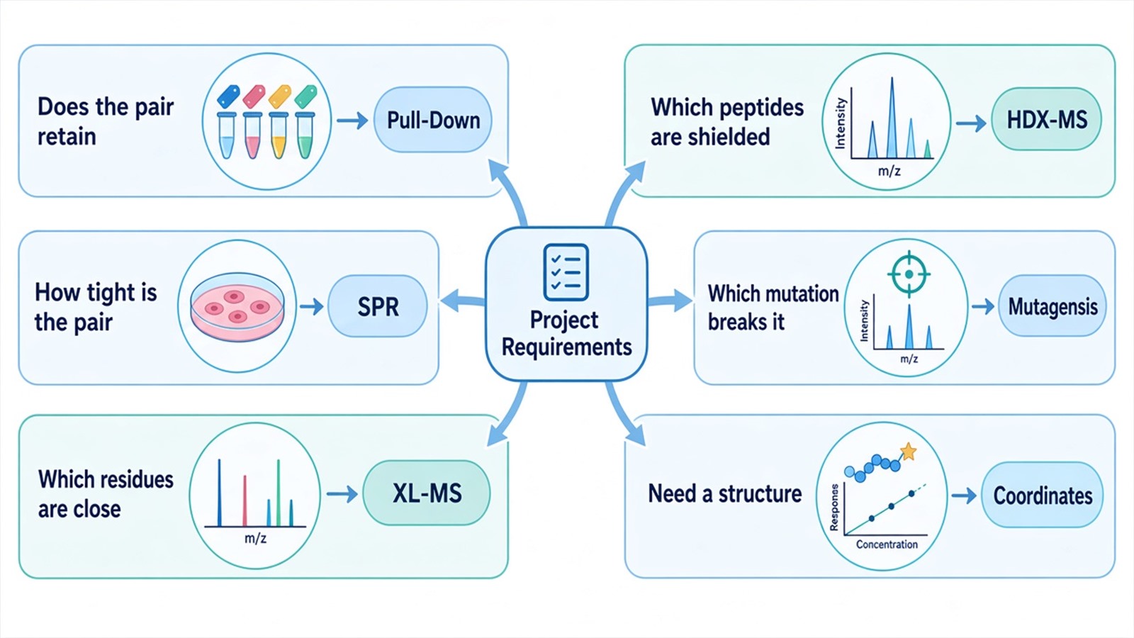
Figure 2. Matching Mechanism Questions With PPI Experiments.
Deliverables depend on the scoped route. Pull-down reports can include gel or MS identification of retained prey versus control constructs. SPR reports can include sensorgrams and fitted constants. HDX-MS reports can include peptide coverage and uptake differences. XL-MS reports can include residue-pair tables. Deliverables do not automatically include a docked atomic model, a cell assay, or a claim that screening hits share the same interface.
To start intake, send the named pair, construct map, available purity, and the mechanism claim to info@MtoZ-Biolabs.com or use the site inquiry form so experiment choice can be confirmed before shipment.
Frequently Asked Questions
1. Can a Co-IP or AP-MS partner list be used as mechanism evidence?
A partner list supports candidate selection. Mechanism evidence begins when a named pair is tested for domain dependence, affinity, interface constraints, or a designed mutant. Co-recovery can be indirect, so the list should not be rewritten as an interface model.
2. How much protein is typically needed to start SPR or HDX-MS planning?
Amounts depend on purity, immobilization or labeling format, and the number of conditions. SPR ligand guidance on common chips is often in the low-microgram-per-milliliter range. HDX-MS needs enough material for free and bound time courses. Exact input should be confirmed after the claim is set.
3. Does an HDX protection change identify the contact residues?
Protection can mark a contact, a distal conformational tightening, or both. Peptide-level maps guide mutagenesis. Residue assignment still needs mutants, crosslinks, or an independent structure.
4. When should site-directed mutagenesis be run before structural MS?
Mutagenesis can precede structural MS when a motif or predicted hot-spot is already specified and a pull-down or SPR assay can test it. Structural MS is more useful earlier when the surface is unknown and a peptide-level map is needed to choose mutants.
5. What should a feasibility email include for mechanism-of-interaction work?
Include the named pair, domain or residue hypothesis, available constructs, estimated purity and buffer, and whether the next need is affinity, HDX-MS, XL-MS, or mutant comparison. Feasibility review checks that package against the claim before preparation is confirmed.
Researchers can align domain, affinity, interface, and mutation tests, then confirm constructs through MtoZ Biolabs Protein-Protein Interaction Analysis intake.
Reference
- S. Jones, J.M. Thornton (1996). Principles of protein-protein interactions. Proc. Natl. Acad. Sci. U.S.A., 93, 13-20. https://doi.org/10.1073/pnas.93.1.13
- J. Homola (2008). Surface plasmon resonance sensors for detection of chemical and biological species. Chem. Rev., 108, 462-493. https://doi.org/10.1021/cr068107d
- J.R. Engen (2009). Analysis of protein conformation and dynamics by hydrogen/deuterium exchange MS. Anal. Chem., 81, 7870-7875. https://doi.org/10.1021/ac901154s
- A. Leitner, M. Faini, F. Stengel, R. Aebersold (2016). Crosslinking and mass spectrometry: an integrated technology to understand the structure and function of molecular machines. Trends Biochem. Sci., 41, 20-32. https://doi.org/10.1016/j.tibs.2015.10.008
