



Proteomics Analysis of Phosphorylation Modifications
Proteomics analysis of phosphorylation modifications examines phosphopeptides across many proteins in a complex biological sample. In LC-MS/MS-based phosphoproteomics, phosphopeptide enrichment and site-localization analysis provide site-level evidence across many proteins. The resulting data can be used for pathway interpretation and for selecting targets for follow-up assays.
Researchers planning proteome-scale phosphorylation analysis can review the MtoZ Biolabs Phosphorylation Quantitative Proteomic Analysis Service for phosphopeptide enrichment and LC-MS/MS-based site identification or quantification.
What Proteomics Analysis of Phosphorylation Modifications Can Report
Proteomics analysis of phosphorylation modifications reports accepted phosphopeptides after phosphate-selective capture, under a declared false discovery rate (FDR) and a localization rule (Mann and Jensen 2003; Olsen and Mann 2013; Beausoleil et al. 2006). The object is the modification on the peptide. A row means that phosphorylation passed the search and, if claimed as a site, the localization setting. It does not mean every copy of the parent protein carries the phosphate, and it does not mean a signaling network was reconstructed.
Serine, threonine, and tyrosine phosphorylations are different modification subclasses. A single plus-80 Da flag in an unenriched protein search is not an analysis of those modifications. TiO2 and IMAC recover an S/T-dominant peptide set (Thingholm et al. 2006). Phosphotyrosine affinity purification recovers a pY-biased set (Rush et al. 2005). Mixing those eluates without recording which capture produced a given row makes the modification class unassignable.
Unmodified peptides from the same protein confirm that the parent sequence was recovered. They are not phosphorylation modifications and they do not finish the analysis. Occupancy, kinase identity, and disease mechanism remain separate claims.
Mass Spectrometry, Immunodetection, and Chemical Probes
1. Mass spectrometry of the modifications
Bottom-up liquid chromatography-tandem mass spectrometry (LC-MS/MS) after class-specific capture is the method that makes the modification the majority analyte. Precursor mass records the increment. Fragments test whether the residue can be named (Beausoleil et al. 2006). Label-free areas, tandem mass tags, and data-independent acquisition change how those modifications are compared. They do not replace capture, and they do not convert an expression proteome into an analysis of phosphorylation modifications.
Phosphotyrosine affinity purification is one capture chemistry. It is not a synonym for mass spectrometry mapping of all phosphorylation modifications. For proteome-scale studies of tyrosine phosphorylation, a dedicated pY fraction is required. The MtoZ Biolabs Tyrosine Phosphorylation Proteomics Analysis Service is that object and should be scoped separately from an S/T metal-affinity map.
2. Antibodies and chemical probes
Immunodetection reports an epitope. Site-specific and pan-class antibodies can be sensitive and, when validated, specific for that reagent. They analyze one advertised modification or motif, not a proteome of phosphorylation modifications. Chemical synthetic probes, phosphate-binding stains, and imaging reagents can visualize phosphate-rich species. Visualization is not proteomics analysis. A probe image does not produce a localized site table, and a site table does not produce a spatial map.
When the study focuses on one named protein’s modifications rather than a proteome-scale modification table, residue-level chemistry is addressed by the MtoZ Biolabs Phosphorylation Site Identification Service.
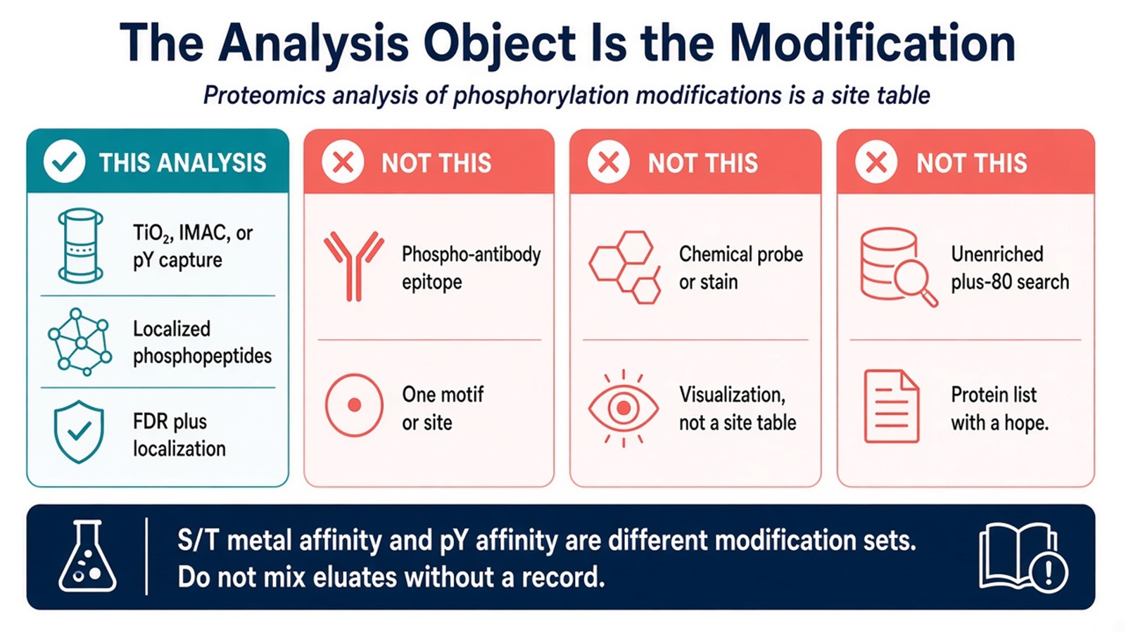
Figure 1. The analysis object is the captured, localized phosphorylation; epitopes, probes, and plus-80 search options answer different sentences.
Biological Interpretation Beyond Phosphosite Identification
1. Signaling-network language
Quantitative phosphoproteomics helps researchers identify phosphorylation changes associated with cellular states and treatments. Large-scale identification can list phosphorylation modifications present under a cellular state. Comparing those modifications across states can list which ones changed. Pathway annotation can organize proteins carrying regulated phosphosites into biological processes, while functional experiments are needed to establish how those changes affect signaling or disease-associated phenotypes.
2. Biopharmaceutical quality and efficacy language
New targets and drug-development strategies can motivate the analysis. They are not default outputs of the modification table. Quality control and efficacy evaluation of a manufactured biologic are a different analysis object: product-centered peptide mapping and intact-mass checks on a defined drug substance, under a characterization plan. A research phosphoproteome of a signaling lysate does not become biopharmaceutical quality control because the title named phosphorylation modifications.
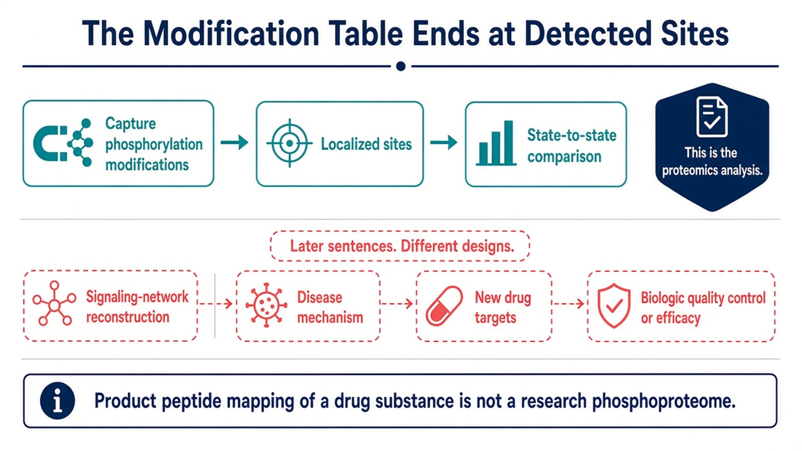
Figure 2. State-to-state modification tables can be compared; reconstructed networks and biologic QC are different analyses.
Factors That Decide Whether the Work Is This Analysis
Planned sentence |
Analysis that can support it |
Avoid interpreting as proteomics analysis of the modifications |
|---|---|---|
Which phosphorylation modifications are present across many proteins |
Phosphate-enriched discovery LC-MS/MS |
An unenriched proteome with phosphorylation as a search option |
Did the same modification change |
Matched quantitative phosphoproteomics |
Protein-group fold change |
Which pY modifications are present |
pY affinity capture plus LC-MS/MS |
A TiO2-only table read as complete phosphorylation |
Is this epitope present |
Phospho-specific antibody |
A proteome of modifications |
Where is phosphate in the cell |
Imaging or a validated probe |
A site table |
Did a signaling network switch |
Modification table plus a cascade readout |
KEGG on the site list |
Does a biologic meet a characterization spec |
Product peptide mapping / intact mass |
A research phosphoproteome |
Phosphatase inhibitors and rapid lysis protect the modifications that any of those analyses depend on. Residual EDTA poisons IMAC and is a sample-handling limit of the metal-affinity analysis, not a signaling finding. Probe work has its own permeability and specificity limits. Antibody work has lot and phosphatase-control limits.
Experimental Design for Phosphoproteomics
Define the object as modifications before the lysate is split. Presence sentences go to class-enriched discovery with a declared S/T or pY capture. Change sentences add a quantitative method and biological replicates. Single-protein modification sentences leave proteome scale. Epitope and probe sentences stay out of the proteomics-analysis claim.
Avoid combining mass spectrometry, immunodetection, and chemical probes as three equivalent phosphorylation-modification detection methods that together constitute proteomics analysis. Mass spectrometry of captured peptides is the proteomics analysis. The other two detect different objects. Do not add a longer signaling-network or quality-control paragraph as a substitute for naming which modifications will be reported.
Low-input pellets should be evaluated before an S/T map and a pY map are both promised. Technical repeats of one enrichment do not replace independent cultures when the analysis will be read as a change of modifications. Match-between-runs can fill identifications and can also move an incorrect modification if tolerances are too open. Record peptide FDR and localization thresholds with the table that will be cited as the analysis.
Interpreting Modification-Spectra Language
A localized phosphopeptide table supports claims about detected phosphorylation under the capture, search, and localization rules used. Calling that table a revealed signaling network, a disease-mechanism map, a new-target engine, or a quality-control record for a biologic restates the subtitle and overstates the analysis. Absence of a modification means the pipeline did not report a usable match. Failed capture, thin fragments, and a pY-only or S/T-only resin can hide modifications that remain in the cell.
Completeness language such as the phosphorylation-modification spectrum of the cell overstates one enriched injection (Olsen and Mann 2013). Clinical diagnostic cutoffs sit outside ordinary research proteomics analysis of phosphorylation modifications unless a separately validated assay framework is in place.
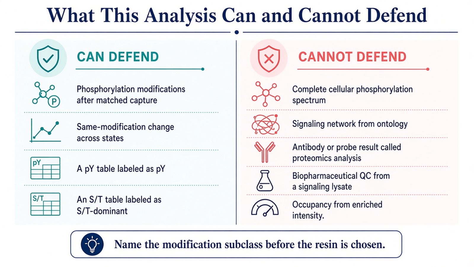
Figure 3. Captured phosphorylation modifications can be defended; reconstructed networks, probe images as proteomics, completeness, and biologic QC cannot.
Frequently Asked Questions
1. Is an expression proteome that lists known phosphoproteins an analysis of phosphorylation modifications?
It reports protein groups. Analysis of the modifications requires captured phosphopeptides and a localization rule.
2. Can a phospho-antibody or a chemical probe replace this proteomics analysis?
They can detect an epitope or visualize phosphate-rich species. They do not produce a proteome-scale modification table.
3. Does phosphotyrosine affinity purification cover phosphorylation modifications in general?
It covers a pY-biased set. Most cellular phosphorylation sits on serine and threonine and needs metal-affinity capture.
4. Does comparing modification tables across cell states reveal a signaling network?
It reveals which modifications were detected or changed. Network mechanism still needs a cascade or functional readout.
5. Can this analysis be used for quality control of a biopharmaceutical?
Product characterization of a defined drug substance is a different design. A research phosphoproteome of a signaling lysate is not that record.
MtoZ Biolabs can provide proteomics analysis of phosphorylation modifications from phosphate-selective enrichment through LC-MS/MS site mapping, including guidance on whether an S/T map, a pY fraction, or a single-protein modification map is the object the title actually names when project samples are available.
If further evaluation of modification-table versus epitope, probe, or product-characterization design is needed, project details can be submitted for a feasibility review.
References
- M. Mann, O.N. Jensen (2003). Proteomic analysis of post-translational modifications. Nat. Biotechnol., 21, 255-261. https://doi.org/10.1038/nbt0303-255
- J.V. Olsen, M. Mann (2013). Status of large-scale analysis of post-translational modifications by mass spectrometry. Mol. Cell. Proteomics, 12, 3444-3452. https://doi.org/10.1074/mcp.O113.034181
- S.A. Beausoleil, J. Villen, S.A. Gerber, J. Rush, S.P. Gygi (2006). A probability-based approach for high-throughput protein phosphorylation analysis and site localization. Nat. Biotechnol., 24, 1285-1292. https://doi.org/10.1038/nbt1240
- T.E. Thingholm, T.J. Jorgensen, O.N. Jensen, M.R. Larsen (2006). Highly selective enrichment of phosphorylated peptides using titanium dioxide. Nat. Protoc., 1, 1929-1935. https://doi.org/10.1038/nprot.2006.185
- J. Rush, A. Moritz, K.A. Lee, A. Guo, V.L. Goss, S.P. Spek, H. Zhang, X.M. Zha, R.D. Polakiewicz, M.J. Comb (2005). Immunoaffinity profiling of tyrosine phosphorylation in cancer cells. Nat. Biotechnol., 23, 94-101. https://doi.org/10.1038/nbt1046
