



PRM mass spectrometry versus ELISA is a protein quantification route choice between peptide-level liquid chromatography-tandem mass spectrometry (LC-MS/MS) and antibody-epitope immunoassay. Parallel reaction monitoring (PRM) isolates scheduled peptide precursors, records high-resolution product-ion spectra, and integrates selected fragment chromatograms on a closed panel (Peterson et al. 2012; Gallien et al. 2012). Enzyme-linked immunosorbent assay (ELISA) quantifies target protein through specific antibody binding and an enzymatic or related readout. The guide below compares the two routes for project selection with workflow, locked parameters, limits, and fit. It is not a price contest, not a claim that PRM is always more accurate than ELISA, and not a disease or drug-development brochure.
Both routes can support relative change across frozen groups when prep and contrast stay matched. Amount claims need different calibration designs: stable isotope-labeled internal standard (SIS) peptides and a matrix curve for PRM when amount is declared (Gerber et al. 2003), versus a protein or recombinant standard curve for many ELISA formats. Peptide uniqueness and epitope availability are separate feasibility gates (Bantscheff et al. 2012).
In MtoZ Biolabs targeted offerings, PRM methods are scoped when the peptide panel is closed and high-resolution scheduling fits the cohort block. Service intake appears in Parallel Reaction Monitoring (PRM) Service.
What the PRM-MS Versus ELISA Decision Covers
Four decision layers belong in the brief and stay separated.
Analyte layer asks whether the report unit is a proteotypic peptide surrogate or an antibody-defined epitope. Shared peptides, isoforms, and PTM sites can break one mapping while the other still works.
Reagent layer asks whether a validated antibody and linear range already exist, or whether proteotypic peptides can be scheduled without antibody generation.
Panel layer asks how many named targets must be remeasured in one study block. PRM schedules closed peptide lists on LC-MS/MS. ELISA multiplex depends on antibody compatibility and assay format, not on peptide cycle time.
Claim layer states relative change versus calibrated amount, and whether clinical cutoff language is in scope. Neither route creates a diagnostic test from a single exploratory table.
Choose PRM-MS versus ELISA by analyte unit, reagent status, and claim design, not by sensitivity slogans alone.
PRM-MS and ELISA Compared on Project Dimensions
Comparison should use decision dimensions, not platform labels alone.
Dimension |
PRM mass spectrometry |
ELISA |
|---|---|---|
Detection unit |
Scheduled peptide precursor and product ions |
Antibody-bound protein epitope |
Typical strength |
Antibody-independent peptide panels; fragment review after acquisition |
Established epitope assay when antibody and curve already work |
Multiplex limit |
Cycle time versus chromatographic peak width |
Antibody cross-talk and format capacity |
Absolute amount |
SIS peptides and matrix curve when declared |
Protein or recombinant standard curve when declared |
Common bottleneck |
Peptide uniqueness, digest parity, matrix interference |
Antibody availability, specificity, and dynamic range |
Intact-protein claim |
Bottom-up surrogate unless intact design is separate |
Epitope readout; may not equal total protein isoforms |
PRM is not multiple reaction monitoring (MRM). MRM and selected reaction monitoring (SRM) pass predefined transitions on a triple quadrupole (Lange et al. 2008). PRM stores a high-resolution product-ion spectrum. Route choice between PRM and MRM after matrix pilot appears in Parallel Reaction Monitoring vs MRM: Which Fits?.
ELISA ease of use does not remove antibody characterization work. PRM instrument complexity does not remove peptide selection or matched pretreatment.
End-to-End Decision and Method Workflow
A practical comparison spine usually runs as follows.
- Freeze the reporting sentence: relative change, calibrated amount, epitope claim, or peptide-surrogate claim.
- List named targets and whether validated antibodies already exist for each.
- For antibody-ready targets, pilot ELISA linearity and specificity in the study matrix when that route is preferred.
- For antibody-limited or multiplex peptide targets, close proteotypic peptides and map each to its protein or site.
- Match extraction or sample handling across contrast arms for the chosen route.
- For PRM, lock inclusion lists, collision energy, resolution, and cycle time on matrix-matched digests; for ELISA, lock antibody lots, dilution series, and plate controls.
- Size biological n plus quality control (QC) appropriate to the route.
- Acquire study and QC measurements; review interference, standard-curve fit, or transition and fragment rules as declared.
- Test only the predefined contrast; archive method versions, reagent lots or panel versions, and software with deliverables.
Steps 1 through 3 decide the route. Steps 4 through 9 produce the quantitative record on the chosen method.
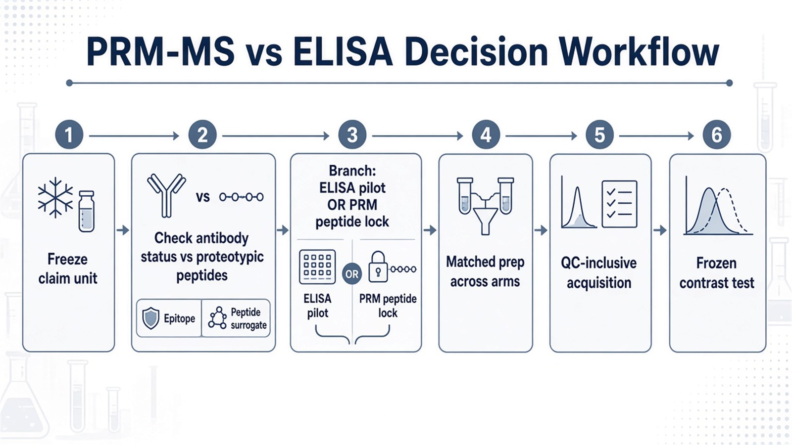
Figure 1. Route choice sits on analyte unit and reagent status; after lock, PRM-MS and ELISA follow separate method spines under the same contrast discipline.
Parameters That Must Be Locked for Each Route
Layer |
PRM-MS parameter to declare |
ELISA parameter to declare |
|---|---|---|
Target map |
Peptide-to-protein or site version |
Epitope and antibody clone or lot |
Calibration |
SIS and curve if amount claimed |
Standard material and curve if amount claimed |
Prep |
Matched digest and load |
Matched sample handling and dilution |
Selectivity QC |
Fragment chromatogram interference review |
Cross-reactivity and spike-recovery notes |
Schedule or plate |
Cycle time versus peak width |
Plate map, replicates, and control wells |
Contrast |
Frozen before peak or OD review |
Frozen before OD review |
Deliverable |
Transition or fragment table, method file |
Curve, OD table, reagent lots |
Limits follow the same table. PRM does not automatically outperform ELISA in every matrix. ELISA does not automatically scale to large closed peptide panels without compatible antibodies. Internal standards improve PRM amount claims only when declared; they are not required for every relative PRM contrast. Peptide-level PRM framing for surrogate readout appears in Targeted Protein Quantification: Achieving Precision at the Peptide Level through PRM Technology.
How to Choose PRM-MS Versus ELISA
Use analyte unit, reagent status, panel size, and claim unit together.
Project need |
Route that often fits |
Recheck before committing |
|---|---|---|
Validated antibody and curve already work |
ELISA |
Force PRM without peptide need |
No antibody; proteotypic peptide available |
PRM-MS |
Assume any peptide equals the epitope |
Multiplex closed peptide panel |
PRM-MS or hybrid with ELISA on key epitopes |
One ELISA kit as full panel |
Amount on named peptides |
PRM with SIS and curve |
Relative PRM peaks as concentration |
Amount on antibody epitope |
ELISA with declared standard |
Peptide PRM as epitope amount |
Isoform or PTM site on peptide sequence |
PRM on modified or unique peptide |
Total-protein ELISA as site amount |
Intact-protein or native complex claim |
Separate design; not peptide PRM alone |
Bottom-up surrogate as intact amount |
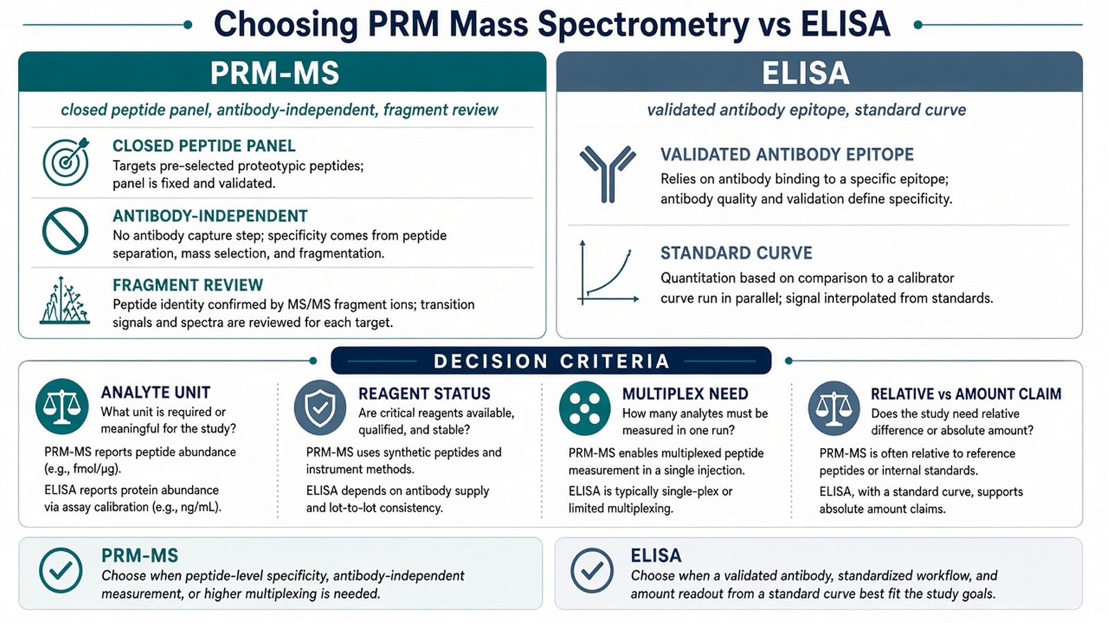
Figure 2. Choose ELISA when epitope reagents already answer the claim; choose PRM-MS when closed peptide surrogates fit better than antibody constraints.
Teams with target lists, antibody status, peptide drafts, and contrast maps can review MtoZ Biolabs Parallel Reaction Monitoring (PRM) Service when PRM is declared in the project record.
Frequently Asked Questions
1. Is PRM mass spectrometry always more sensitive than ELISA?
No. Sensitivity depends on matrix, peptide or epitope behavior, and method lock. Matrix-matched pilots decide which route meets the declared claim.
2. Does PRM require internal standards for every study?
No. Relative contrasts can run light-only when that claim is declared. SIS and a curve become necessary when peptide amount units are required.
3. Can ELISA and PRM report the same biological change?
Sometimes they agree on direction, but peptide surrogates and antibody epitopes are different analytes. Bridging studies are needed before treating the two tables as interchangeable.
4. Can one PRM method replace a multiplex ELISA panel?
Only when proteotypic peptides exist for each target and cycle time still samples peaks adequately. Antibody-defined epitopes without peptide surrogates stay on immunoassay routes.
5. When should projects keep both routes?
When some targets have strong antibodies and others need peptide-level or PTM-site readout, a hybrid design can assign each target to the matching method under one contrast map.
MtoZ Biolabs can discuss PRM versus ELISA scoping when target map, reagent status, and claim unit are documented.
If further evaluation of PRM versus MRM, SIS calibration, or peptide-versus-epitope bridging is needed, project details can be submitted for a feasibility review.
Reference
- A.C. Peterson, J.D. Russell, A.A. Klammer, P.F. Doubleday, H. Liu, J.N. Anderson, M.J. MacCoss (2012). Parallel reaction monitoring for high resolution and high mass accuracy quantitative, targeted proteomics. Mol. Cell. Proteomics, 11, 1475-1488. https://doi.org/10.1074/mcp.O112.020271
- S. Gallien, E. Duriez, A. Crone, M. Kellmann, T. Moehring, T. Domon (2012). Targeted proteomic quantification on quadrupole-orbitrap mass spectrometers. Mol. Cell. Proteomics, 11, 1709-1723. https://doi.org/10.1074/mcp.O112.019232
- V. Lange, P. Picotti, B. Domon, R. Aebersold (2008). Selected reaction monitoring for quantitative proteomics: a tutorial. Mol. Syst. Biol., 4, 222. https://doi.org/10.1038/msb.2008.61
- S.A. Gerber, J. Rush, O. Stemman, M.W. Kirschner, S.P. Gygi (2003). Absolute quantification of proteins and phosphoproteins from cell lysates by tandem MS. Proc. Natl. Acad. Sci. U.S.A., 100, 6940-6945. https://doi.org/10.1073/pnas.0832254100
- M. Bantscheff, S. Lemeer, H.U. Haab, B. Kuster (2012). Quantitative mass spectrometry in proteomics: a critical review. Anal. Bioanal. Chem., 404, 939-965. https://doi.org/10.1007/s00216-012-5892-4
